Knee X-Ray and MRI
1/34
There's no tags or description
Looks like no tags are added yet.
Name | Mastery | Learn | Test | Matching | Spaced | Call with Kai |
|---|
No analytics yet
Send a link to your students to track their progress
35 Terms
Grade I = minimal osteophytes w/ no problems w/ space or cartilage
Define the Kelgren Lawrence grade. How do you know?
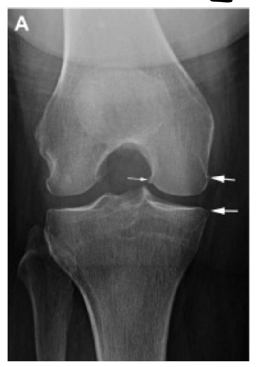
Grade II = 1 definite osteophyte w/ NO space narrowing
Define the Kelgren Lawrence grade. How do you know?
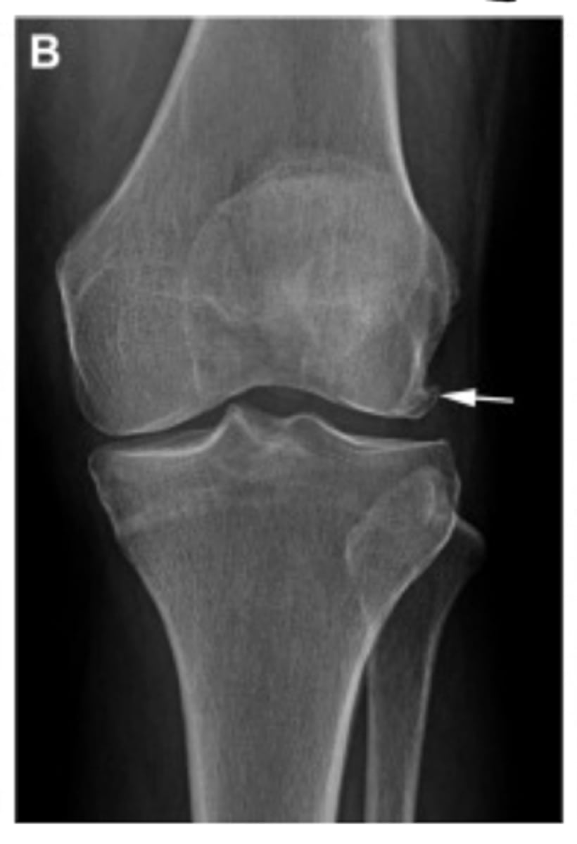
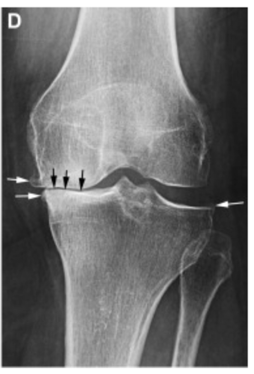
Grade III = marginal osteophytes w/ space narrowing + sclerosis & multiple osteophytes
Define the Kelgren Lawrence grade. How do you know?
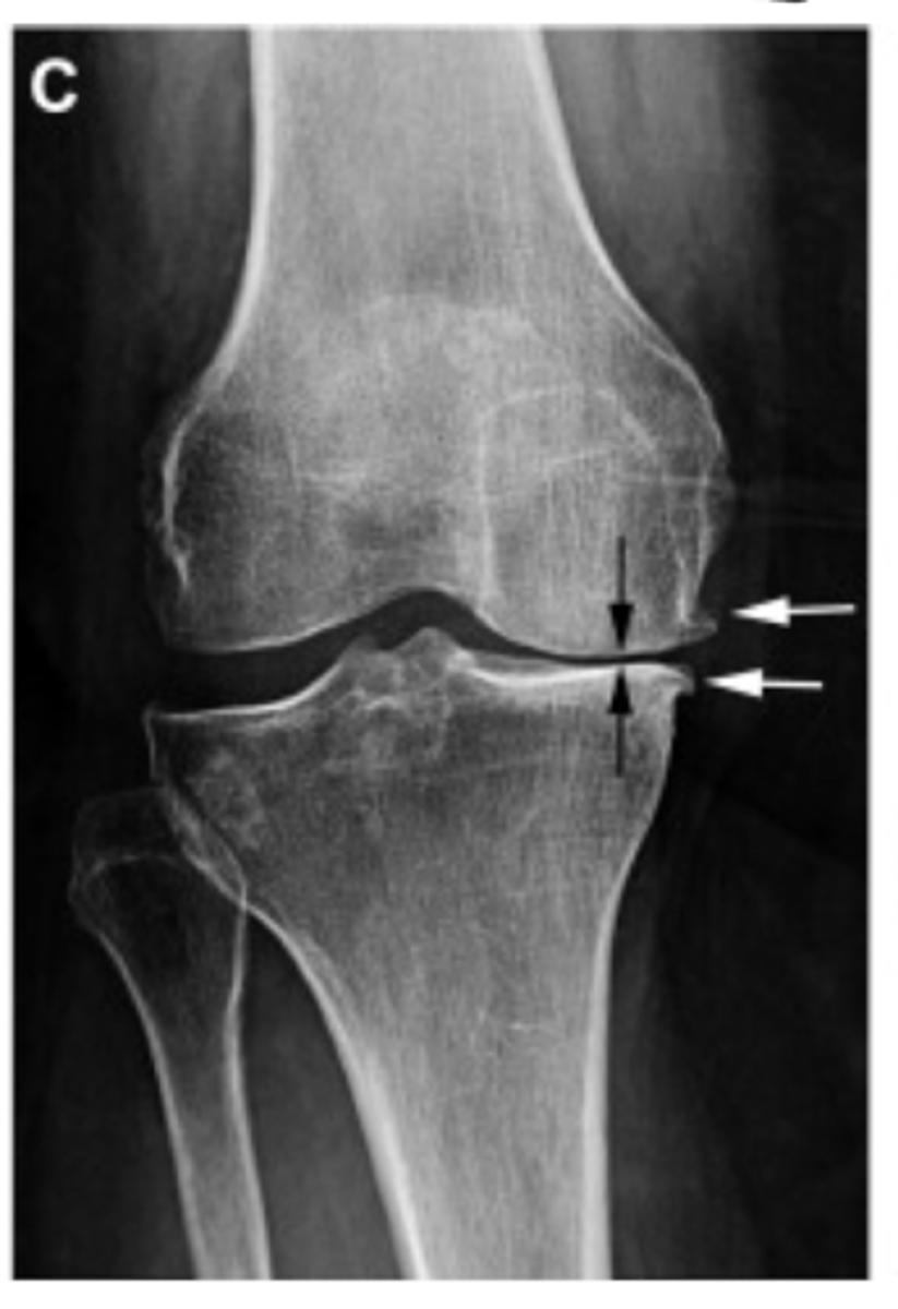
Grade IV = obliteration of joint space (bone on bone)
Define the Kelgren Lawrence grade. How do you know?
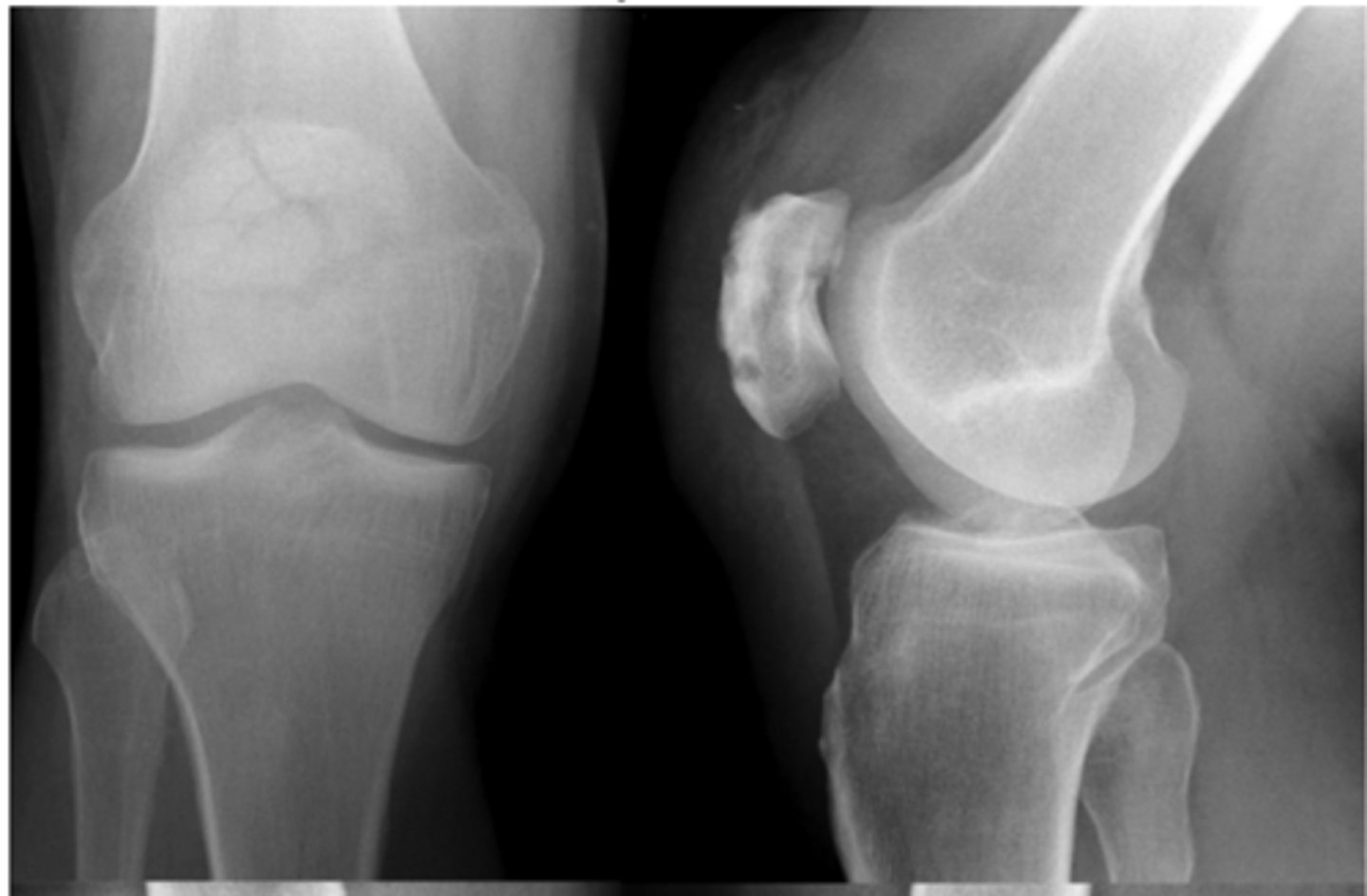
Comminuted patellar fracture
Define the pathology.
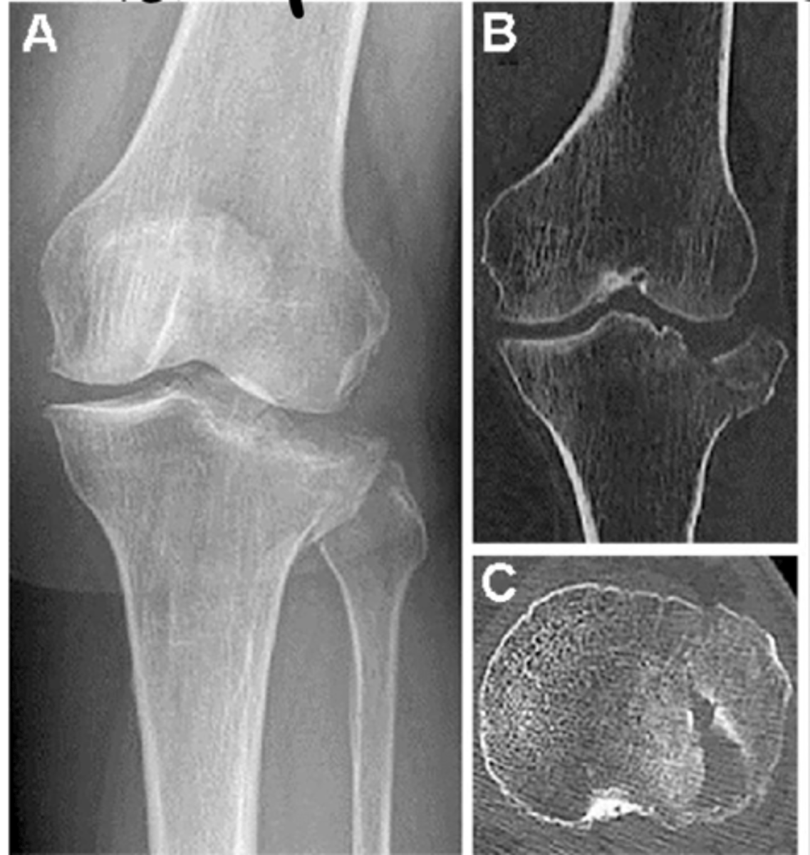
Tibial plateau fractures
Define the pathology.
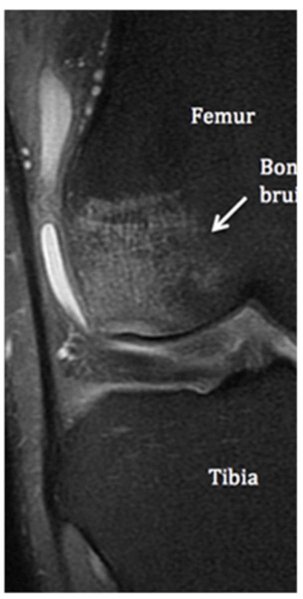
Sub-chondral edema/bone bruise (HUGE influence on rehab!!)
Define the pathology.
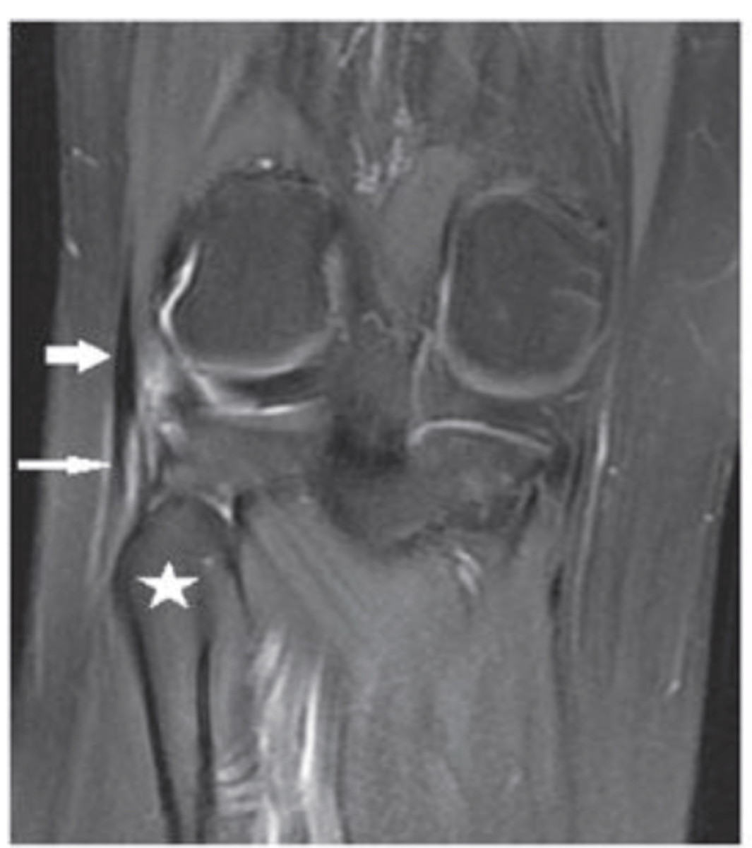
Synovitis surrounding the joint
Define the pathology.
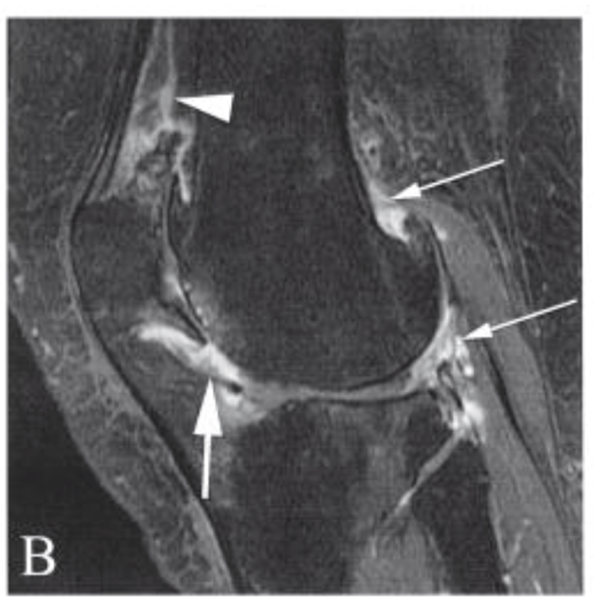
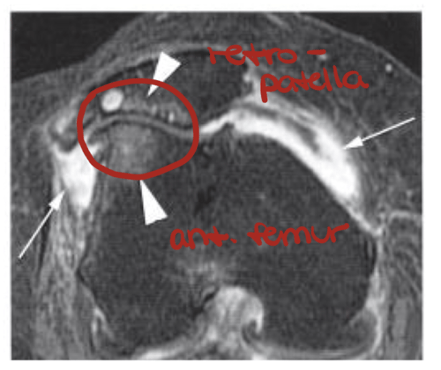
Bone edema/bone bruise
Define the pathology.
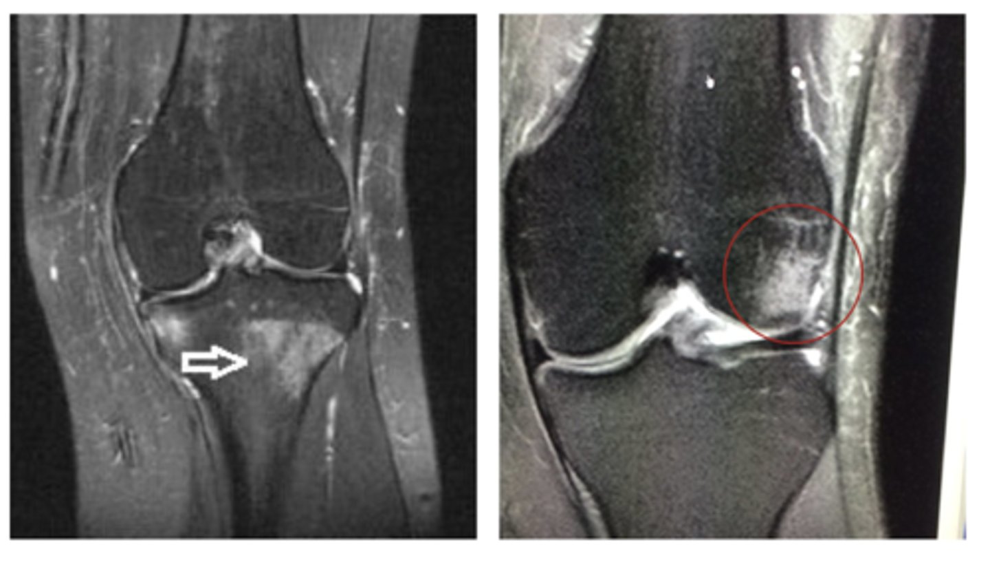
Bone edema/bone bruise
Define the pathology.
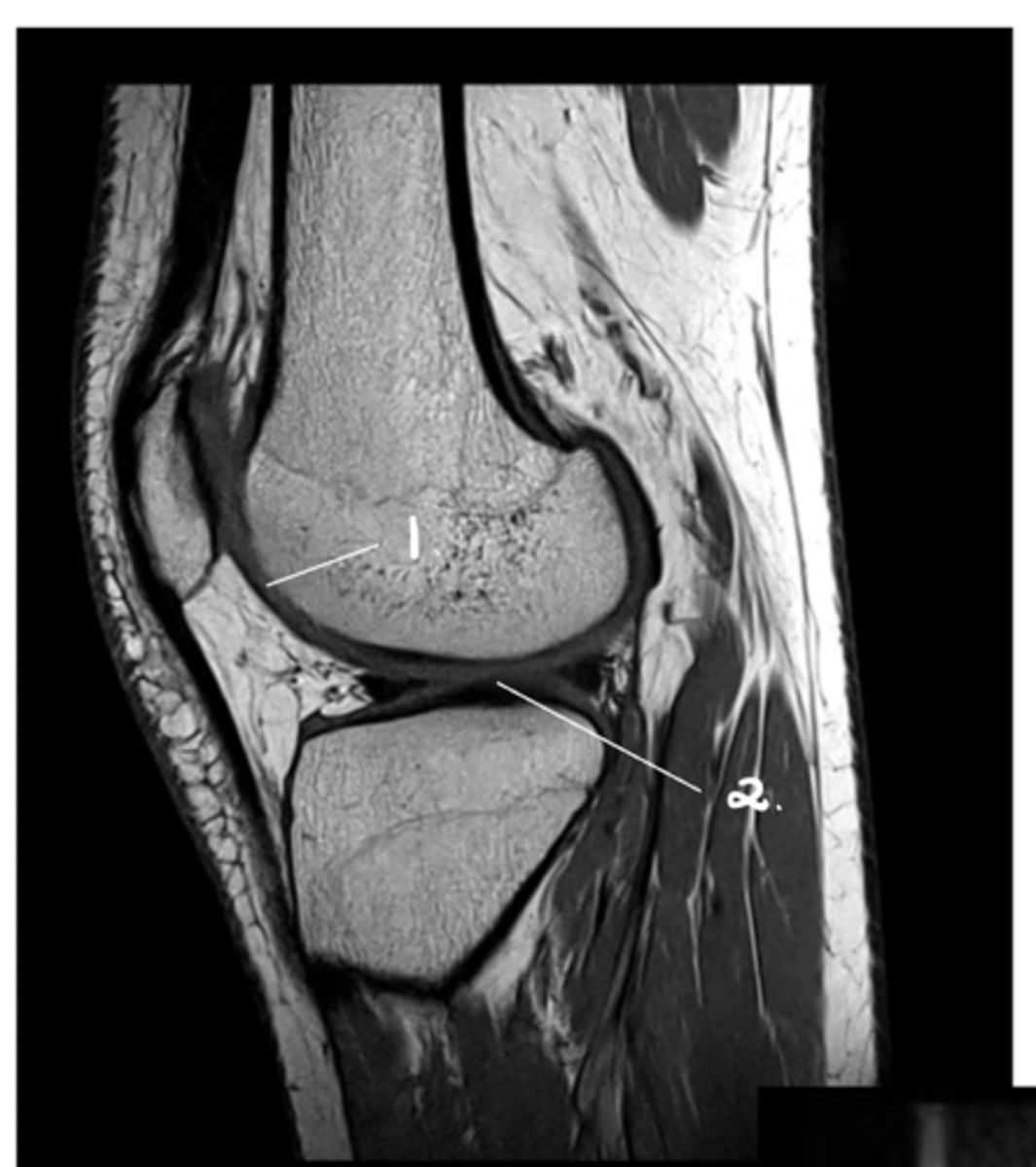
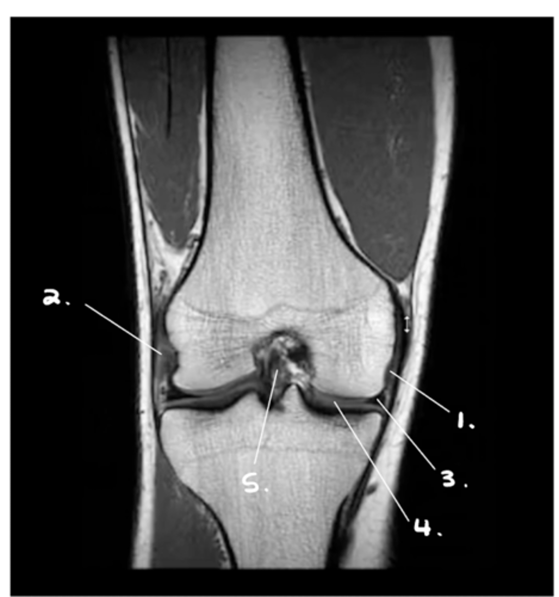
1. Hyaline cartilage (all of the grey)
2. Meniscus (looks kind of like a thickening of the cartilage from this angle)
Label 1 & 2.
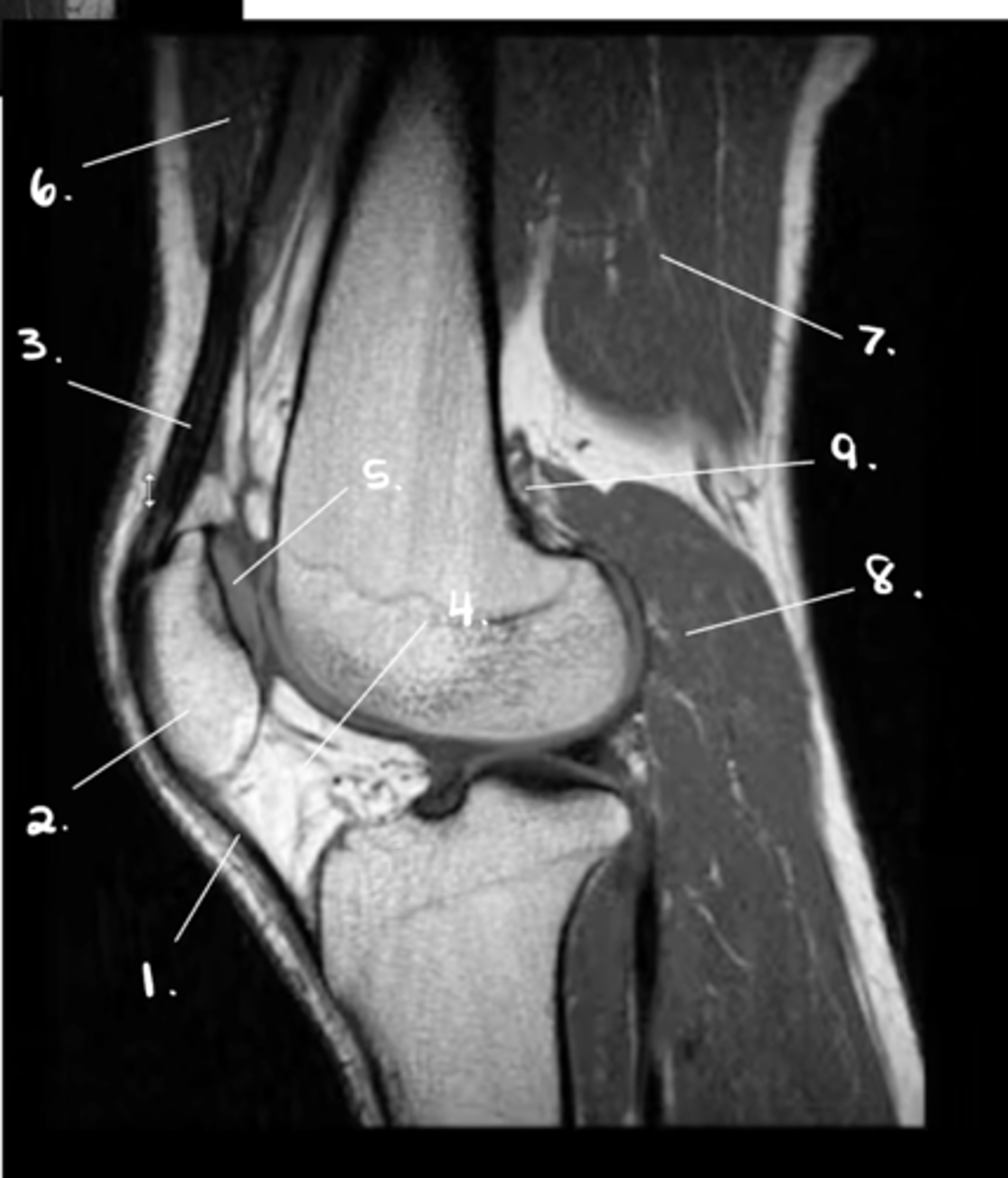
1. Patellar tendon
2. Patella
3. Quadriceps tendon
4. Hoffa's fat pad
5. Retro-surface cartilage of patella
6. Quadriceps
7. Hamstrings
8. Gastroc.
9. Poplitus
Label 1-9.
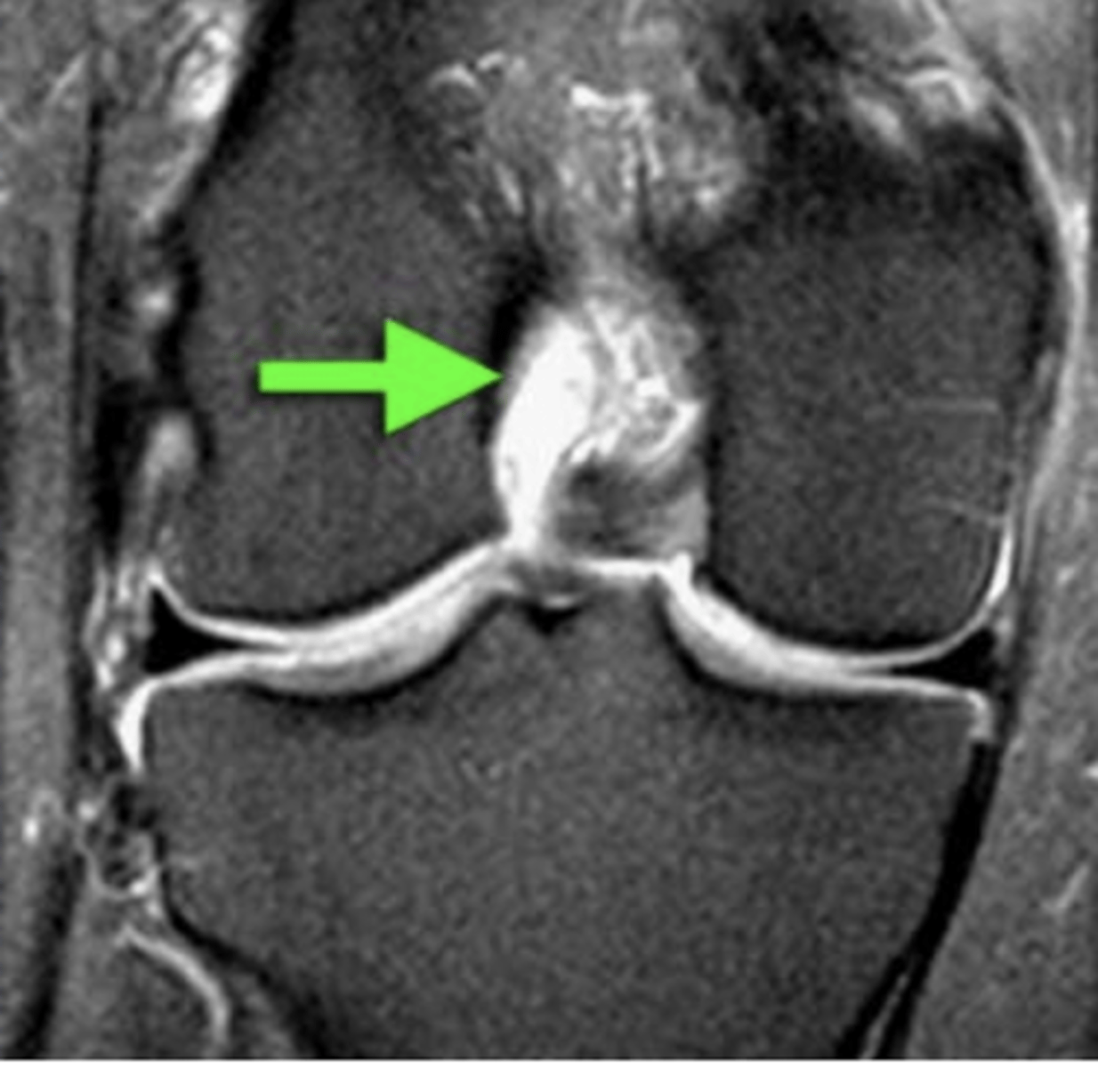
ACL (always goes inferior --> superior in a posterior direction)
Which ligament can you see in this sagittal view?
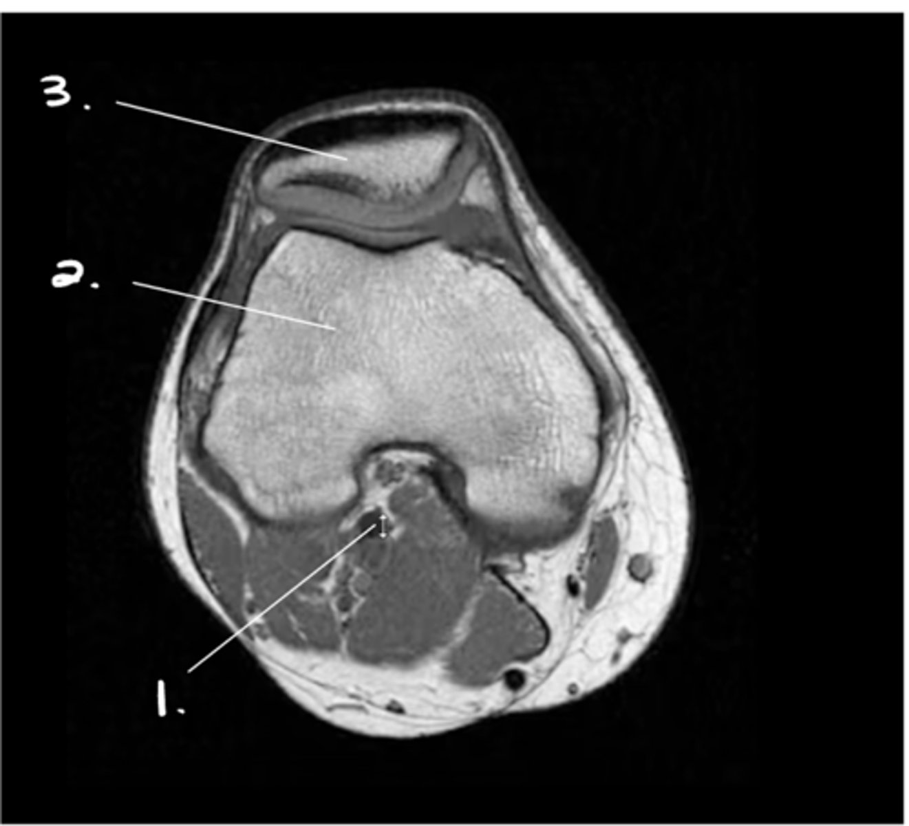
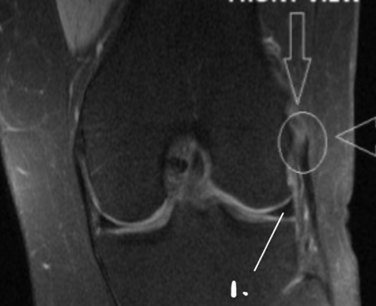
1. Popliteal artery
2. Femur
3. Patella
Label 1-3.
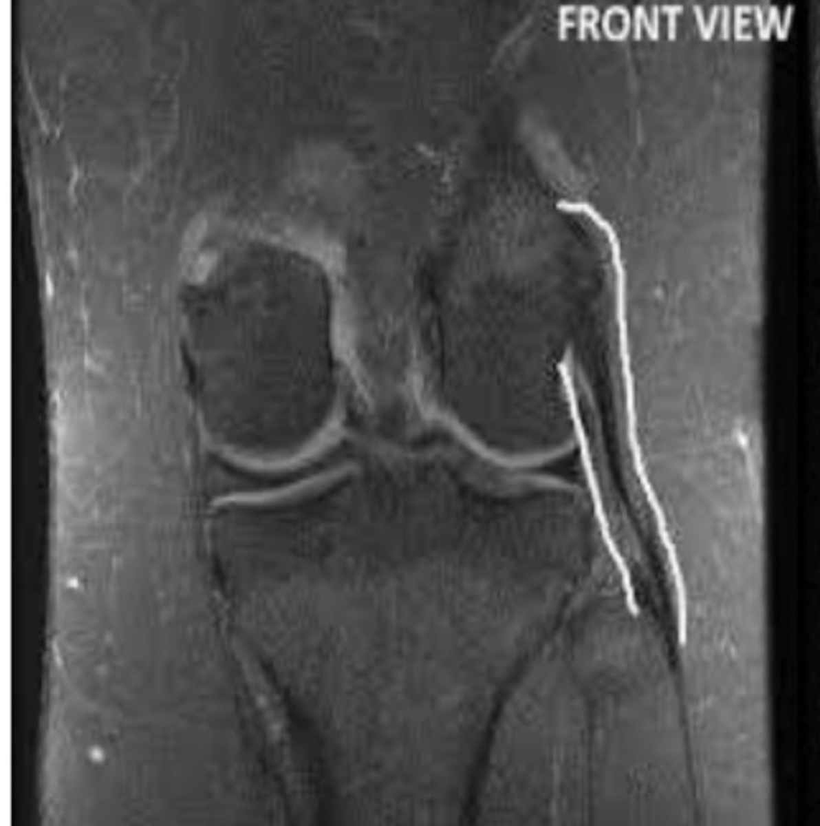
Medial meniscus
Label this structure.
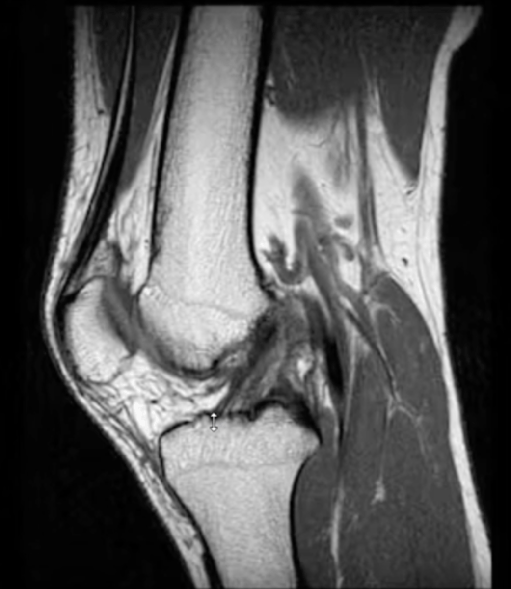
1. MCL
2. LCL
3. Medial meniscus
4. Hyaline cartilage of tibia & femur
5. ACL
Label 1-5.
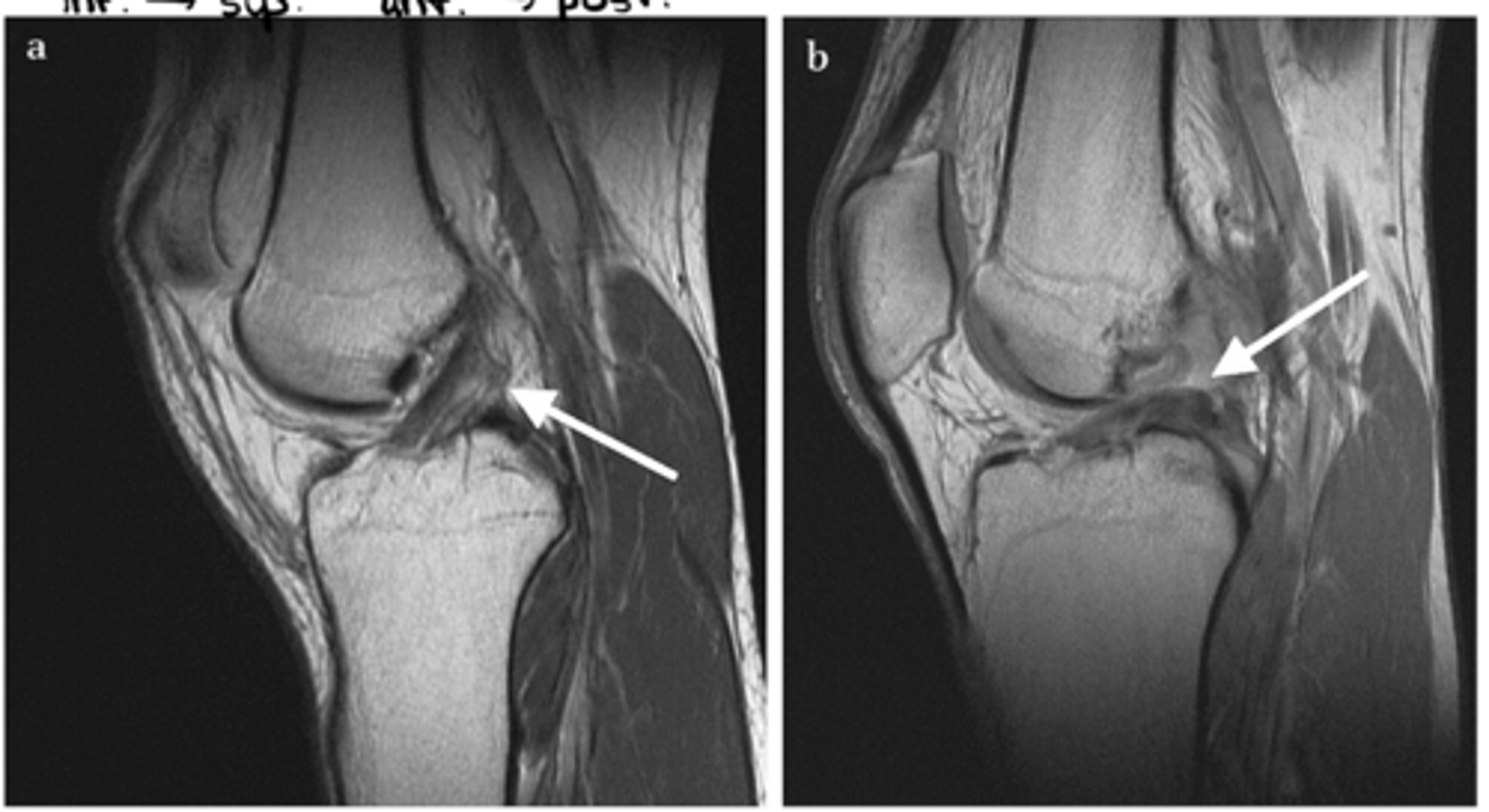
A = healthy ACL running inferior & anterior to superior & posterior
B = fully torn ACL (since the ligament is laying down flat on the tibia, we know that it is a full tear...if it were a partial tear, it would just appear wavy)
What is the difference between image a and b?
Complete tear of the ACL (right down the middle)
Define the pathology.
Complete tear of the ACL
Define the pathology.
Tear of the ACL
Define the pathology.
Tear of the ACL
Define the pathology.
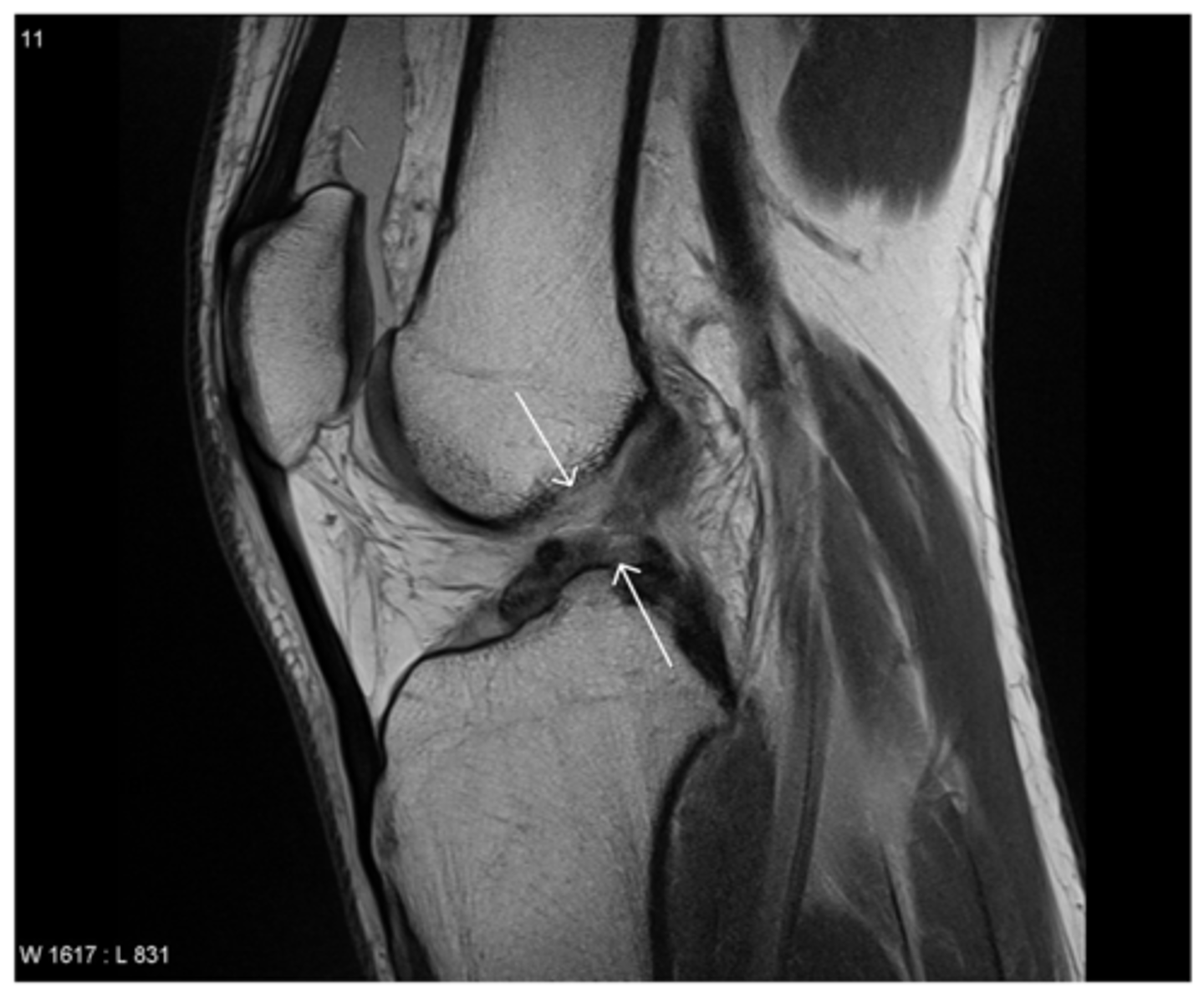
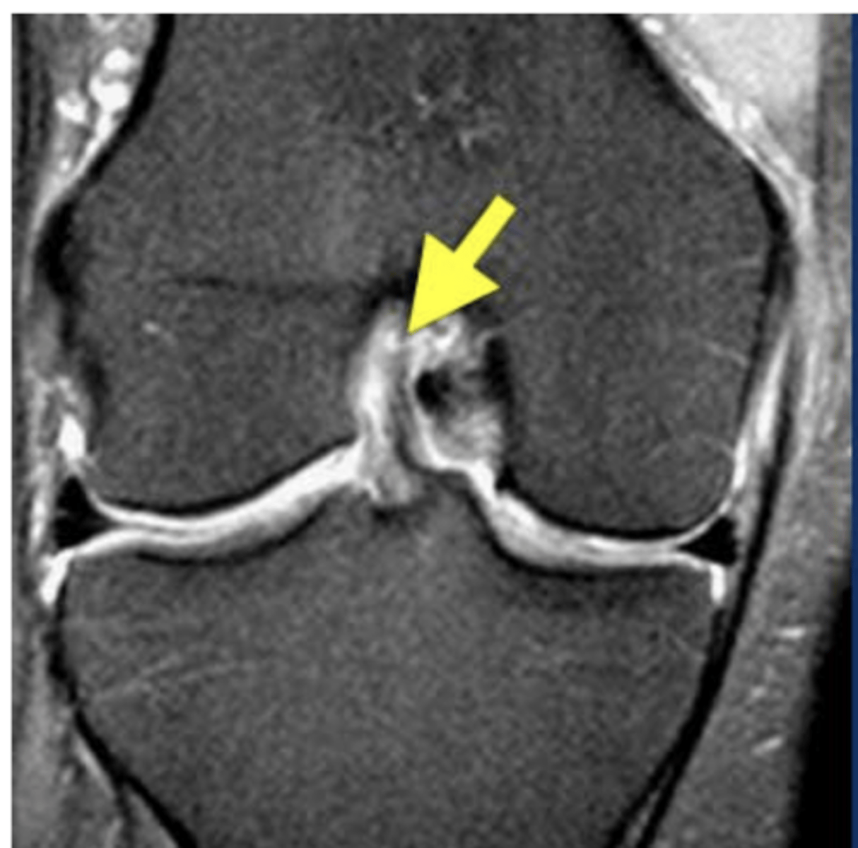
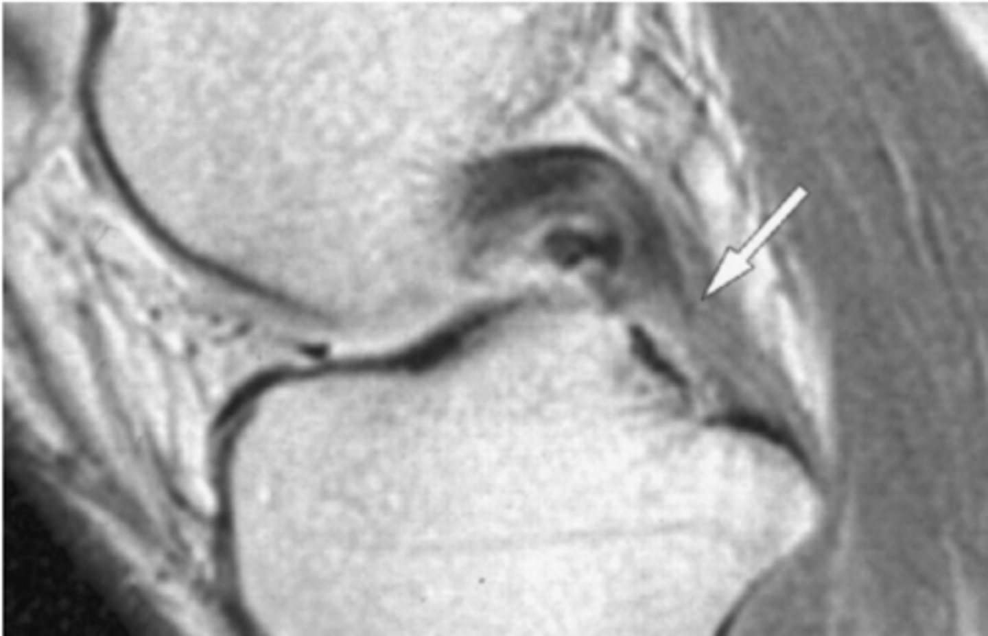
PCL (runs from posterior & inferior --> to anterior & superior)
Which structure is the white arrow pointing too?
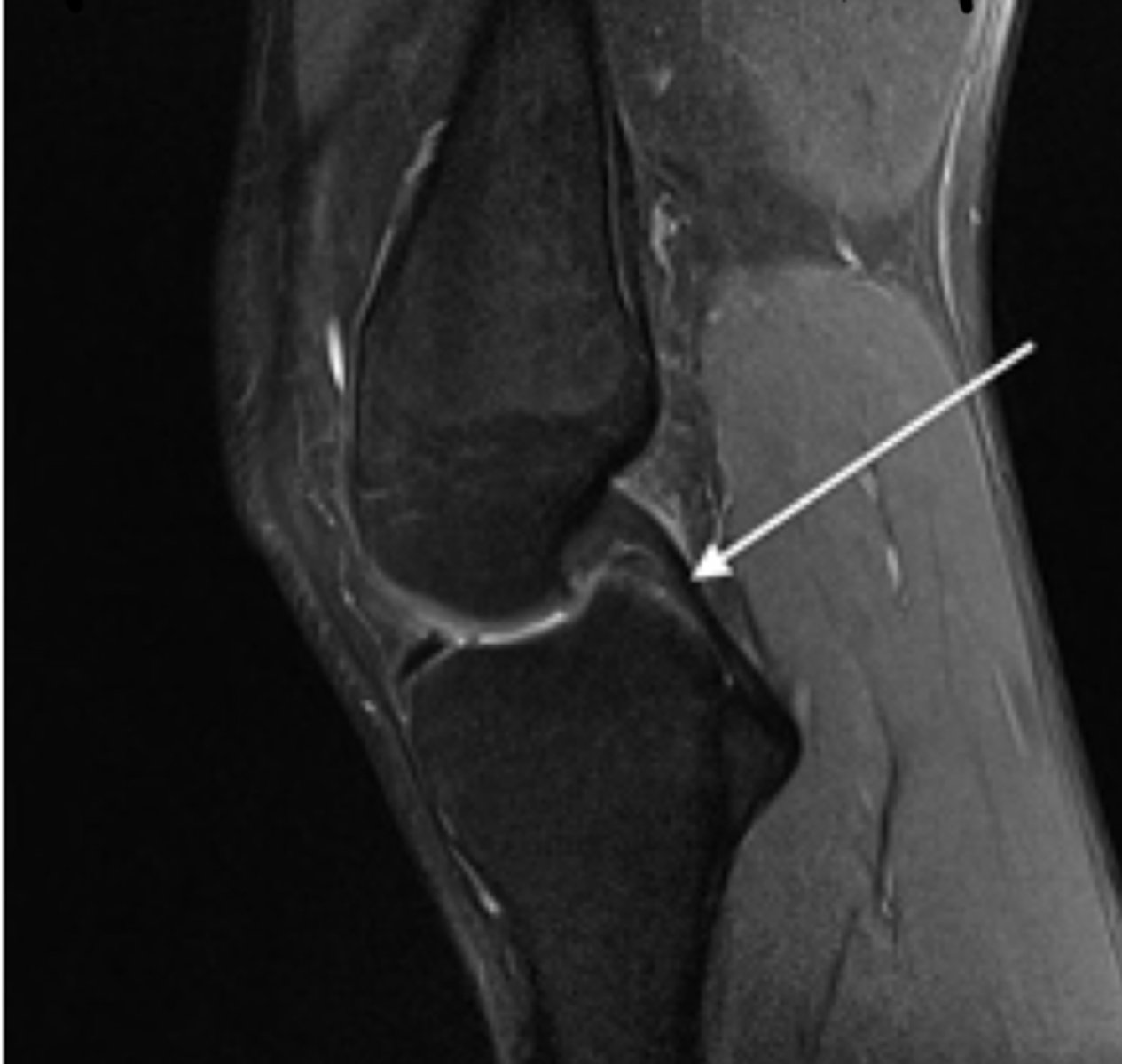
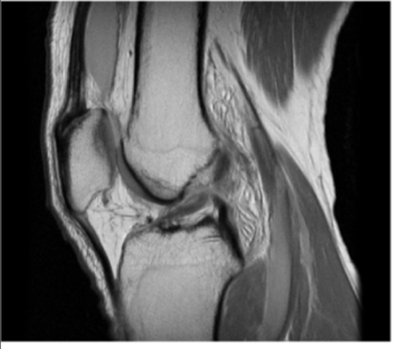
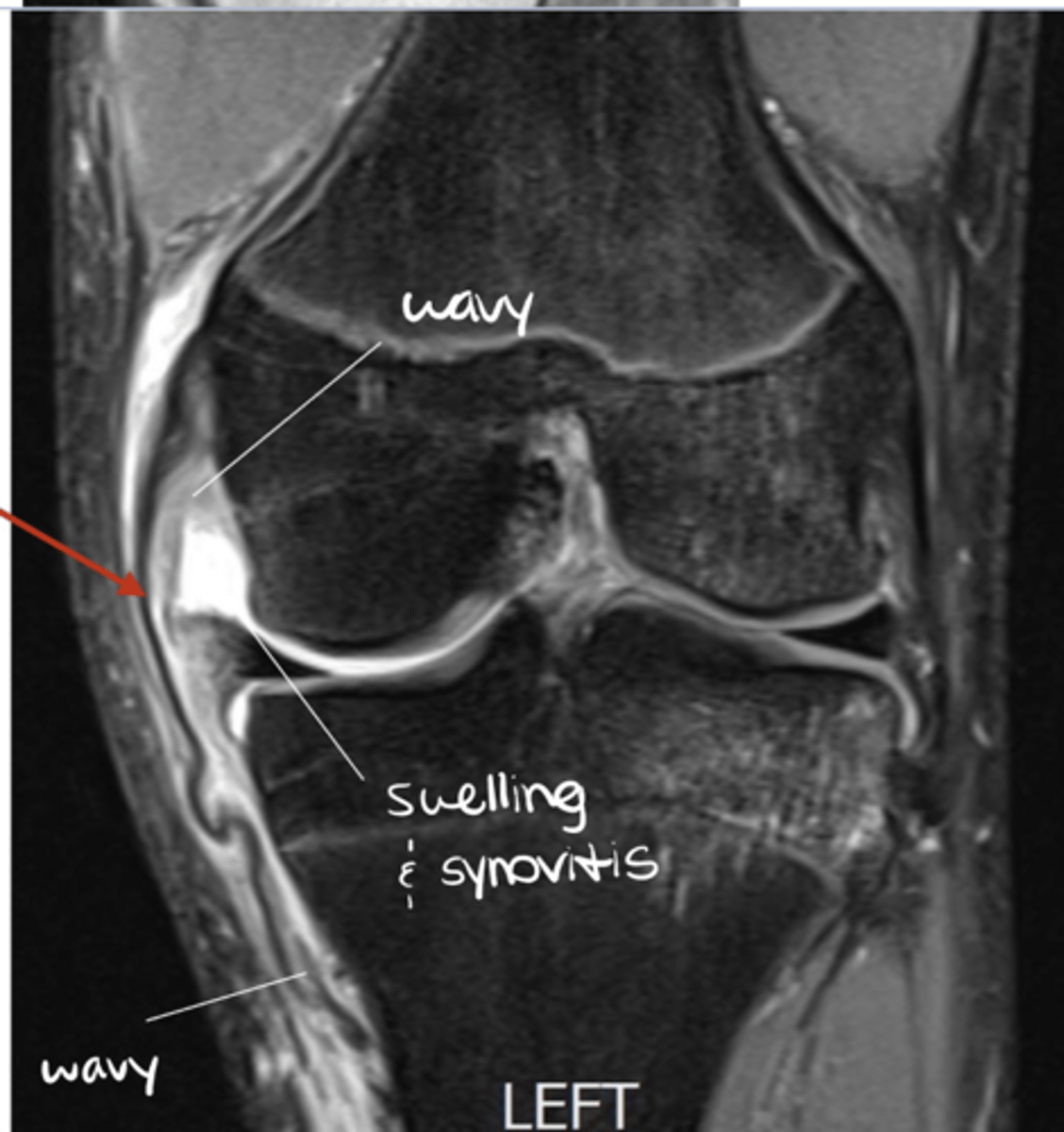
Tear of the PCL -- normally, ligaments are pulled taut and appear straight; if they are torn, they appear wavy and/or curved over
Define the pathology. How do you know?
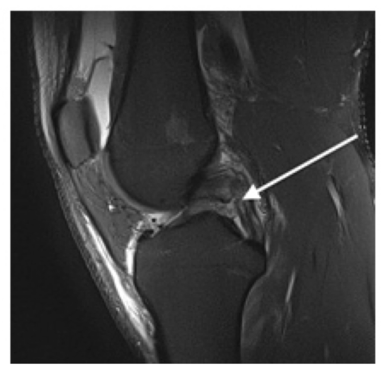
Tear of the PCL -- when ligaments curve like this and/or get wavey, it is a dead give-a-way that a tear is present
Define the pathology.
Grade III MCL tear
Define the pathology.
GOTCHA!! This is a NORMAL LCL!!
Define the pathology.
Torn LCL
Define the pathology. (#1 is just pointing to the meniscus)
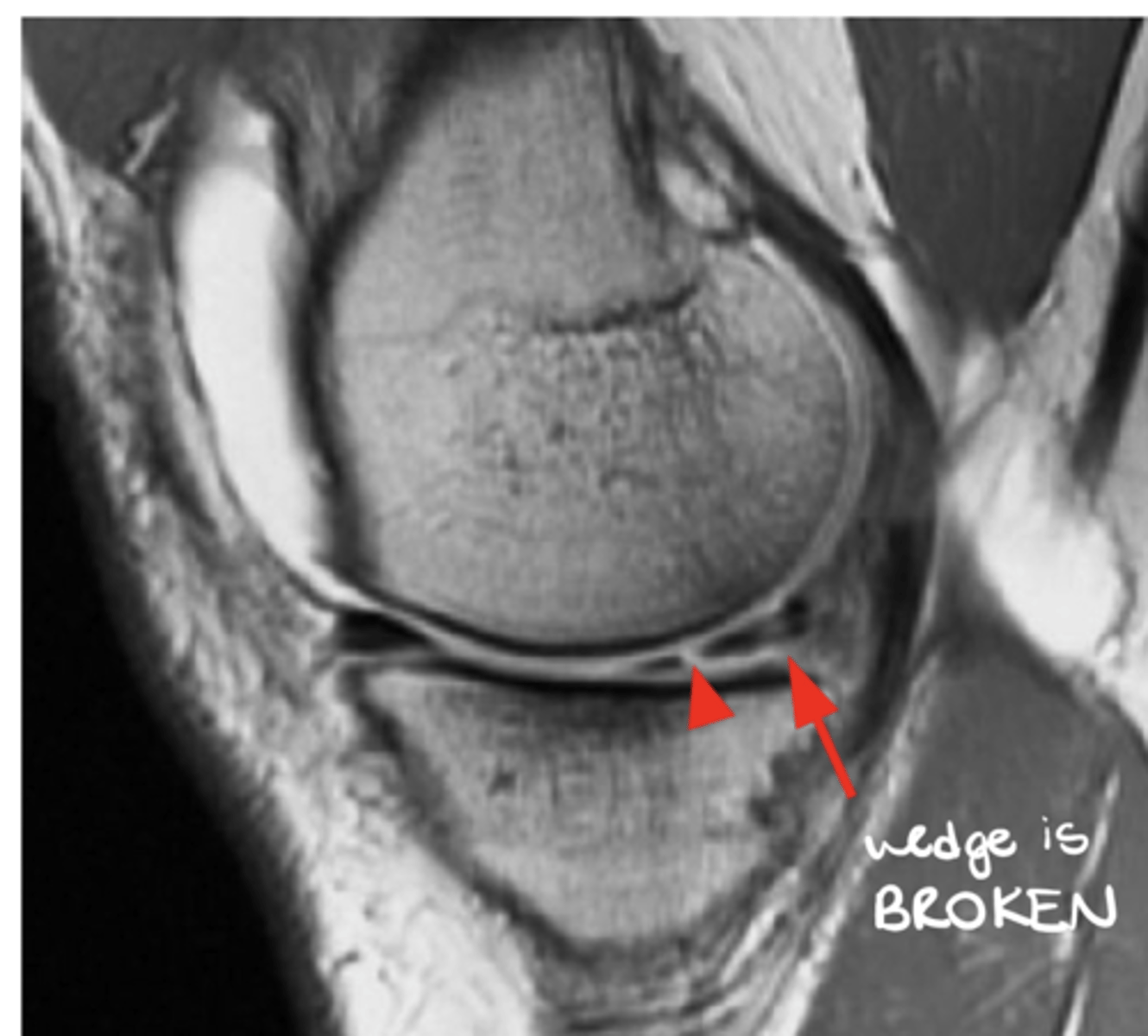
Meniscus tear (the meniscus should look like a wedge)
Define the pathology.
Meniscus tear
Define the pathology.
Patellar tendinopathy ("Jumper's knee")
Define the pathology.
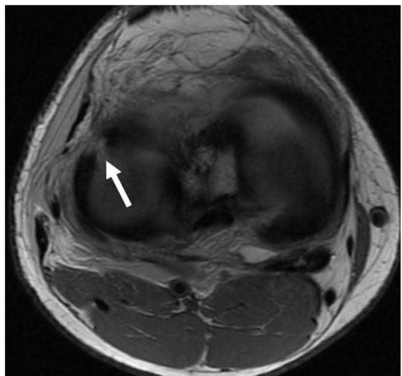
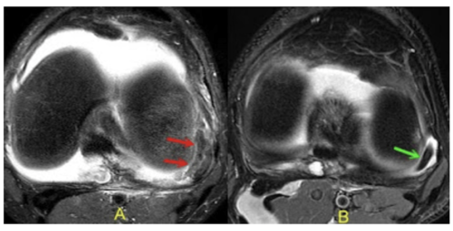
IT band-related lateral knee pain (cyst on the left image, some signal change on the right image)
Define the pathology.
Distal biceps femoris tendinopathy
Define the pathology.
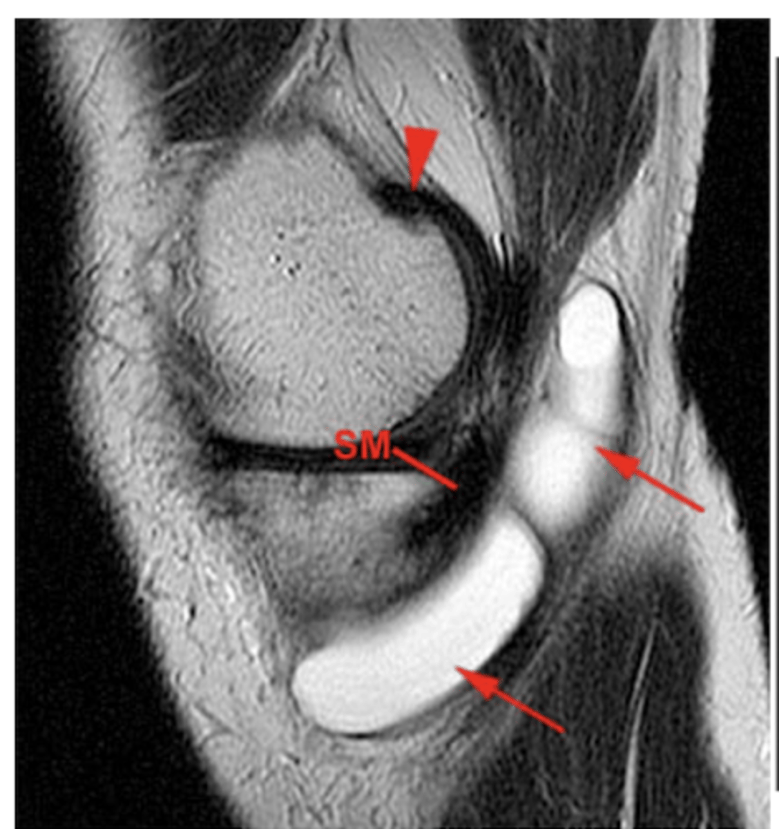
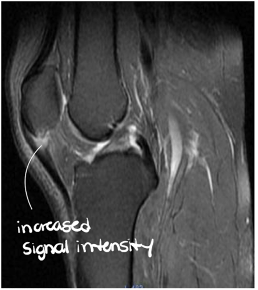
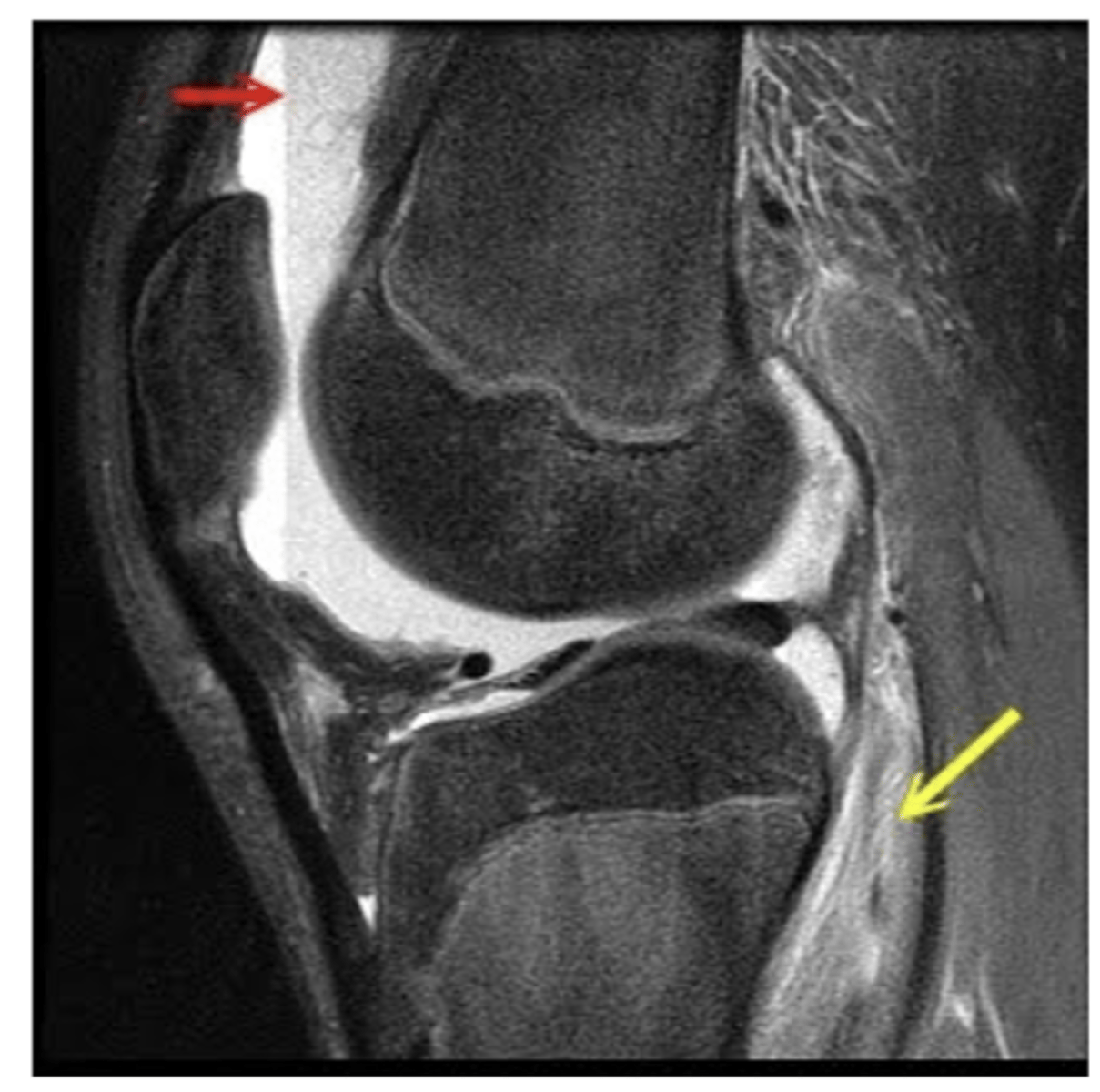
Popliteus injury
Define the pathology.
Popliteus injury
Define the pathology.
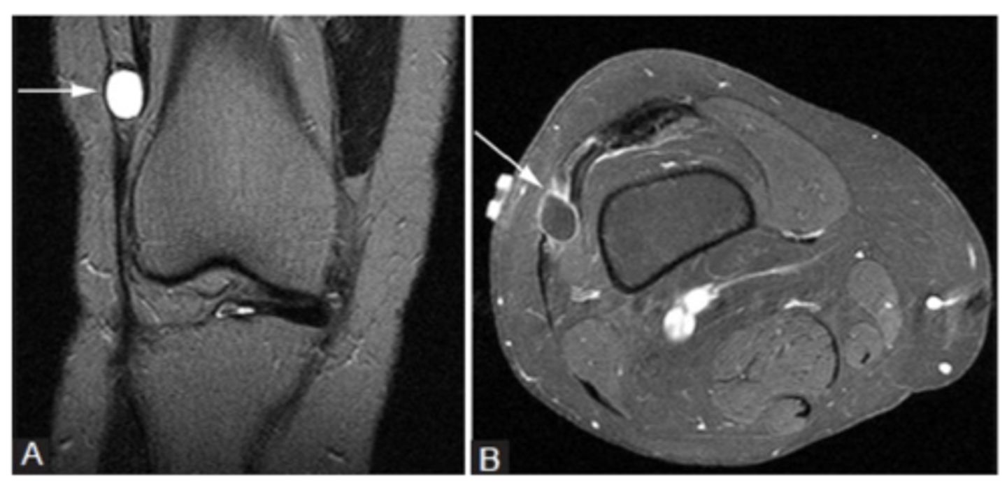
Pes anserine bursitis
Define the pathology.