visual processing: the visual pathway and processing and reflexes
1/18
There's no tags or description
Looks like no tags are added yet.
Name | Mastery | Learn | Test | Matching | Spaced | Call with Kai |
|---|
No analytics yet
Send a link to your students to track their progress
19 Terms
what does our visual system detect?
visible light (400-700nm)
how are images formed by refraction?
refraction is teh bending of light as it passes from one medium to another with a different refractive index (e.g air to water)
when light rays pass through a refracting surface (like a lens) they change direction so they can be brought to a focus (convergence) or diverge from a point
the cornea and lens refract incoming light
light rays bend so they converge on the retina
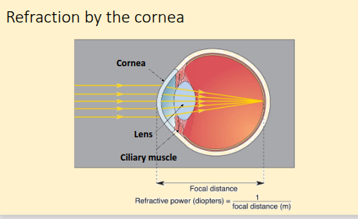
how does refraction by the cornea work?
light enters the eye as parallel rays
cornea (clear curved outer layer at front of eye) does ~70% of refraction
change in refractive index from air to cornea is large so light bends sharply
cornea has fixed shape so refractive power is constant
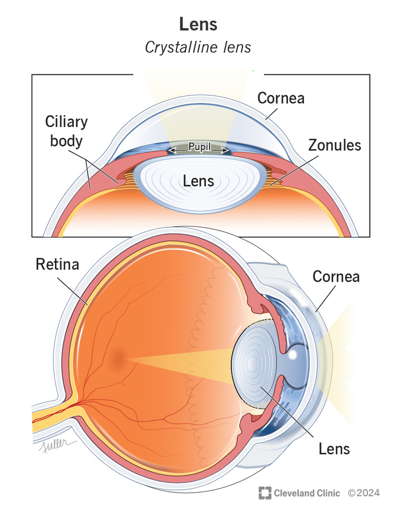
the lens provides ~30% refraction and is flexible as curvature changes through ciliary muscle action
when near object, ciliary muscles contracts and lens thickens, becoming more curvered = more refraction = focus moves forward (accomodation)
what is the structure of the eye’s focusing system?
cornea = clear, curved front surface of eye, avascular (no blood vessels), made up of 5 layers
lens = behind the iris and in fronf of the vitreous body, suspended by zonular fibres, bioconvex and avascular
zonular fibres = think, fibrous strands that connect the lends capsule to the ciliary body, when ciliary muscles contract, fibres loosen = lens rounds and focuses on close objects
what is focal distance?
the distance from the refracting surface to where light rays meet (the focal point)
what is refractive power?
how strongly the eye bends light. measured in diopters (D)
P = 1/focal distance (metres)
a normal eye has a total refractive power of ~60 diopters
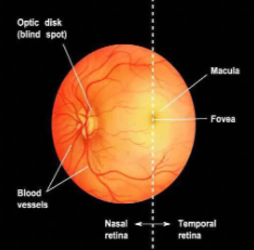
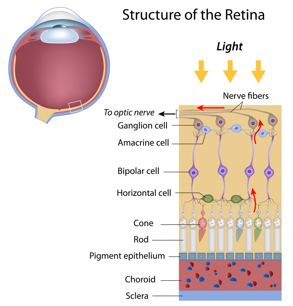
what is the structure of the retina (when viewed through an ophthalmoscope)?
retina is the light sensitive-neural layer at the back of the eye that converts light into neural signals
it is divided relative to the fovea into the nasal retina and temporal retoma
the temporal retina contains the macula and fovea
the macula is a small, yellowish area which has a high density of cones that are responsible for sharp colour vision, it contains the fovea (centre) and the parafoveal region
macular pigment (yellow) helps filter blue light and protects photoreceptors
the fovea is the point of greatest visual acuity containing only cones and no rods, it provides sharp, detailed central vision
the nasal retina contains the optic disc (blind spot) where the optic nerve exits the eye
it is a blindspot as there are no photoreceptors
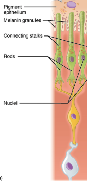
what are rods?
long and cyndrical
~120 million rodes in the human retina
all contain rhodopsin
form convergent networks (many rods to 1 bipolar cell)
found in peripheral retina
high light sensitivity (respond in dim light)
monochromatic
low visual acuity due to convergence
low temporal resolution = slow response
easily saturates in bright light
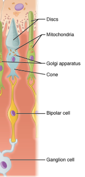
what are cones?
short and conical
~6 millions cones in retina
photopsins (red, blue green)
1 cone synapses to 1 bipolar cell
concentrated in fovea and macula
low light sensitivity (requires bright light)
trichromatic
high visual acuity especially in fovea
high temporal resolution = faster response
what is the structure of the retina?
photoreceptors are located in outermost layer and convert light into electrical impulses by reducing glutamate release (phototransudction)
horizontal cells connect neighbouring photoreceptors laterally in te outer plexiform layer and this defines edges (lateral inhibition)
bipolar cells receive input from photoreceptors and horizontal cells (inner nuclear layer), have on bipolar cells that activate when liht increases (-glutamate), off active when light dereases (+glutamate)
amacrine cells are found in the inner plexiform layer and connect bipolar cells to ganglion cells and modulate signalss using GABA/glycine = time dependent processing
ganglion cells are in the ganglio cell layer and receive input from bipolar and amacrine cells, their axons form the optic nerve
what is the distribution of photoreceptors in the retina?
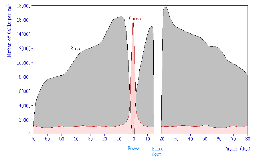
what is the difference in receptive fields of rods and cones?
cones have small receptive fields and this means each cone responds to light from a very limited area of the retina
this provides a high spatial resolution, precice colour vision but at the cost of reduced sensitivity to rods as there is less sumaation
what is the effect of lateral displacement in the fovea?
in the fovea cells like ganglion, bipolar and blood vessels are pushed aside (laterally displaced) so light has a direct path to the cone
this means there is less scattering and distortion as light doesnt have to pass through additional layers
with a direct path, the image projected onto the foveal cones is much sharper
this reduces blurring
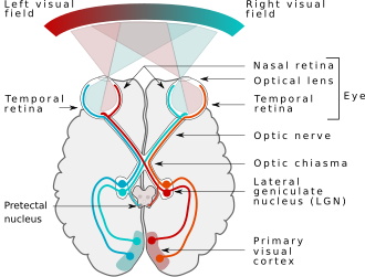
what is the ascending visual pathway?
each eye sees both a left and right visual field which is processed by the contralateral hemisphere of the brain
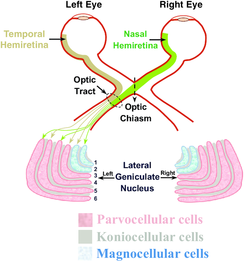
light from left visual field hits nasal retina of left eye and temporal retina of the right eye/light from right visual field hits nasal retina of the right eye and temporal retina of the left eye
at the optic chiam (formed by optic nerve), fibres from the nasal retina cross to the opposite side but temporal fibres stay the same = partial decussation = info from left visual field of both eyes go to right hemisphere
chiasm forms the optic tract which carries info from the same visual field (left or right) from both eyes
this goes to the lateral geniculate nucleus (LGN) which fans out into optic radiations to the primary visual cortex (V1) - fibres from upper retina travel via parietal lobe (cuneus), fibres from lower retina travel via temporal lobe (meyers loop)
what are the layers of the LGN?
6 layers
layers 1-2= magnocellular
layers 3-6= parvocellular
between these are koinocellular layers
magnocellular layers have large receptive fields, high temporal resolution and low spatial resolution, they receive input mostly from rod dominated pathways = detect movement, broad outlines and rapid changes in visual field
parvoceullar layers have small receptive fields, low temporal resolution and high spatial resolution, they receive input from cone-dominated pathways and detect shape, fine texture and colour
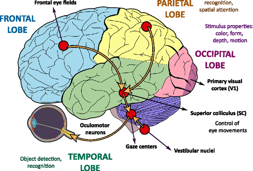
what is the role of the superior colliculus?
intergates vision with other sensory inputs
controls saccadic eye movements (rapid shifts in direction of gaze to quickly move eyes from point of focus to another)
activate neurons controlling neck muscles which is important for head movements in response to a visual stimulus
SC is critical for fast, reflexive responses to visual stimuli, like turning your head to a sudden movement
what is the role of the suprachiasmatic nucleus?
hypothalamus
SCN uses light information to synchronise internal biological clocks with day-night cycle
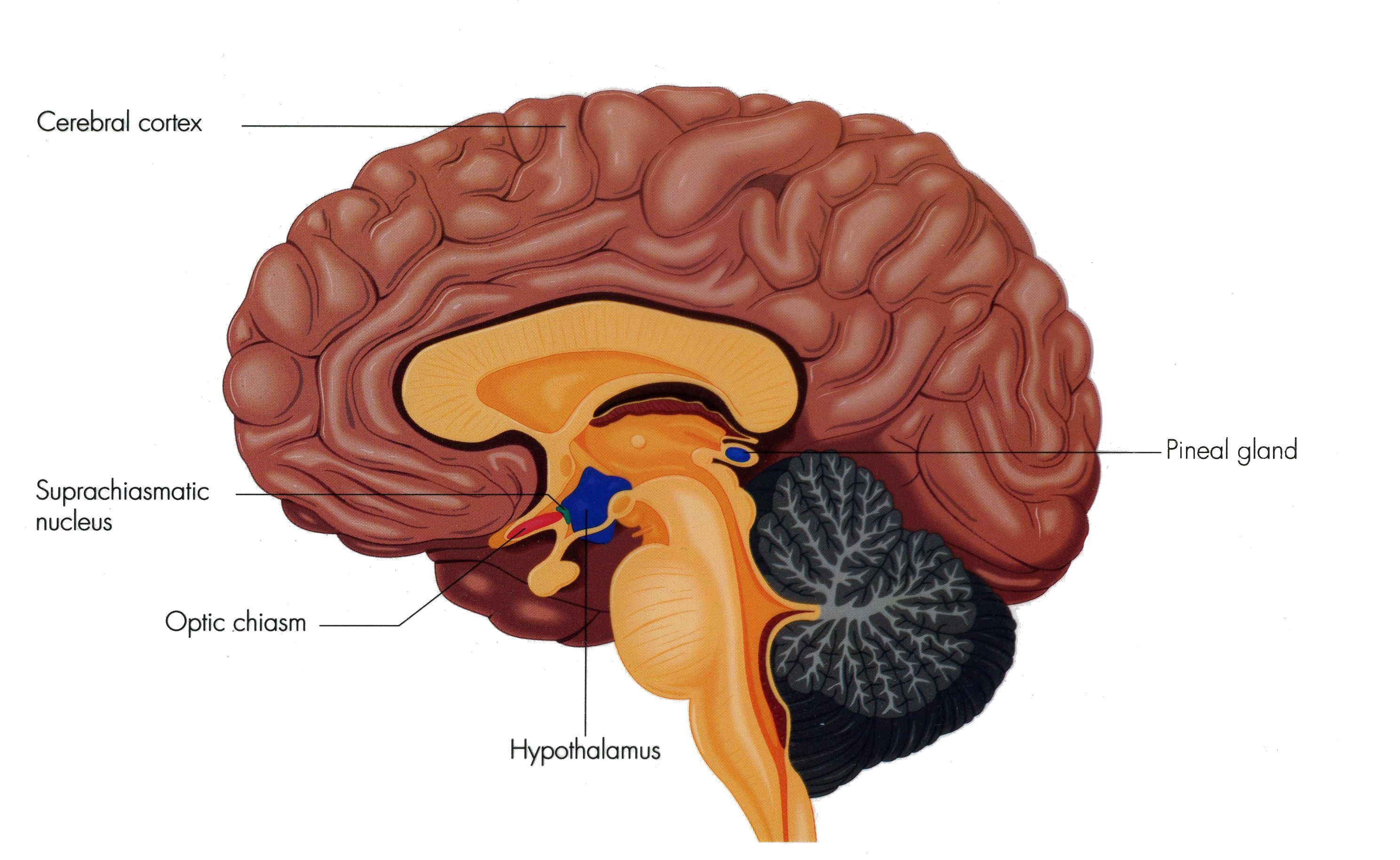
what is the role of the pretectal nucleus in the midbrain?
mediates the pupillary light reflex (pupil constrcition in response to light) and lens accomodation reflexes
outputs to the edinger-westphal nucleus = parasympathetic fibres of the oculomotor nerve = constrict pupillae muscles
therefore responsible for automatic pupil and lens adjustments to light intensity

what is the role of the frontal eye fields?
region of middle frontal gyrus
voluntary control of eye movements, especially sacacdes (quick jumps of the eyes) and tracking objects
important for visual attention, conscious, goal-direted eye movements e.g following a moving ball