VT 111 Lec. 5 Special Senses
1/42
There's no tags or description
Looks like no tags are added yet.
Name | Mastery | Learn | Test | Matching | Spaced |
|---|
No study sessions yet.
43 Terms
The Special Senses
Taste
smell
hearing
balance (equilibrium)
vision
Sense Organs
Are extension of the CNS that allow it to monitor what is going on inside & outside of animal
All have specialized sensory nerve endings (dendrites) called sensory receptors
Taste
Gustatory sense
Chemical
Receptors
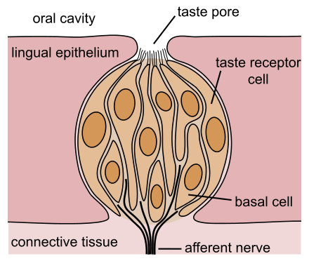
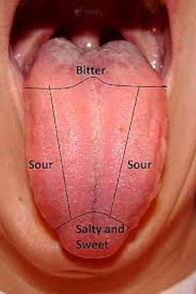
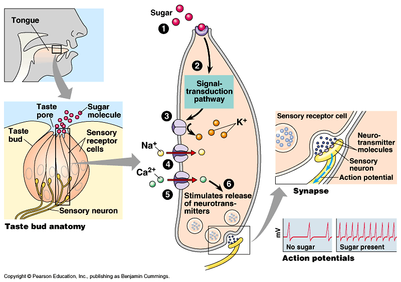
Taste buds detect chemicals dissolved in saliva
Located in small bumps or papillae on tongue, palate and back of mouth (throat – pharynx); tiny rounded structures made up of gustatory & supporting cells
Each taste bud has 50-100 sensory receptor cells
Modified dendrites (hairs) of cells project up into taste pores – tiny openings on surface of taste buds
Molecules or ligands are spread around the mouth in saliva and come into contact with dendrites
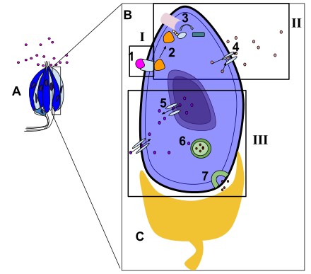
Example of signal transduction of sweet taste
A: taste bud
B: one taste cell
C: neuron
1. glucose
2. receptor
7. NTs

Smell
Olfactory sense
Chemical
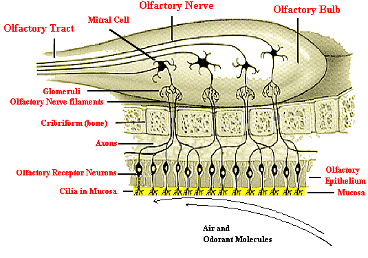
Olfactory epithelium covers turbinates (bony structures) in nasal passages
Large surface area
Humans: 5 cm2
Dogs: up to 170 cm2
Hairs of cells immersed in layer of mucus
Molecules are dissolved in mucus -> contact sensory processes -> nerve impulses sent to brain ->interpretation of smells
Chemoreception: Taste/Smell
Taste/smell in animals
Fish, amphibians, and reptiles have a very acute sense of chemoreception through their external nares
Birds have poor sense of smell
Vomeronasal organs:
chemoreceptor for smell located in the mouth of animals
detects heavy moisture-borne odors
reptiles, especially snakes (their forked tongue)
mammals, in the dorsal lip
Involved in social and reproductive communication byway of phermones
Hearing and Equilibrium
Auditory sense
Mechanical
Ear anatomy
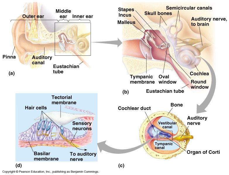
External
Funnel that collects sound wave vibrations and directs them to eardrum
Middle
amplifies and transmits vibrations from eardrum to inner ear
Inner
sensory receptors (convert mechanical vibrations to nerve impulses); as well sensors for equilibrium
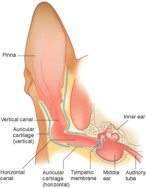
Divisions of the hearing apparatus: external ear
Extends from pinna and auditory canal to tympanic membrane
Conducting zone
Pinna
Acts as funnel
May be mobile for sound detection
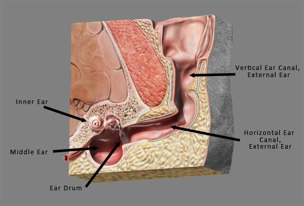
External auditory canal
Soft membrane-lined tube
L-shaped in most domestics
Outer vertical portion
Inner horizontal portion
Carries sound waves to tympanic membrane
Tympanic membrane (eardrum)
Thin connective tissue membrane stretched across opening between external auditory canal to middle ear cavity
Sounds waves cause sympathetic vibration
Divisions of the hearing apparatus: middle ear
Middle ear: extends from tympanic membrane to oval window.
Separated from external ear by tympanic membrane; separated from inner ear by membranes that cover the windows of cochlea
Hollowed out area of temporal bone filled with air & lined by soft tissue membranes
Contains the 3 bony ossicles link tympanic membrane with cochlea of inner ear (receptors for hearing are located)
Outermost bone: Malleus (hammer) – attached to tympanic membrane; forms synovial joint with middle bone
Middle bone: Incus (anvil) forms joint with stapes
Stapes (stirrup) – attached to oval window of cochlea
Space opens into pharynx via eustachian tube
Allows equalization of air pressure on both sides of eardrum
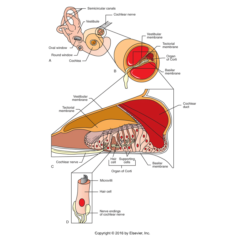
Divisions of the hearing apparatus: inner ear
Inner ear: extends from oval window to include vestibulocochlear apparatus
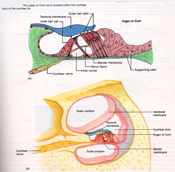
Cochlea: snail-shell shaped spiral cavity in temporal bone
Organ of Corti (organ of hearing) in cochlear duct filled with endolymph (fluid)
Hair cells (receptor cells of hearing) rub together to generate nerve impulses -> travel to brain = sound interpretation
Supporting cells
Tectorial membrane
lies on top of hair (modified dendrites) of hair cells
U-shaped tube filled with perilymph (fluid)
Ascending – vestibular canal
Descending – tympanic canal
Ends: oval window and round window
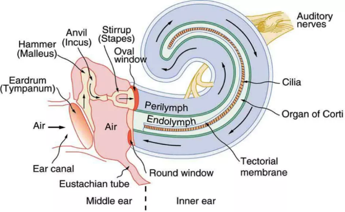
Process of sound reception
Sound waves enter outer ear; air pressure vibrates tympanic membrane
Tympanic membrane moves bony ossicles that move oval window
Oval window vibrates, causing pressure waves in perilymph
Pressure waves get transferred to endolymph in cochlear duct
These waves move the tectorial membrane bending the villi of the hair cells
Process of Hearing
Pressure wave causes basilar membrane to vibrate up/down
Since cilia is imbedded in tectorial membrane, cilia bend and deform the apical membrane
Na+ channels open in receptor cells to result in generator potential
If enough generator potentials → AP
Each depolarized hair cell releases neurotransmitter to excite an associated neuron
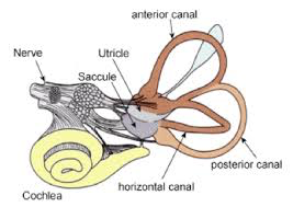
Equilibrium
Monitored by parts of the inner ear called the vestibule and semicircular canals
Vestibule
Between cochlea and semicircular canals
Composed of saclike spaces: utricle and saccule
Continuous with cochlear duct – filled with endolymph; surrounded by perilymph
Inside: sensory epithelium (macula) consisting of hair & supporting cells covered by gelatinous matrix with otoliths (crystals of calcium carbonate)
Semicircular canals
On other side of vestibule from cochlea
Canals are at right angles to one another
Filled with endolymph surrounded by perilymph
Ampulla contains receptor (crista ampullaris)
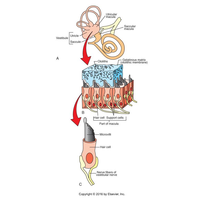
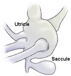
Vestibule: Utricle and Saccule
Utricle is for detection of linear acceleration or position in a horizontal plane
Saccule is for detection of linear acceleration or position in a vertical plane
In the macula (sensory receptor), gravity causes the gelatinous matrix with otoliths to put constant pressure on the sensory hairs when the head is still
Movement bends hairs which generate nerve impulses
Linear motion
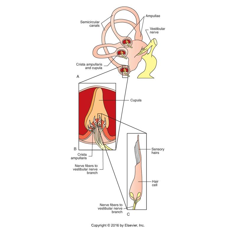
Semicircular canals
Each canal has a endolymph filled membranous tube surround by perilymph
Continuous with other inner ear structures
Canals are at right angles (3)
Near utricle end → widened area called ampulla
Contains receptor structure
Crista ampullaris
Cone shaped area of supporting cells and hair cells with processes sticking up into gelatinous structure (cupula)
No otoliths → no weight
Functions as a float and moves with endolymph
During movement, inertia causes endolymph to lag behind
Pulls on cupula and bends hairs → nerve impulse
Rotary motion
The vestibular system senses rotary motion of the head with the semicircular canals, and linear motion and the position of the head with the vestibule
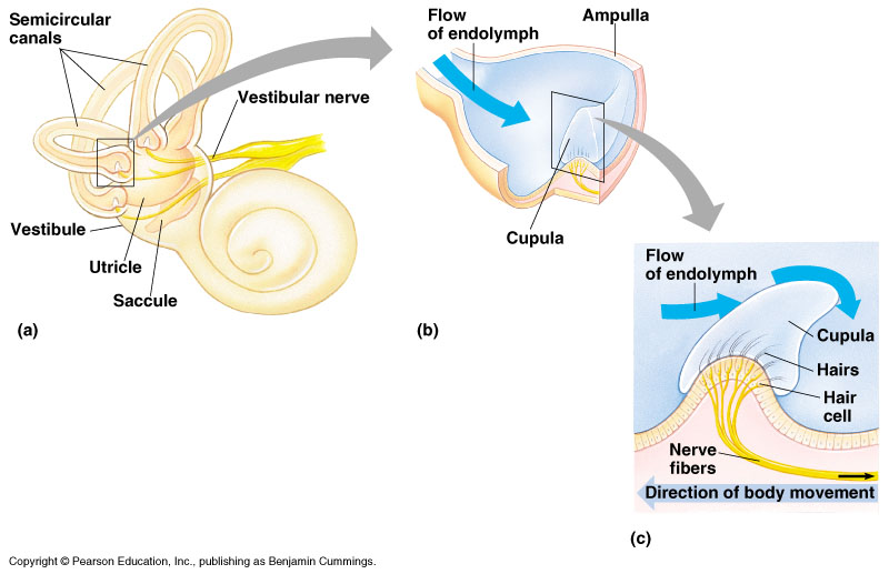
The Sense of Motion
The head moves
Fluid movement lags in one plane of semicircular canals
Fluid movement pulls on cupula
Hairs are bent
Nerve impulse is generated
Brain receives information about motion of the head
Vestibular Syndrome in Dogs
Common clinical signs:
Circling or falling to one side
Pronounced head tilt
Nystagmus—the rapid and involuntary oscillating movement of the eyeballs.
Facial drooping may occur if there is a tumor or inflammatory disease of the inner or middle ear
Treatment:
Supportive care
IV fluids
Anti-nausea/motion sickness medications
Maropitant (Cerenia), meclizine (Dramamine)
Sedatives – if severely ataxic
Antibiotics – if inner ear infection is present
+/- Corticosteroids – not generally
Vision
Eye components:
Function is to help form an accurate visual image
Function is not to detect the image
Photoreceptors:
Located in a single layer of cells in the retina
Function is to detect the image
Generate visual nerve impulses
Mammalian eye anatomy
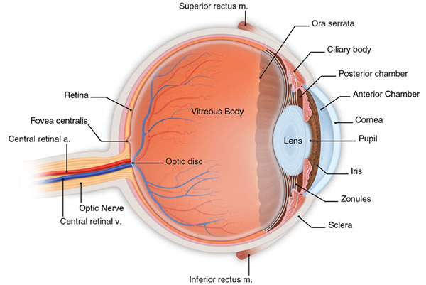
Eyeball - Outer Fibrous Layer
Cornea:
Transparent; admits light to interior of the eye
Orderly arrangement of collagen fibers
No blood vessels; many pain receptors
Sclera:
The “white” of the eye
Limbus
Junction of the cornea and sclera
Eyeball - Middle Vascular Layer
Choroid:
Lies between sclera and retina
Consists mainly of pigment and blood vessels
Tapetum lucidum is highly reflective area in rear of eye
Iris:
Pigmented muscular diaphragm
Controls amount of light that enters the posterior part of the eye
Opening in center of iris = pupil
Ciliary body:
Ring-shaped structure behind the iris
Contains tiny muscles that adjust shape of the lens to allow near and far vision
Eyeball - Inner Nervous Layer
Retina:
Lines the back of the eye
One component of the fundus
Contains rods and cones, the sensory receptors for vision
Compartments of the Eyeball
Aqueous compartment:
Located in front of the lens
Subdivided by iris
Anterior chamber
Posterior chamber
Contains clear, watery fluid = aqueous humor
Produced in posterior chamber by cells of ciliary body
Drained by canal of schlemm
Vitreous compartment:
Contains clear, gelatinous fluid = vitreous humor
Fills whole back of eyeball behind lens and ciliary body
The Lens of the Eye
Soft, translucent layers of fibers
Elastic and biconvex
Front surface in contact with aqueous humor; back surface in contact with vitreous humor
Helps focus a clear image on the retina through accommodation process
Formation of a Visual Image
4 refractive media in the eye help form a clear upside-down image on the retina
Cornea, aqueous humor, lens, vitreous humor
Brain inverts image
Conscious mind sees everything right-side up
Functional Aspects
Accommodation: the process of focusing light on the retina for close up & distance vision. Ciliary apparatus is contractile.
Relaxation of ciliary muscle moves lens posteriorly, increasing tension on suspensory ligaments → pulls lens flat for long distance vision
Contraction of ciliary muscle causes lens to move forward, loosening tension on ligaments → lens forms a ball for close up vision*
eyestrain
Humans have very strong powers of accommodation
Carnivores have mediocre close up vision
Herbivores have poor close up vision
Extraocular Structures
Conjunctiva:
Thin, moist, transparent membrane
Covers front portion of eyeball
Bulbar conjunctiva
Bulbar = globe
Lines interior surfaces of eyelids
Palpebral conjunctiva
Palpebral = eyelids
Conjunctival Sac:
Space between bulbar and palpebral portions of conjunctiva
Eyelids:
Upper and lower folds of skin, lined by conjunctiva
Lateral and Medial Canthus:
Corners where the eyelids come together
Extraocular Structures continued…
Tarsal Glands = Meibomian Glands:
Their tiny openings are found along eyelid margin
Produce waxy substance to prevent tears from overflowing onto the face
Eyelashes:
Prominent on upper lid of most animals
Third Eyelid = Nictitating Membrane:
T-shaped plate of cartilage covered by conjunctiva
Located medially between eyelids and eyeball
No muscle attachment; passive movements
Lymph nodules and accessory lacrimal gland on ocular surface
Extraocular Structures continued…
Lacrimal Apparatus:
Structures that produce and secrete tears and drain tears away from the surface of the eye
Lacrimal glands are the primary source of tears
Tear Drainage System:
Lacrimal puncta
Lacrimal sacs
Nasolacrimal duct
Eye Muscles:
Small, skeletal muscles
Attach to sclera
Capable of wide range of movements
Tears
Liquid film that moistens and protects the surface of the eye
3 main layers of tears:
Inner mucous layer – from cells in conjunctiva
Contains antibacterial substances
Middle tear layer – from lacrimal glands and accessory lacrimal glands of the third eyelid
Keeps the cornea moist
Outer oily layer – from tarsal or meibomian glands
Reduces evaporation of underlying tear layer
Prevents tears from flowing over the lid margin
Unique Structures in Animals
Nictating membrane: found in all domestic mammals
3rd eyelid situated ventromedially, between eyelids and globe
“flips” across the eye to protect it
Consists of a T-shaped plate of cartilage covered by conjunctiva
Contains lymph nodules and a tear gland
Passive movement
Tapetum lucidum:
mirror like pigmentation of choroid
Increases light gathering ability of retina
Improves night vision
Found in domestic animals
Except: swine and humans
Retina
Lines most of the vitreous compartment
Made up of layers of neurons:
Pigment layer (outer layer)
Photoreceptors (layer)
Rods
More sensitive to light
Cones
Sensitive to color and detail
Fovea centralis (primates) - lots of cones to perceive great detail
Neurons relay nerve impulses from photoreceptors to to optic nerve
Bipolar (layer) cells
Ganglion (layer) cells
Nerve fiber layer (inner layer)
Nerve fibers converge at optic disc → leave eye to form optic nerve
“Blind spot” – no photoreceptors
Rods & Cones
Rods:
High sensitivity
Better in low light
B/W vision
Low visual discrimination
Highest concentration in peripheral area of retina
Cones:
Low sensitivity
Color vision
High visual discrimination
Primates: High concentration in central area of retina called the fovea centralis
Clearest area of vision
Reading
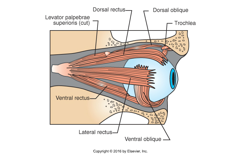
Extraocular Eye Muscles
Lateral Rectus
Dorsal Rectus
Medial Rectus
Ventral Rectus
Dorsal Oblique
Ventral Oblique
General Types of Stimuli
Mechanical
Touch, hearing, balance
Thermal
Hot, cold
Electromagnetic
Vision
Chemical
Taste, smell
Visceral Sensations
Miscellaneous category of interior body sensations
Vague and poorly localized
Hunger
Thirst
Visceral stretch
GI tract
Urinary system
Touch and Pressure
Tactile Sense
Sensation of something in contact with the surface of the body
Pressure
Sensation of something pressing on the body surface
Different touch and pressure receptors produce sensations of light contact, deep pressure, vibration, or hair movement
Temperature
2 types of temperature receptors
Superficial
Receptors in the skin
Detect changes in skin temperature
Central
Receptors in hypothalamus
Monitor temperature of the blood
Pain
Pain Receptors = Nociceptors
Widely distributed inside and on surface of the body
Not present in the brain
May be simple nerve endings or more specialized structures to detect mechanical forces, temperature, etc.
Purpose: protect body from damage
Pain: 4 processes contribute to sensory pain
Transduction –conversion of stimulus to nerve impulse occurs at sensory nerve ending
Transmission – impulse moves up the sensory nerve fibers to spinal cord
Modulation – occurs at spinal cord, can amplify or suppress impulses thru synapses between neurons in dorsal horn. Can be influenced by drugs
Perception – conscious perception occurs in cerebral cortex
Pain Classifications
Superficial—affecting skin (& subcutaneous areas) and body surface
Deep—involving muscles & joints
Visceral – relating to internal organs
Acute – sharp and intense
Chronic – dull and aching
Proprioception
Ability to sense where your limbs/body parts are
Operates primarily at subconscious level
Stretch receptors located in muscles, tendons, ligaments and joints capsules keep CNS informed about movement of limbs, etc
Evaluation: curling animal’s foot and seeing how long it takes them to correct the position