Bone Neoplasms and joint disease
1/42
Earn XP
Description and Tags
Path I
Name | Mastery | Learn | Test | Matching | Spaced | Call with Kai |
|---|
No analytics yet
Send a link to your students to track their progress
43 Terms
in dogs what is the most common bone tumour, and are they B or M
osteosarcoma, malignant, primary tumours
what are secondary tumours of the bone
metastasis
what is the common site for osteosarcoma, and generally do they cross the joint?
distal to the radius and proximal to the humerus - away from the elbow
distal femur and proximal tibia - towards the knee
they do not cross the joints
in what dog breed do we see osteosarcomas in, and where do they arise
giant breeds - arise in the endosteum (deep in the medulla)
distal radius is the common location
malignant till proven not

name the three form of oseosarcomas from left to right
lytic, sunburst, mass
describe the beahviour of osteosarcomas
highly magligants and early metastasis to the lungs
short survual time of P
can metastasize to regional lymphnodes, but its rare
what is Codman’s triangle
elevation of the periostium, generally new bone formation, this means we will not see the mass if we sample here, need to sample past it, can be see on Rads
not always related to a neoplasm
when sampling for an osteosarcoma explain the sampling process
need to sample at least 3 sites
sample the metaphysis (not the diaphysis)
need to go deep into the medullary cavity
choose the lytic lesions - do not sample codman’s triangle
what is the ddx for osteosarcoma and how to confirm
hemangiosarcoma
use histo to confirm!
what is the second most common tumor in bones and who do we generally see it in
chondrosarcoma, in medium breeds, middle age dogs, involves the flat bones and the turbinates
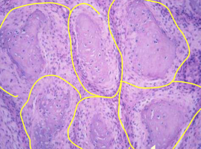
where are chondrosarcomas most commonly seen and what do they look like grossly and histo
axial bones, end of bones, grey/blue looking in appreance, on histo can see chondrocytes
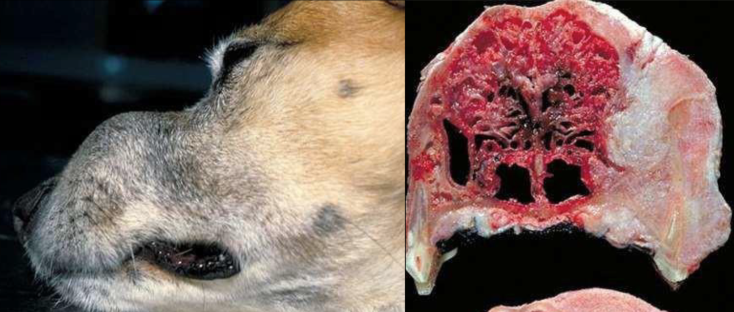
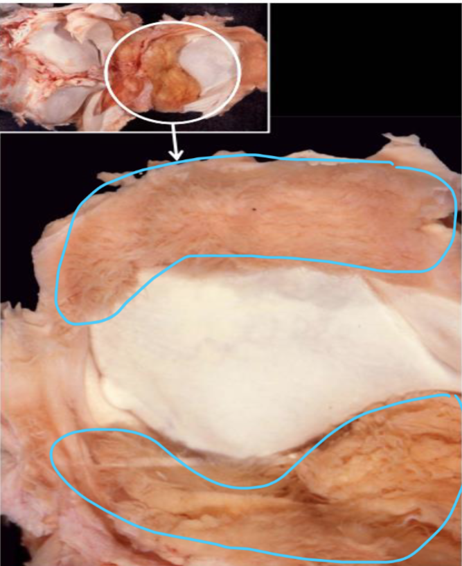
what is this
multilobar tumour of bone, slow growing and locally invasive, affects flat bones like the skull, sometimes ribs and pelvis
can invade to the brain
flat bone and locally invasive
what is the number one bone tumour in cats
skeletal fibrosarcoma
skeletal fibrosarcomas, where do they originate from, what bones, will they cross the joint space
can be seen esp in prev vx site
usually from the periosteal surfaces
can me from the metaphysis of long bones and look like osteosarcomas
these WILL cross the joint space
what is different about Maxillary fibrosarcomas
in dogs
looks begnign on histo but is locally invasive!
what is this
maxiallary fibrosarcoma, imp u give description of the location as it will liik begnin in histo, but its nit
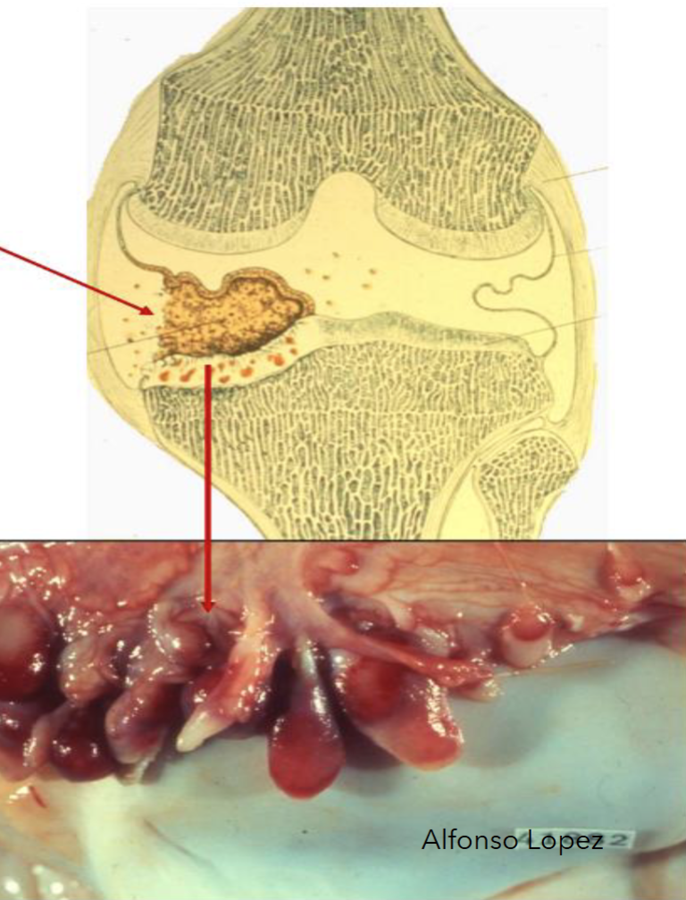
what is plasma cell myeloma - multiple myeloma
multiple lytic lesions in bone marrow esp vertebral bodies and ribs
what are some “markers” asscoaiated with multople myeloma
the neoplastic plasma cells will produce immunoglobulins, increase plasma protein (monoclonal gammopathy)
10% cases we will see hypercalcemia (poor prog)
describe the hypercalcemia of maliganancy
humoral hypercalcemia/pseusohyperparathyroidisim
caused by a release in PTH-like hormone produced by the cancer cell
this will act like PTH and cause the release of Ca into the blood stream, additionally actual PTH will decrease since Ca is increasing, but the PTH like will not
we will see osteoclasts resorption of bone, increased Ca and in the kidneys and GI
osseous invasion by sqaumous cell carcinoma is common in the ____ of cats and ____ of dogs
digits of cats and facial bones of dogs
osteoma
non maliganat
do not invade adj bone
often found in the skull
ossifying fibroma
maxilla and mandibles of cattle and horses
intramedullaty begnins lesions, but issues with compression of the adj bone
osteochondromas
herediatry
supposed to be begning but because of the location they have issues with compressing adj surfaces
can progress to malignancy
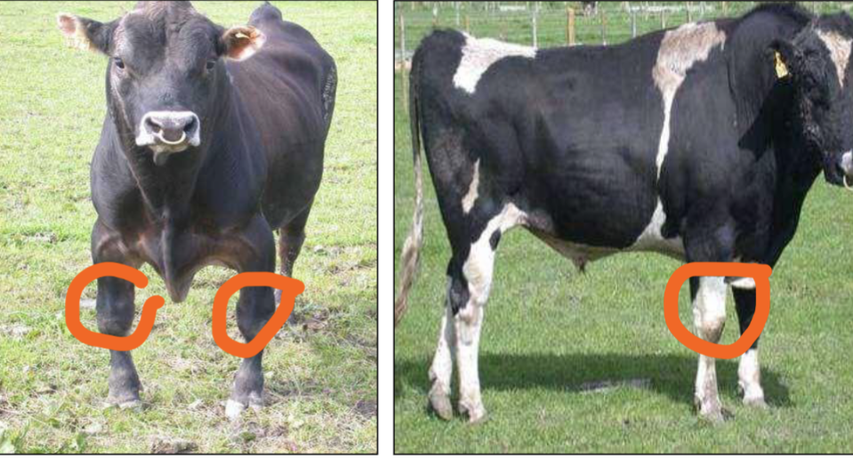
what is the most common joint tumor in dogs and what breed does it affect
histiocytic sarcoma
rottweilwers
histiocytic sarcoma, what is the cell of origin, what joints,
macrophage cell lineage, elbow and stifle, sometimes can invade the bone in advanced cases
what is the second most common joint tumour in dogs, who does it occur in, age
synovial myxoma, large breed and middle aged dogs - bobermen and labs
can occur in cats
synovial myxoma, where does it occur, and progression
in a single joint, stifle and digit most common
slow growing and can see CS b4 dx
begnin tumour but because its in the joint can cause bone lysis on both sides
joint sarcoma Dx
exclude histocytic sarcoma
need immuno to dx
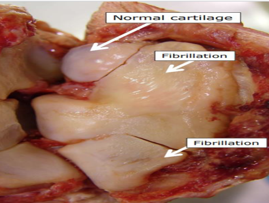
what is this and describe it
its fibrilation which is partial loss of the cartilage in a joint causd by the loss of peptidoglycants, additionally will see increased water content and a dull apperance of the cartiglage
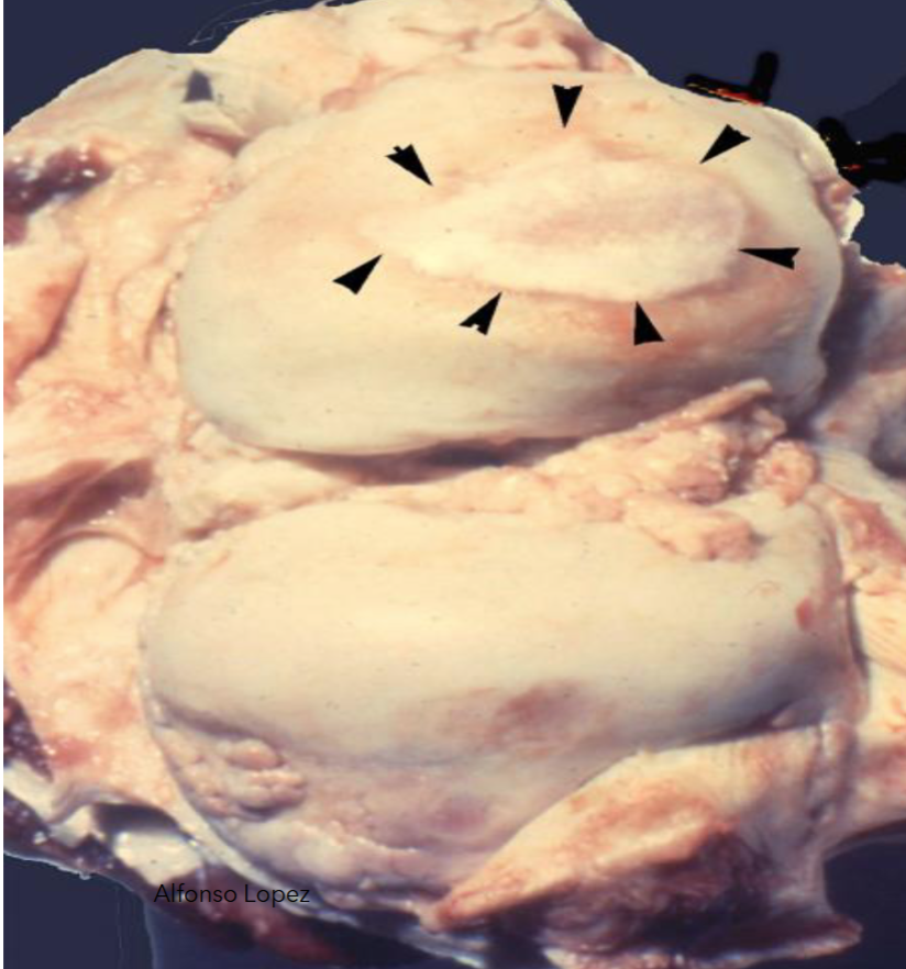
what is this and how it it caused
ebrunation - comeplete loss of the joint cartilage, can reveal the subchondral bone
these lesions are irreverable
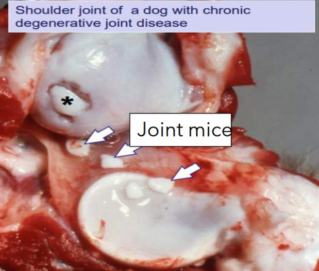
what are these
joint mice - they are viable and often cont to grow fragments of cartilage that float in the joint space
these arrise when cartilage degenerates and detached from the subchondral bone
some remain viable and become joint mice feeding off the synovial fluid
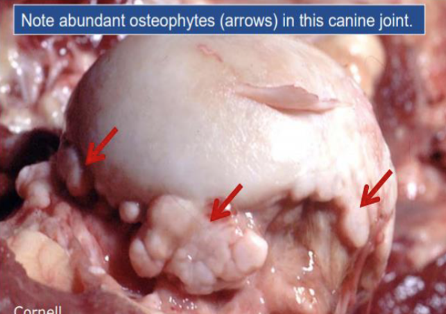
what is this
osteophytes - geneally a response to skeletal stress
lateral outgrowths of bone that are fromed by chondrification (meaning cartilage is layed doen first then its ossifiied)
osteophytes vs enthersophytes
both are responses to skeletal stress
the enthesophyte is a bony spur at the tendon or liagment insertion point on a bone growing in the direction of the natural pull of the tendon/ligmant involved
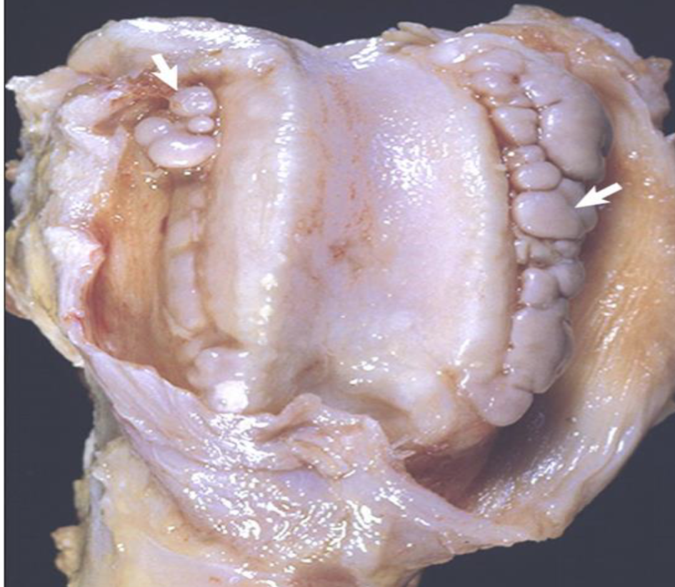
what is this and desribe it
synovial villous hyperplasia - common and non-specific rxn to synovial injury, the synovial memebrane becomes velvety in appreance because it becomes covered in many villous projections
waht is this
pannus - formation of granulation tissue on the synovial membrane or eroded articular cartilage
the granulation tissue will often undergo chondral or osseous metaplasia
what is primary DJD
degenrative joint disease
no apparent cause, can be use/work related
mild degenrative changes in large weight bearing joint and often incidental on necropsy
what is secondary DJD
often more severe than primary
underlying abnormality in joint or the suppoting strucutr that leads to the degeneration of the joint
what are some predisposing factors of 2ndary DJD
trauma to ligaments
disease eg. Legg Cavle Perthe
septic arthritis
persisitnat hemarthrosis (bleeding into the synovial cavity)
what is this
DJD clinical apperance
Intervertebral disc disease Hansen type I
common in chondrodystrophic breeds - pekanisese,cocker, beagle
degeneration and mineralization of the nucleus polposuses with degenration of the inner layers of the annulus
assocaited with fibroblast growth factor 4 restrogene
affects all the discs - results in spinal compression
Intervertebral disc disease Hansen type II
all breeds
AKA senile degenrative disc disease
partial rupture with buldging into the vertebral cancal
the nuclus pulposus is dry ans degenerated but not mineralized
usually affect single disc unlike type I
can leads to DJD, nerve impingement and spondylosis
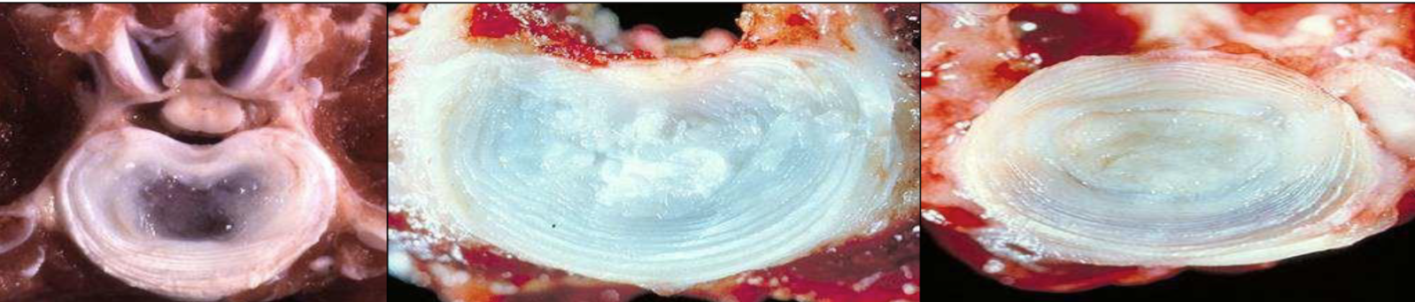
name the following and why
normal
type I - because we see the mineralization
type II - desiccation and degeneration but no mineralization