Muscular System Notes
1/42
There's no tags or description
Looks like no tags are added yet.
Name | Mastery | Learn | Test | Matching | Spaced | Call with Kai |
|---|
No study sessions yet.
43 Terms
Muscular System
The system responsible for movement in the body, where muscles contract to produce movement.
Insertion
The point where a muscle is attached to a movable bone.
Origin
The point where a muscle is attached to an immovable bone.
Prime Movers (Agonists)
Muscles most responsible for producing a certain movement, such as the biceps brachii in forearm flexion.
Antagonists
Muscles that oppose or reverse a certain movement, such as the triceps brachii in forearm flexion.
Synergists
Muscles that assist the prime mover, such as the brachioradialis and brachialis in forearm flexion.
Fixators
A type of synergist that immobilizes the muscle’s origin bone to increase the prime mover’s effectiveness.
Cardiac Muscle
Striated muscle tissue of the heart that contracts involuntarily to pump blood.
Smooth Muscle
Non-striated muscle tissue found in the walls of visceral organs that contracts involuntarily.
Skeletal Muscle
Striated muscle tissue attached to bones that contracts voluntarily to produce movement.
Myofibrils
Organelles that make up most of muscle cells, composed of long, striated units called sarcomeres.
Sarcomeres
The contractile unit of muscle, separated by Z discs.
Myofilaments
Thread-like organelles within muscle fibers, consisting of actin (thin) and myosin (thick) filaments.
Epimysium
Connective tissue that wraps around the entire muscle.
Perimysium
Connective tissue that surrounds each fascicle (bundle of muscle fibers).
Endomysium
Connective tissue that covers an individual muscle fiber.
Sliding Filament Model
The mechanism of muscle contraction where myofilaments slide past each other, causing sarcomeres to shorten.
Action Potential
A large change in membrane potential that spreads rapidly over long distances within a cell, triggering muscle contraction.
Neuromuscular Junction
The connection point where a motor neuron meets a muscle fiber, including the synaptic cleft.
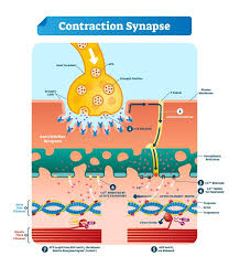
Step one
A muscle contraction starts in the brain where signals are sent along the motor neuron (yellow ball)

Step two
The impulse travels down the membrane and into the axon terminal of a neuron where it causes calcium to be released from the sarcoplasmic reticulum. T tune green and calcium dark blue.

Step three
Calcium binds to a structure on the actin that causes it to change shape. The DNA like strands are myofilaments.

Step four
The change in shape allows myosin to form cross bridges between he actin and the myosin heads. Myosin red.
Myosin
The thick filaments contain the contractile protein ________.
Actin
The thin filaments contain the contractile protein ________.
Sarcomeres
Muscles contract because ____________ contract when the myofilaments slide past each other.
myofilaments
Muscles contract because sarcomeres contract when the _____________ slide past each other. The smallest part of the muscle build.
Actin, tropomyosin, troponin.
Myofibrils are thin filaments called ________ that contain ___ and ___.
Sarcomeres
The functional unit of a muscle fiber.
Electrical impulses, contract.
The nervous system uses _________________________ to connect with skeletal muscles and signal for them to _______.
Resting membrane potential
The electrical charge difference across the membrane of a muscle cell at rest.
Myofibril
-Thread-like organelles of the muscle fibers
-Structured in long, striated units called sarcomeres
second smallest part of the muscle
Muscle fiber
-Long, thin muscle cells
-Each is covered by sarcoplasmic reticulum, which transmit an impulse to the muscle fiber
Middle of the muscle compound
Fascicle
Bundles within the muscles. Second biggest in the muscle compound.
Why can't myosin touch actin initially?
Tropomyosin blocks actin, preventing myosin from binding without ATP and access.
How does myosin gain access to actin?
A neuron signals an action potential, releasing Ca+2 from the sarcoplasmic reticulum. Ca+2 binds to troponin, which moves tropomyosin out of the way, granting access.
How does myosin get energy to bind to actin?
Myosin grabs ATP and breaks off a phosphate, releasing energy needed for the power stroke, leading to muscle contraction.
What happens during muscle relaxation?
ADP unbinds from myosin, allowing fresh ATP to bind. Myosin releases actin until new ATP is broken down to restart the cycle. Ca+2 unbinds from troponin, and tropomyosin blocks actin again.
When do muscle contractions occur?
Only when activated or stimulated by the nervous system.
How does the nervous system signal skeletal muscles to contract?
It uses somatic motor neurons to connect with muscles and send contraction signals.
What are excitable cells?
Neurons and muscle cells that respond to external stimuli by changing their resting membrane potential.
What creates the large change in membrane potential?
The movement of ions through ion channels.