Dental Tech weeks 6 & 7
1/104
There's no tags or description
Looks like no tags are added yet.
Name | Mastery | Learn | Test | Matching | Spaced | Call with Kai |
|---|
No analytics yet
Send a link to your students to track their progress
105 Terms
Adaptation
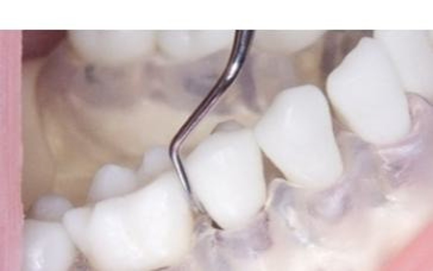
The positioning of the first 1-2mm of the workings ends lateral surface into contact w/ the tooth.
What are the three working ends' sections (what's different between curet and sickle?)
Leading-third, Middle-third, Heel-third. The leading-third for curet is a toe-third and for sickle is a tip-third.
How to find the correct working end of an instrument for posterior teeth
The lower shank will be parallel to the distal surface and the functional shank goes 'up & over' the tooth (V goes towards the D).
How does the face of sickle scalers and curets compare to the lower shank
The face of a curet and a sickle scaler are perpendicular to the lower shank.
How do Anterior sickles typically look?
Often single ended; may have 2 different sickles if double ended.
How do Posterior sickles typically look?
Usually two sickles paired on a double-ended instrument w/ mirrored working ends.
How to achieve correct angulation for sickle scaler for anterior and posterior teeth
Achieved by tilting the lower shank toward the tooth surface & creating an angulation of 70-80°.
Where is the "getting ready zone" for Sickle scaler use on anterior teeth
Begin at the midline and work toward the proximal surface, then turn and lead with the tip in opposite direction.
Where is the "getting ready zone" for Sickle scaler use for posterior teeth?
Begin at distofacial line angle & work back toward the distal surface, then turn and go forward from distofacial line angle toward the facial and mesial surfaces.
What is the correct angulation for supragingival calculus removal
The face-tooth surface angulation is an angle between 45° & 90°, however between 60° & 80° is ideal for the cutting edge to bite into the calc. & fracture the deposit.
What are the three types of forces used for calculus removal
Pinch Pressure, Stabilization, Lateral Pressure.
What is Pinch Pressure
The pressure exerted from fingers in modified pen grasp while holding the instrument handle.
What is stabilization in terms of force
The act of preparing for an instrument stroke by locking the joints in finger and pressing fingertip against surface to gain control (fulcrum).
What is Lateral Pressure
Created by applying pressure w/ index & thumb inward against instrument handle.
When will lateral pressure be applied
Will apply lateral pressure prior to & during instrumentation strokes.
How much lateral pressure should be applied during calculus removal strokes
firm lateral pressure for calculus removal
How much lateral pressure should be applied during root debridement strokes
Less lateral pressure than calculus removal strokes, but still firm
What is "Burnishing", what can it lead to?
Removing only the outer layers of calc. deposits, this causes biofilm to continue living and makes it more difficult to remove when they are smooth.
What should the angulation be during insertion, what is this angle called?
the face-tooth surface angulation should be between 0° and 40°. This is called the closed angle
Stroke width & types during instrumentation
Stroke width will be 1-2 mm. Strokes can vertical, oblique or horizontal, which will lead to a crosshatch pattern
What do Root debridement strokes remove
Subgingival biofilm, residual calculus or surface irregularities from the root surface
What angulation is needed for root debridement strokes
Requires an angulation between 60-70°
Universal curets
Used to remove small & medium sized calculus deposits from the crowns and roots of anterior and posterior teeth.
How is the technique of adapting a universal curet different for anterior teeth
The lower shank of a universal curet will lie across the tooth surface when the correct working end is selected.
Where can universal curets be used
On posterior and anterior tooth, both supragingivally and subgingivally
What is the "Acquired pellicle"
A thin acellular tenacious film of proteins, glycoproteins, carbohydrates and lipids between the tooth surfaces & the oral environment.
When does the pellicle begin forming
Begins to form within minutes after brushing and will be fully formed within 30-90 minutes.
Where is the pellicle the greatest?
Varies in thickness but is greatest near the gingival margin.
What is the purpose of the pellicle?
It protects against acid demineralization, it has some antimicrobial function & keeps surfaces moisturized
What is the negative of the pellicle
It aids in adherence of microorganisms
What is Dental biofilm
A dense nonmineralized complex mass of bacteria colonies.
What is the matrix of the dental biofilm composed of?
The matrix is composed of polysaccharides & proteins that protect the biofilm from the host's immune system.
Biofilm detection methods
With direct vision (stained biofilm picks up extrinsic stains, thick biofilm appears dull with matted fur appearance & thin biofilm is translucent and may need a disclosing agent to see), use of explorer or probe, use of a disclosing agent or using biofilm index clinical record.
Biofilm formation - Step 1
Pellicle Formation = Pellicle provides glycoproteins for microorganisms to adhere to.
Biofilm formation - Step 2
Initial Adhesion = Planktonic bacteria begin attaching to the pellicle and begin to secrete a substance (consisting of Polysaccharides, Glucans and Fructans) that helps filamentous bacteria bind to the pellicle.
Biofilm formation - Step 3
Maturation = More bacteria are attaching and forming microcolonies, maturation occurs by 72 hours.
Biofilm formation - Step 4
Dispersion = The bacteria and colonies are mature and they spread and colonize new areas as the bacteria convert to motile forms.
What is the composition of early (days 1-2) biofilm
Consists of primarily gram-positive cocci (days 1-2)
What is the composition of biofilm after 7 days of being undisrupted
gram-negative anaerobic bacteria is favored
When does gingivitis become clinically evident
10-21 days after biofilm is left undisrupted
Composition of dental biofilm
80% water and 20% microorganisms and matrix that are organic and inorganic
What are the organic elements within dental biofilm
The polysaccharides, they are metabolized by S. Mutans and produce glucans & fructans. Also Proteins, these bind with the glucans and further
support biofilm growth
What are the inorganic elements within biofilm
Calcium, Phosphorus and Fluoride
Are calcium & phosphorus more concentrated in saliva or biofilm?
More concentrated in biofilm
How long does peak fluoride concentration last after it is in the biofilm
The peak lasts for 3-6 hours and then it returns back to its baseline
What can biofilm accumulation initiate?
Dental caries, periodontal infection & the formation of calculus.
What microorganisms initiate the caries process?
Streptococcus Mutans and Streptococcus Sobrinus
What microorganisms help progress a carious lesion
Lactobacilli
Acid formation begins how long after a cariogenic substance comes into contact with the biofilm
immediately
What is the critical pH for demineralization?
5
How long does it take for the biofilm to restabilize its pH
at least 1-2 hours
What is the stabilized pH of biofilm?
6.2 - 7.0
What does "Cariogenic" mean?
producing or promoting the development of tooth decay
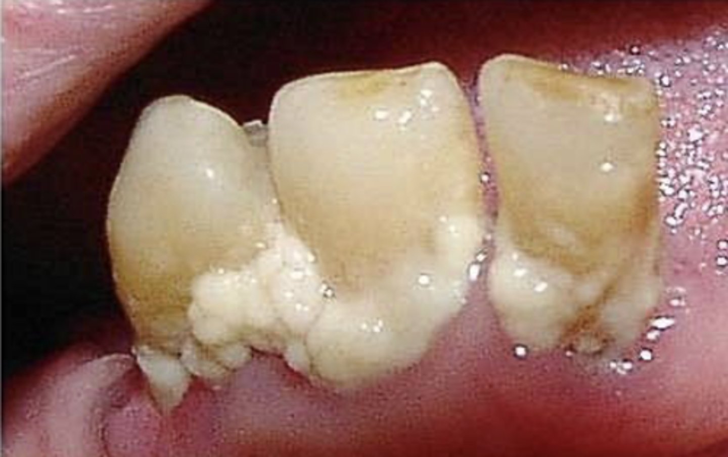
What is Materia Alba and how is it removed?
A soft & white cottage-cheese resembling deposit, it is easily removable.
What is materia alba made up of?
It is made up of living & dead bacteria, desquamated epithelial cells, disintegrating leukocytes, salivary proteins and food debris.
What is calculus
It is hard & tenacious mineralized dental biofilm, filled with crystals of various calcium phosphates and is covered in a layer of nonmineralized biofilm.
What is calculus filled with?
Crystals of various calcium phosphates
What are the common locations for calculus? What is significant about these spots?
Lingual Surfaces of mandibular anterior teeth and buccal surfaces of maxillary molars, they are adjacent to salivary glands
What way does calculus form and what proves this?
Calculus forms in layers that are parallel to the tooth, incremental lines prove this
What do incremental lines prove
Calculus forms in layers that are parallel to the tooth surface.
What is the composition of Calculus
Composed of inorganic & organic elements, with percentages based on the deposit.
What is the organic content of calculus
Lipids & proteins and various microorganisms, desquamated epithelial cells, leukocytes, and mucin.
What is the inorganic content of calculus
Calcium, phosphorus, carbonate, sodium, magnesium, trace elements such as copper, zinc, strontium, fluoride, and calcium phosphate crystals.
What is the percentage of inorganic content of Enamel?
96%
What is the percentage of inorganic content of Dentin?
65%
What is the percentage of inorganic content of Cementum & Bone?
45-50%
What is the percentage of inorganic content of calculus?
Ranges from 58-80%
Where do mineralization centers form in the oral cavity
Form within the biofilm
How long does it take for mineralization centers to be left undisturbed for before they unite & touch
for 24-72 hours
Mineralization first occurs within the _____________ and the __________ provide the matrix for depositing minerals.
inter-microvial matrix & filamentous microorganisms
Where does Supragingival Calculus receive its minerals?
from saliva
Where does Subgingival calculus receive its minerals from?
From sulcular fluid
What increases the amount of sulcular fluid? In turn, what does that increase lead to?
Inflammation increases the amount of sulcular fluid and in turn provides more minerals for the calculus.
What is the average time for mineralization of calculus? How long for rapid/slow calculus growers?
12 days, but only 10 days for rapid growers and 20 days for slow growers
Half of the mineralization of calculus occurs within the _______
Occurs within the first two days
Heavy calculus formers have higher salivary levels of......
Higher salivary levels of calcium and phosphorus
Light calculus formers have higher salivary levels of...
Have higher salivary levels of parotid pyrophosphate
When calculus is attached to the pellicle, it is ______ to remove
Easy to remove
When calculus is attached to minute irregularities, it is ________ to remove
Becomes difficult to remove
When calculus is directly attached to the tooth surface, it is _________
More prone to fracturing while scaling which will leave behind calculus crystals and encourage biofilm formation
How can dental stains be classified?
By their location & their source
What are the two types of "location" stains?
Extrinsic stains and Intrinsic stains
What are extrinsic stains
Stains on external surface, can be removed by brushing, scaling, polishing
What are intrinsic stains
Stains due to changes of structural composition or enamel thickness, cannot be removed but whitening may help with discoloration
What are the two types of "source" stains?
Exogenous and Endogenous
What are exogenous stains
Stains that develop from sources outside the tooth, can become extrinsic or intrinsic
What are endogenous stains
Stans that develops within the tooth, always intrinsic (like dentin discolorations)
What can cause endogenous Intrinsic stains
Occur from traumatized teeth that do not have root canal therapy
Genetic examples of intrinsic stains
Amelogenesis imperfecta & dentinogenesis imperfecta
What can cause Exogenous Intrinsic Stains
Can occur from restorative materials, like amalgam or endodontic therapy
Area specific curets
The cutting edges are curved and have a tilted face (these are our graceys)
Working cutting edge of area specific curets
Only the lower cutting edge is used for calculus removal
Gracey 5/6
Used on anterior teeth, requires complete flipping of the handle to use each working end
Gracey 13/14
Used only for the distal surfaces of posterior teeth
Gracey 15/16
Used for mesial surfaces of posterior teeth
What are the 4 types of scoring methods
Individual assessment score
Clinical trial
Epidemiological survey
Community surveillance
Individual Assessment Score is used for
can be used to measure the amount / condition of an oral disease
Clinical Trials are used for...
to determine the effectiveness of an item on prevention, progression or control of a disease
Epidemiological surveys used for....
to provide info on trends/patterns of oral health & diseases
Community surveillance is uses...
uses a selected population to identify factors that affect oral health