Chapter 11: Membrane Structure
1/59
There's no tags or description
Looks like no tags are added yet.
Name | Mastery | Learn | Test | Matching | Spaced | Call with Kai |
|---|
No analytics yet
Send a link to your students to track their progress
60 Terms
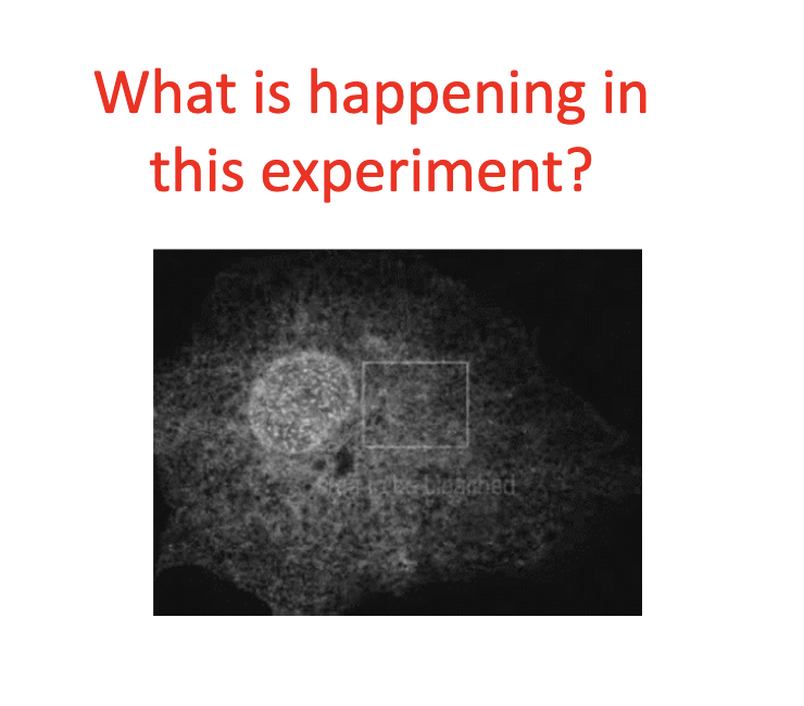
What’s happening in this experiment? Why is it significant?
What is the cell membrane made of?
-2 closely apposed sheets of lipid molecules (approx 5nm AKA 50 atoms) thick
-Includes proteins inserted into the sheet (proteins act as channels, transporters, sensors, and receptors)
Eukaryotes have _____ membranes in addition to plasma membranes.
internal membranes
-internal membranes enclose intracellular compartments and form various organelles, including the ER, golgi, endosomes, and mitochondria
The lipid bilayer of the cell membrane has what reaction to most water soluble molecules?
serves as a permeability barrier
What are the 3 major classes of membrane liquid molecules?
1). Phospholipids
2). Sterols
3). Glycolipids
The lipid bilayer is ______.
fluid
Individual lipid molecules are________.
able to diffuse within their own monolayer. They CANNOT spontaneously flip from one monolayer to another.
The 2 monolayers of the cell membrane__________________.
have different lipid compositions
—> these different compositions reflect the diff functions of the two faces of the cell membrane
How do cells that live at different temperatures maintain their membrane fluidity?
They modify the lipid composition of their membranes
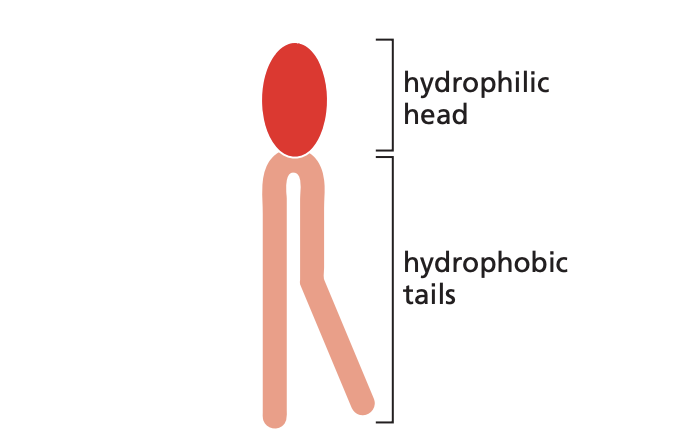
Amphipathic
Having both hydrophobic and hydrophilic regions
—> will have 2 charges and a polar plus a nonpolar end
example: phospholipid, detergent molecule
Bacteriorhodopsin
Pigmented protein found in the plasma membrane of Halobacterium halobium; pumps protons out of the cell and is fueled by light energy.
Cell cortex
Specialized layer of cytoplasm within inner plasma membrane.
this area is rich in actin filaments in animal cells
cholesterol
Short, rigid lipid molecule present in large amounts in the plasma membranes of animal cells, where it makes the lipid bilayer less flexible.
detergent
Soapy substance used to solubilize lipids and membrane proteins.
fat droplet
Large cluster of hydrophobic fats or oils that forms inside the cell
Glycocalyx
Protective layer of carbohydrates on the outside surface of the plasma membrane formed by the sugar residues of membrane glycoproteins, proteoglycans, and glycolipids.
Membrane domain
Functionally and structurally specialized region in the membrane of a cell or organelle; typically characterized by the presence of specific proteins
membrane protein
A protein associated with the lipid bilayer of a cell membrane.
Phosphatidylcholine
Common phospholipid present in abundance in most cell membranes; uses choline attached to a phosphate as its head group.
Phospholipid
most abundant / major type of lipid molecule in many cell membranes
—>have a phosphate-containing,hydrophilic head (either polar or nonpolar) linked to a pair of hydrophobic fatty acid tails
-composed of two fatty acid tails linked to one of a variety of phosphate containing polar groups.
diacylglycerides with small functional head groups linked to the glycerol backbone by phosphate ester bonds. phosphate head is hydrophilic. four most common ones found in mammal cell membranes: choline, ethanolamine (neutral), serine (-), inositol (neutral)--sugar head group can be further phosphorylated
saturated
an organic molecule that has NO double or triple bonds
unsaturated
an organic molecule that contains one or more double or triple bonds between its carbon atoms.
What is the composition of the cell membrane?
-lipids and proteins are held together by non-covalent bonds
-membranes also contain carbohydrates
-the ratio of proteins to lipids can vary based on membrane types
What are the four main types of membrane lipids?
Phospholipids (phosphoglycerides)
Sphingolipids
Glycolipids
Cholesterol
Sphingolipids
ceramides formed by the attachment of a carbohydrate or sugar sugar molecule (amino alcohol to long hydrocarbon tails)
one of the 4 main types of amphipathic membrane liquids, common in neuronal cells
has a sphingosine backbone and amino alcohol with a long hydrocarbon tail
SM = sphyingomyelin
Glycolipids
lipids formed by the attachment of a carbohydrate or sugar molecule
Glycolipids reside in the outer leaflet of the bilayer, often in lipid rafts
*One of the 4 types of amphipathic membrane lipids
Cholesterol
a smaller and less amphipathic lipid component of plasma membranes, only found in animals
amphipathic structural component of plasma membranes. has a polar hydroxyl head and nonpolar hydrocarbon tail.Tail: hydrophobic steroid-hydrocarbon (nonpolar)
Head: hydroxyl (polar)
**One of the 4 types of amphipathic membrane lipids
What are the four most common phospholipids in animal cell membranes?
Choline (neutral charge)
Ethanolamine (neutral)
Serine (negative)
Inositol (neutral, becomes negative when phosphate groups are added)
Experimental evidence of the lipid bilayer
measurement of lipid content as a function of membrane surfce area: Gorter & Grendel extracted lipids from RBCs and found that the lipids were 2x the surface area of the blood cells--bilayer!
Membrane Composition
lipid and protein components bound together by non-covalent bonds. membranes contains carbs, and their protein/lipid ratios vary among membranes.
How would you test the function a given membrane
lipid to the physical properties of biological
membranes?
Reconstitute lipid specific liposomes and test functionality.
What factors affect bilayer fluidity?
lipid head group composition (head groups can pack differently)
fatty acid chain length (long chains decrease fluidity)
fatty acid chain saturation state (saturated decreased fluidity and makes the membrane thicker)
temperature (colder temps have less fluid membranes)
cholesterol concentration (both; increases fluidity of inner portion of leaflets and decreases the fluidity of outer portion of leaflet). helps to stabilize the bilayer by preventing phase separation of lipids into lateral arrays of lipids with similar freezing temps.
Describe the mobility of lipids in membranes (both versions)
no covalent bonds anchor the phospholipid position. they exchange positions with neighbors (10^7/sec). could diffuse the length of an animal cell in 20 sec
phospholipid flips from one leaflet to another every 2 weeks (less common than #1) Serine is the phospholipid that does this the most
Experimental methods for assessing Lipid Mobility?
electron spin resonance: measurements of labeled lipid analogs; detects rotation, flexural rigidity of fatty acid tails, and flip flops
fluorescence recovery after photobleaching (FRAP) of fluorescent lipid probes: this detects lateral diffusion and rate of lipase cleavage of inner leaflet lipids; flip flop. Label plasma membrane with fluorescently-tagged lipid and photo bleach--wait for the fluorescent recovery.
How would you experimentally demonstrate that cholesterol is critical for plasma membrane integrity?
-Culture mammalian cells in medium from
which cholesterol has been removed
-Inhibit the enzyme responsible for cholesterol
synthesis (or select for mutant cells lacking the
enzyme)
-Assay cells lacking cholesterol for resistance to
sheer stress (e.g. pipeting cells up and down)
or to hypotonic stress—cells lacking cholesterol
readily lyse under these stressed conditions.
What are the asymmetric properties of bilayer lipids?
inner and outer membrane leaflets have different lipid compositions. provides different physico-chemical properties appropriate for different interactions. membrane lipids move easily within a leaflet but only rarely flip flop.
an example of lateral asymmetry of bilayer lipids are lipid rafts: Glycolipids, sphingomyelin, cholesterol, as well as selective lateral sorting of membrane proteins can be concentrated in relatively detergent resistant patches of membrane called lipid rafts. Rafts are largely defined based on biochemical studies; in vivo
demonstration of their presence is controversial. rafts are saturated in cholesterol and sphyingomyelin and they widen the membrane.
How can you test for transbilayer asymmetry?
First determine total lipid composition of a membrane
preparation (e.g. rbc plasma membrane)
2. Take intact rbcs and digest outside of membranes with
specific lipases (lipases are enzymes that chews up
lipids)
Determine levels of digested/modified lipids
compared to total membrane.
Fluid-Mosaic Model
core lipid bilayer exists in a fluid state, capable of movement (unless restrained). mosaic proteins form a mosaic of particles penetrating the lipids.
Explain and describe the experimental evidence for the existence of the lipid bilayer
measurement of lipid content as a function of membrane surfce area: Gorter & Grendel extracted lipids from RBCs and found that the lipids were 2x the surface area of the blood cells--bilayer
Does bilayer thickness vary? If so, how and why?
varies with lipid composition; certain transmembrane proteins concentrate/laterally segregate into membranes of a particular thickness.
Membrane Proteins
not required for membrane structure; no specific protein is a component of all membranes--specific to the type of membrane. inner mitochondrial membrane is only 76% protein and myelin is 18%.
Membrane proteins can be peripheral and integral.
plasma-membrane proteins can be anchored to the bilayer by covalently linked hydrocarbon groups.
Peripheral membrane proteins
intact with membrane surface (phospholipid head groups) or with exposed regions of integral membrane proteins. extractable with high or extremely ionic strength, divalent cation (Ca or Mg) chelation, or high pH (low H+). bound to either side through direct lipid interactions or to the cyto/exoplasmic domain of integral proteins.
Integral membrane proteins
embedded in the phospholipid bilayer
two types of integral membrane proteins: one or more peptide transmembrane domains (alpha helices or beta pleated) or lipid anchorage (fatty acylation, GPI). non-embedded domains on one or both sides of the membrane. detergent required for the extraction from the membrane (mild non-ionic detergent). single, multiple or partial spanning, lipid linked
Glycophorin
has a single transmembrane alpha-helical domain.
has 23 hydrophobic amino acids
transmembrane domain forms homodimer.
What is the most common membrane-spanning domain?
hydrophobic alpha helix. is is often flanked on one or both sides of the bilayer by charged or polar amino acids.
helical transmembrane domains of multi-spanning proteins often have polar residues oriented on one side of the helix. This allows for channel formation.
there are a subset of generally membrane pore-forming proteins that span the membrane at beta sheets folded into a beta barrel.
Anatomy of an Integral Membrane Protein
extracellular/lumenal domain can be glycosylated and stabilized by S-S bonds.
How to experimentally determine membrane
protein topography/organization within
the bilayer plane
Freeze fracture TEM of biological membranes: Rapid
freeze cells or tissue in IN2 chilled solvent (e.g.
Freon; isopentane, reduces ice crystals)
1. Place specimen on -150*C specimen stage in
vacuum evaporator.—evacuate chamber
2. Cleave frozen specimen with cooled microtome
blade; for etched samples, raise temp. to 115*C to sublime away ice to expose
cytoplasmic structure and unfractured
membrane surfaces
3. Shadow specimen with metal and carbon; thaw
and remove underlying tissue with bleach; view
replica in TEM
Heavy metal shadowing of specimen for EM: vacuum evaporator for coating specimens with metal (platinum) or carbon. These specimens have been shadowed from a fixed angle. Better resolution obtained if specimen is rotated during shadowing.
Freeze-fracture analysis of the lateral topography of integral membrane proteins: fracture a cell in two portions (E face--external--is sunken in and P face--cytosolic side--is outward). etching occurs when ice is sublimed away to reveal the cytoplasm and surfaces of membranes. etching depth is limited due to limited ice formation and pertubation of subcellular structure.
What are methods to analyze protein lateral mobility?
Redistribution of cell surface antigens after mouse-human cell fusion (Frye & Edidin, ATP didn't affect the membrane, so passive; no new proteins were needed to make it move because they removed glutamine that helped the new proteins to enter the membrane) make primary antibodies and secondary antibodies. use secondary antibodies for immunolocalization and immunoblot analysis. the mechanism that yielded their results was the movement, or "diffusion" of the antigen in the plane of the membrane.
Lectin (multivalent sugar-binding proteins that bind to specific sugars on glycoproteins; can be used fluroescently for localization of specific types of glycoproteins and as tags for FRAP and capping assays) or antibody-infused patching and capping of cell surface proteins: membrane proteins have fluorescent probe. the antibody or lectin induced patching (clustering) and capping of cell surface proteins demonstrates induced lateral movement; you can visually see.
Direct visualization of membrane protein mobility by FRAP and FLIP analysis: use monovalent fluorescent lectins or fluorescently tagged monovalent antibodies to specific proteins to fluroescently tag integral membrane proteins.
FRAP/FLIP visualization
FRAP targets one area to bleach of the cell and measures that area's fluorescence return. sharp drop and gradual lifting up on the graph.
FLIP uses continuous bleaching where you bleach one area and measure another; wait for the bleaching to bleach all of the cell. The graph looks like a skating half-pipe.
Why do some proteins recover quicker than others?
protein mobility may be limited by attachment to cytoskeleton in cytoplasm or extracellular proteins outside of the cell (Lodish, 1990). a subset of major membrane proteins in RBCs are tethered to underlying membrane cytoskeleton.
Methods to restrict the lateral mobility of integral membrane proteins?
lateral clustering, extracellular matrix binding/clustering of IMPs, cytoskeletal restraint of IMPS, and cell adhesion mediated clustering of receptors (two cells together).
lateral segregation of membrane proteins occurs on the surface of sperm, and you can visualize by using antibodies to 3 different sperm membrane proteins.
Transport of Substances Across Cellular Membranes can occur via these 2 methods
Passive diffusion and protein-assisted transport
passive diffusion
method of transporting materials into or out of cells down the concentration gradient and without energy. transports gases, all small nonpolar molecules, and sometimes water. rate of diffusion is proportional to its conc. gradient across the membrane and its hydrophobicity.
protein-assisted transport
transport of ions, sugars, amino acids, and sometimes water need assistance of transport proteins. ATP-powered pumps, channels, and carriers are examples of integral proteins that function as these transport proteins. ATPases use energy from ATP hydrolysis to pump mostly ions across a membrane up the concentration gradient--active transport. 3 examples of pumps: sodium/potassium ion pumps to maintain a low sodium and high potassium concentration (3 Na for every 2 K), hydrogen ion pumps to maintain low pH inside lysosomes and vacuoles, and maintenance of intracellular osmolarity so that pumps can pump out ions that flow into the cell due to their greater concentration of charged molecules in order to prevent the cell to continuously swell with water.
Channel proteins
form a protein-lined tunnel through the membrane; allows for gated transport of water and specific ions down their concentration gradient; molecules move in a single file at 100 million per second.
Carrier/Transporter proteins
bind only 1 to a few molecules then undergoes conformational change, bound molecules transported across the membrane. molecules move across at 100-10,000/second. this form is called facilitated diffusion. 3 types:
uniporters: transports one molecule at a time down the conc. gradient (glucose from ECF in animals)
symporters: transports 2 molecules and ions in the same direction
antiporter: transports 2 molecules in different direction
Uptake of Glucose
proper uptake of glucose requires epithelial cells lining small intestines to have different transport proteins on basal surface than on apical surface (polarized).
on the apical surface, import of glucose is up the conc. gradient and is coupled to import of sodium ion down the conc. gradient by glucose symport protein.
basal surface contains Na/K pump which maintains low cytosolic sodium ion conc., also contains uniport protein that transports glucose down the conc. gradient out of the cell eventually into the blood.
How would you experimentally characterize membrane proteins?
Isolate membranes of interest using methods previously discussed.
2. Extract peripheral proteins with a combination of treatments that differentially
solubilize different classes of peripheral proteins:
e.g. High salt, low salt, high pH, low pH, chelation of divalent cations.
3. Extracted membranes, enriched in integral membrane proteins are then
treated with a mild, nonionic detergent (many to choose from) to solubilize
membrane proteins, hopefully in their native state. Membrane proteins can
then be purified by methods discussed previously.
4. Purified membrane proteins can be reconstituted into liposomes for functional studies.
What are nonionic detergents used for?
to isolate membrane proteins in a "native" conformation.
Reconstitution of an Integral Membrane Protein into Liposomes
solubilize with a nonionic detergent, add detergent solubilized lipids, remove detergent and hope that membrane protein will properly insert into bilayer.