13 - Diencephalon
1/56
There's no tags or description
Looks like no tags are added yet.
Name | Mastery | Learn | Test | Matching | Spaced | Call with Kai |
|---|
No analytics yet
Send a link to your students to track their progress
57 Terms
parts of the diencephalon
epithalamus, subthalamus, thalamus, hypothalamus
nuclei of the dorsal thalamus
afferent and efferent connections
where is the diencephalon located?
mostly hidden from view between the cerebral hemispheres
2% of central nervous system (by weight)
nearly all connections between the cerebral cortex and subcortical structures including the diencephalon travel through the _____.
internal capsule
what part of the diencephalon includes the pineal gland?
epithalamus
what are the borders of the diencephalon?
superior: body of the lateral ventricle
laterally: internal capsule
inferiorly: subarachnoid space
caudally: plane through posterior commissure
rostrally: anterior commissure
hypothalamic sulcus
divides alar plate into superior and inferior portion, differentiates thalamus to hypothalamus
the diencephalon surrounds what ventricle?
3rd ventricle
what does the epithalamus include?
pineal and habenular nuclei
pineal gland is a midline, unpaired structure situated just rostral to the superior colliculi
resembles a pinecone in shape
what is the pineal gland made of? what does it secrete?
connective tissue containing glial cells and pinealocytes, no true neurons
secretes a hormone derived from serotonin - melatonin (during darkness)
what is the pineal gland important for?
regulation of circadian rhythm, sleep wake cycles, involved in seasonal cycles
pineal tumors sx
hydrocephalus in early findings, various deficits in eye movements and pupillary reactions, pineal tumors can cause changes in sexual development
what is the clinical significance of pineal gland?
accrues calcium at about 17 years, opaque in X-rays and shifts in pineal position can indicate expanding masses
where is the habenula?
epithalamus
caudally at the base of the stalk connecting pineal gland to diencephalon is the posterior commissure, rostrally is the habenula
underlying each habenula is habenular nuclei
habenula function and where does it project to?
receives input from basal nuclei and limbic system
project to the reticular formation in the brainstem
how many habenula?
2
(1 pineal gland)
what does the habenula release? what does decreased activity of habenula result in?
biogenic amines from the reticular formation and thought to play a role in assigning reward value to stimuli
habenular nuclei send messages to dopamine and serotonin cells of the brainstem, increase their activity based on how well an individual enjoyed a certain stimulus
lack of activity of the habenula and its projections to the reticular formation may play role in depression
subthalamus nucleus function
receives input from motor areas of cerebral cortex, projects to substantia nigra, reciprocally connected with the globus pallidus, very connected to the basal nuclei
what happens with vascular lesion to subthalamic nucleus?
hemiballismus - involuntary rapdi and forceful movements of contralateral UE
what is prerubral area (Forel’s Field H)?
main length between striatopallidal system and thalamocortical network
connects pathway of basal nuclei and thalamus/cortex
what is the zona incerta? what are its functions?
small mass of gray matter intervening between the subthalamic nucleus and thalamus
involved in sensory-motor programming and cognitive processes related to attention towards important stimuli
primary functions of the subthalamic area?
regulation of movement (basal ganglia)
sensorimotor integration
some cognitive processes
what is the thalamus? how much does it make up diencephalon? boundaries?
pair of large, egg-shaped nuclear masses with a posterior appendage
make up about 80% of diencephalon
extends anteriorly to interventricular foramen, superiorly to the floor of the lateral ventricle, inferiorly to the hypothalamic sulcus, posteriorly overlaps midbrain
what information relays in the thalamus?
all sesnsory pathways (except for olfaction)
many of the anatomical circuits used by cerebellum, basal nuclei, limbic structures
what is the internal medullar lamina?
thin, curved sheet of myelinated fibers in thalamus
divides most of the thalamus into principle cell groups - anterior, medial, lateral, intralaminar nuclear groups
these are midline thalamic nucleus just superior to the hypothalamus sulcus
what is attached to the caudolateral portion of the thalamus?
medial and lateral geniculate bodies
lateral group of thalamic nuclei
composes bulk of thalamus and is further divided into dorsal and ventral tiers

parts of the dorsal tier of lateral nuclei
lateral dorsal nucleus - cingulate gyrus
lateral posterior nucleus - parietal lobe
pulvinar - visual function, eye movements

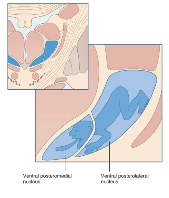
what is in the ventral tier of the thalamus?
VA - motor related
VL - motor-related
VP - includes VPL and VPM which are somatosensory info
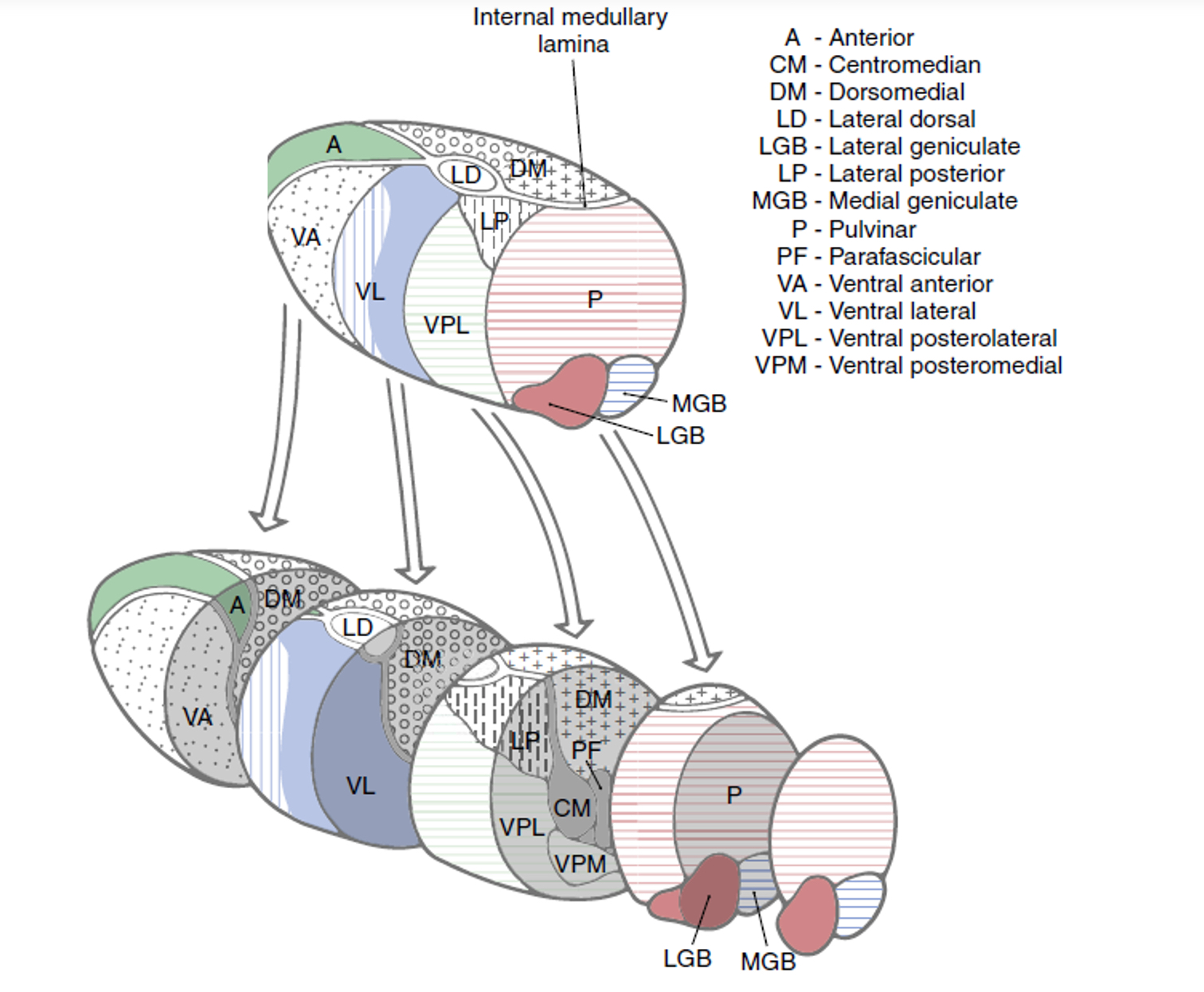
parts and functions of the geniculate nuclei?
lateral geniculate nucleus - afferents from retina via optic tract and projects to the primary visual cortex
medial geniculate nucleus - afferent auditory input via brachium of inferior colliculus and projects to the primary auditory cortex

where are the geniculate nuclei located?
posterior to the ventral tier nuclei and inferior to the pulvinar, protrude posteriorly alongside the midbrain
functions of the thalamus
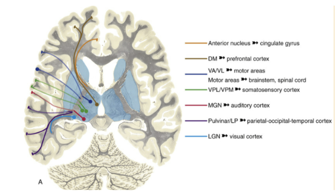
specific or relay nuclei - subcortical structures → cerebral cortex
process, integrate and relays information realted to sensory, motor and limbic system
anterior nucleus
limbic system - cingulate gyrus
dorsomedial nucleus
orbitofrontal cortex
frontal cortex mostly
lateral dorsal nucleus
input to limbic cortical
pulvinar
eye movements, visual function
ventral anterior, ventral lateral
motor
ventral posterior (VPL, VPM)
somatosensory, information from sensory contralateral side of body (VPL), medial lemniscus, spinal trigeminal fibers (VPL), principal trigeminal fibers (VPM)
the somatosensory is organized somatotopically and by function
what is the motor relay nuclei pathway?
cerebellum → VL → M1, M2, SMA
basal nuclei → globus pallidus → VA → prefrontal cortex, frontal eye field
basal nuclei → globus pallidus → VL → SMA, premotor area
limbic relay
hippocampus connects to mammillary bodies
mammillothalamic tract to the anterior nucleus
connections to the cingulate gyrus
internal capsule radiates up to:
thalamocortical
corticothalamic
cortex → subcortical
pathway of cortex to thalamus
corona radiata → internal capsule
cortex associated with parts of thalamus
anterior and posterior limb of internal capsule
anterior - contains the fibers interconnecting the anterior nucleus and the cingulate gyrus, most of those interconnecting the dorsomedial nucleus and prefrontal cortex
posterior - contains fibers interconnecting VA and VL with motor areas of the cortex. contains corticospinal and corticobulbar fibers and the somatosensory fibers projecting from VPL/VPM to the postcentral gyrus
what is internal capsule genu?
transition zone between the anterior and posterior limb and contains some corticobulbar fibers to CN motor nuclei
hypothalamus role
controlling internal environment
modulates ANS, receptors sense and respond to temperature, osmolality, hormones, produces releasing factors to regulate hormone production, releases neurohormones into circulation includes satiety center
hypothalamus parts
3 sections
lateral hypothalamic area - cardiorespiratory function and regulation of food and water intake
medial zone - “meat and potatoes” of hypothalamus
periventricular zone - includes neurons that border ependymal surfaces of the third ventricle
chiastmatic region of medial zone
chiasmatic region - preoptic, supraoptive, paraventricular, anterior and suprachiasmatic nuclei
regulate hormone release, circadian rhythm, body temperature and heat loss mechanisms
tuberal region of medial zone
dorsomedial, ventromedial and arcuate nuclei
ventromedial - satiety center
arcuate - hormonal regulation
mammillary region
posterior nucleus and mammillary nuclei
projects tot eh anterior thalamic nuclei via mammillothalamic tract
neurons of posterior nucleus involved in elevation of blood pressure, pupillary dilation, shivering or body heat conservation
mammillary nuclei are involved in control of reflexes associated with feeding and memory formation
blood supply to the thalamus
posteromedial branches of the posterior cerebral artery and posterior communicating artery
including thalamogeniculate and thalamoperforating artery
thalamogeniculate a supplies
ventral and posterolateral and posteromedial part of thalamus
thalamoperforating a supplies
anterior portion of thalamus and choroid plexus
symptoms of a loss of blood flow to the thalamus and internal capsule
contralateral hemiparesis combined with hemianesthesia
branches of posterior cerebral artery - thalamic pain syndrome and “Pusher syndrome”
thalamic pain syndrome
affects 25% of patients that suffer a stroke to the thalamus region
most commonly implicated artery is the thalamogeniculate artery
sx: contralateral sensory loss, astereognosis, progresses to burning, scalding, usually vauge, allodynia (pain from touch), delayed gradual onset of pain sx
Pusher syndrome
often damage to right side of brain
while sitting or standing pushes away from not injured side until they fall towards hemiparetic side - no proprioception on that side, thinks leaning over is upright