Cerebral Cortex, Thalamus, and Pathways - Lecture
1/61
Earn XP
Description and Tags
Gross Anatomy
Name | Mastery | Learn | Test | Matching | Spaced | Call with Kai |
|---|
No analytics yet
Send a link to your students to track their progress
62 Terms
What is the thalamus?
The thalamus is a small, egg-shaped structure in the brain that acts as a relay station for most sensory information (except smell). It processes and transmits most sensory information to the cerebral cortex for interpretation.

What is the Lateral Geniculate Nucleus (LGN)?
The LGN is a thalamic nucleus that serves as a visual relay. All visual information from the retina, via the optic tracts, travels to the LGN for processing before being sent to the primary visual cortex in the occipital lobe.
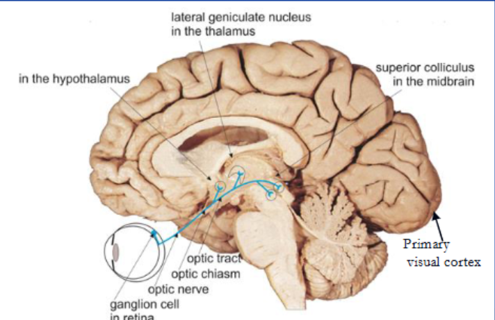
What are the superior colliculi?
Superior colliculi are paired dorsal midbrain structures involved in processing optical stimuli, orienting attention, and coordinating eye and head movements.
Where is the primary visual cortex located?
The primary visual cortex is located in the occipital lobe.
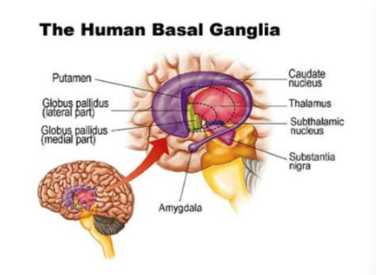
What are the basal ganglia?
The basal ganglia are a subcortical collection of interacting clusters of cell bodies, involved in reward, emotional, and motor circuits.

What are the components of the basal ganglia?
Caudate, Putamen, Nucleus Accumbens, Globus Pallidus, Substantia Nigra, Subthalamic Nucleus.
How do disorders affecting the basal ganglia impact patients?
Disorders like Parkinson's disease can significantly disturb motor control, including eye movements, affecting a patient's ability to scan and focus effectively.
Parkinson’s is caused by a diminished substantia nigra
What is the nucleus accumbens?
The center of joy in the CNS. It helps explain how motivation and emotion influence a patient's visual attention, learning, and therapeutic engagement.
What is the limbic system?
The system that influences emotions, the visceral responses to those emotions, motivation, mood, and sensations of pain and pleasure.
It gives "meaning" to what the eyes see, integrating raw visual data with emotions, memory, and behavior.
What do the somatosensory pathways consist of?
Ascending pathways from the body to the postcentral gyrus/somatosensory cortex in the cerebral cortex.
What are the main sensory tracts in somatosensory pathways?
Posterior column/medial lemniscus system (DC/ML)
Anterolateral system (ALS) (Spinothalamic tract)
Spino-cerebellar tract
Trigeminal sensory system
What is the posterior-column medial lemniscus system (DC/ML) responsible for?
Fine discriminative touch, including touch, pressure, proprioception, and vibration perception
Enables us to "read" raised letters with our fingertips or describe the shape and texture of an object without seeing it.
What is the anterolateral system (ALS)/ Spinothalamic tract responsible for?
Pain, temperature, and crude touch
What are the spino-cerebellar tracts responsible for?
Proprioception
What is the trigeminal sensory system responsible for?
Transmission of tactile, proprioceptive, pain, and temperature information from the head to the cerebral cortex, cerebellum, and reticular formation.
What are the types of sensory receptors?
Mechanoreceptors
Nociceptors
Thermoreceptors
Proprioceptors
What are the types of mechanoreceptors?
Merkel's disks, Meissner's corpuscles, Ruffini endings, and Pacinian corpuscles.
What are nociceptors?
Free nerve endings that detect pain and temperature
What are thermoreceptors?
Specialized nerve endings that detect temperature
What are proprioceptors?
Found deep in muscles and joints, these receptors sense the position and movement of body parts, providing the brain with information about body awareness.
What is the role of first-order neurons?
Carry signals from sensory receptors to the central nervous system (CNS).
Cell body location: For the body, dorsal root ganglion; for the face, trigeminal ganglion.
What is the trigeminal ganglion?
The trigeminal ganglion is the cell body location for first-order neurons from the face.
What is the role of second-order neurons?
Relay the signal from the first-order neuron to the thalamus.
Location of synapse with 1st order neuron:
Body: Dorsal horn of spinal cord or medullary nuclei.
Face: Trigeminal sensory nuclei in the brainstem.
What is the role of third-order neurons?
Transmit the signal from the thalamus to the cerebral cortex.
For the body: Ventral posterolateral (VPL) nucleus to cerebral cortex.
For the face: Ventral posteromedial (VPM) nucleus to the cerebral cortex.
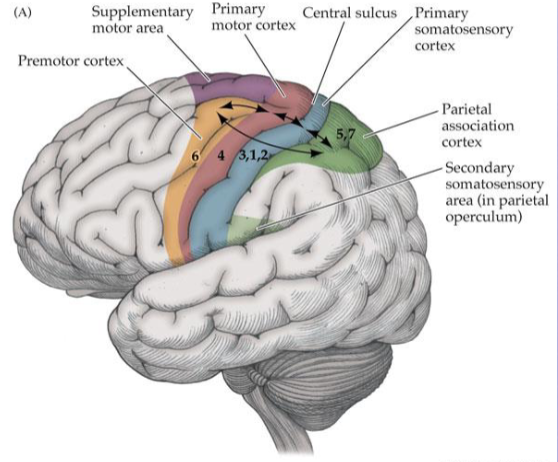
What is the progression in the somatosensory cortex?
Primary somatosensory cortex → secondary somatosensory cortex (parietal operculum) → parietal association cortex (posterior parietal lobule 5 & 7).
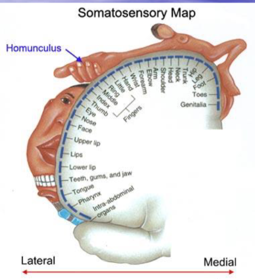
What is a homunculus?
A homunculus is a representation of the body in the somatosensory cortex.
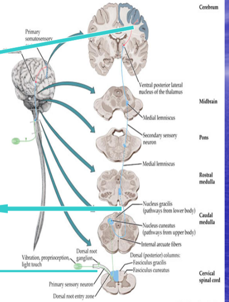
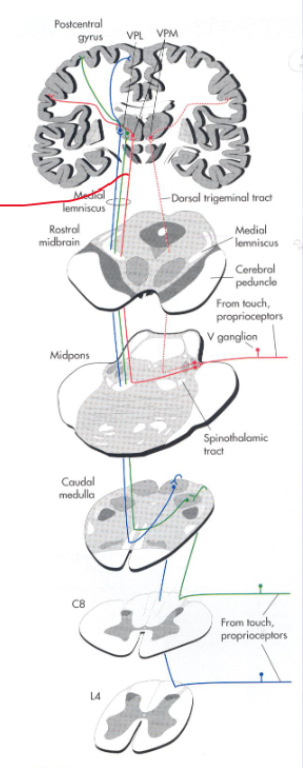
Describe the discriminative touch system (Dorsal column/Medial lemniscus).
Primary or 1st order neuron.
Crossing point: 2nd order neuron.
3rd order neuron: Leaving the VPL Nucleus of the thalamus and ending in the postcentral gyrus.
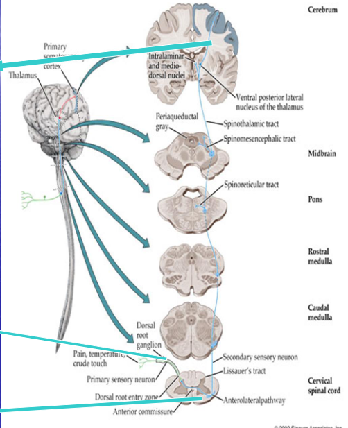
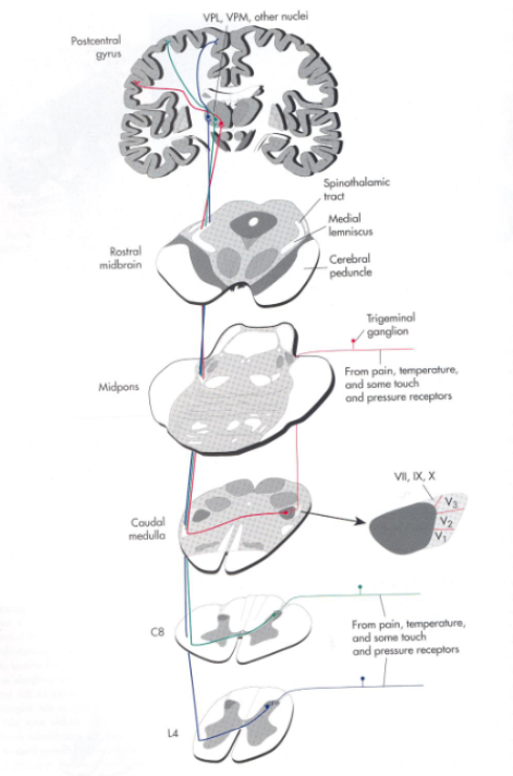
Describe the pain and temperature system (Anterolateral/spinothalamic system).
Primary or 1st order neuron.
Crossing point: 2nd order neuron.
3rd order neuron: Leaving the VPL Nucleus of the thalamus and ending in the postcentral gyrus.
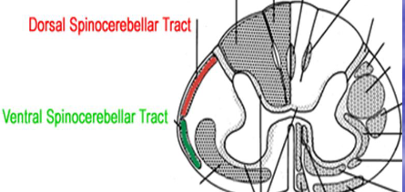
What is the spinocerebellar tract?
Proprioceptive system
Has muscle spindles = major stretch receptor within muscles.
Has golgi tendon organs and joint afferents that monitor stresses and forces at tendons and joints.
Primarily targets the cerebellum → needs minute-by-minute feedback on what the muscles are doing
Arises from primarily the Aα (largest and fastest axons) afferents entering the spinal cord.
What are A alpha nerve fibers?
Proprioception and motor fibers, have very fast conduction velocity
What are A beta nerve fibers?
Pressure and touch fibers
What are A delta nerve fibers?
Fast pain detecting fibers
Why do sensory fibers cross when going to the cerebral cortex?
Because the cerebral cortex operates on a contralateral (opposite side) basis.
Where does the discriminative touch system cross?
High in the medulla.
Where does the pain system cross?
Low in the spinal cord
Where do proprioceptive system fibers cross? (spinocerebellar tract)
The go straight to the cerebellum and they do not cross, because the cerebellum works ipsilaterally

What is the trigeminal sensory system responsible for?
Responsible for transmission of tactile, proprioceptive, pain, and temperature information from the head to the cerebral cortex, cerebellum, and reticular formation.
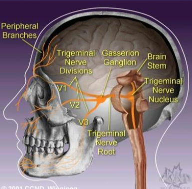
What are the three main branches of the trigeminal nerve?
Ophthalmic (V1), Maxillary (V2), Mandibular (V3).
Cell bodies in Trigeminal (Semilunar, Gasserian) Ganglion, which is the cranial nerve equivalent of a dorsal root ganglion.
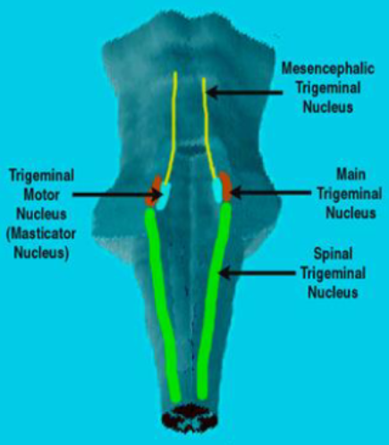
What are the 4 nuclei associated with the trigeminal system?
1. Mesencephalic nucleus. 2. Sensory nucleus. 3. Motor nucleus. 4. Spinal nucleus.
Describe the pathway for touch and proprioception in the trigeminal system.
1st afferents: Trigeminal Ganglion.
2nd order neurons: Main sensory nucleus. Path: Medial lemniscus, dorsal trigeminal tract.
3rd order neuron: VPM to postcentral gyrus. Ventral Trigeminal Tract.
Describe the pathway for pain and temperature in the trigeminal system.
1st afferents: Trigeminal Ganglion.
2nd order neurons: Spinal Trigeminal nucleus. Path: Spino-thalamic tract.
3rd order neuron: VPM to postcentral gyrus.
What is the corneal blink reflex?
Tests trigeminal nerve function.
Mediated by poly-synaptic connections in the brainstem between the trigeminal (CN V) and facial (CN VII) nerves.
Can be impaired by lesions anywhere in this circuit.
What is trigeminal neuralgia (TN)?
Excruciating, lightning strikes of facial pain, typically near the nose, lips, eyes, or ears.
No sensory loss.
Most often in the distribution of Maxillary or Mandibular branches.
Usually unilateral.
Onset age is after 40, More common in women. Unknown etiology.
What are reflexes?
Actions performed involuntarily in response to impulses sent to the central nervous system (spinal cord)
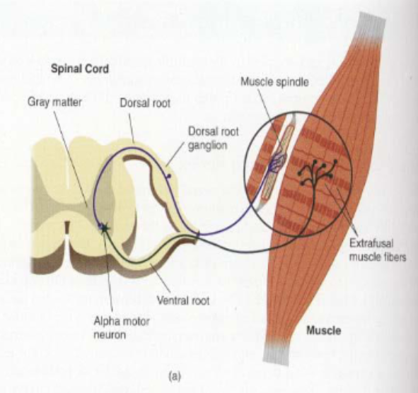
What is the stretch reflex?
Elicited in response to stimuli to tendons.
Normally, when a specific area of the muscle tendon is tapped with a soft rubber hammer, the muscle fibers contract.
Monosynaptic.
Ex: patellar reflex
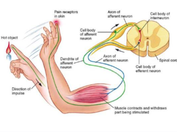
What is the withdrawal reflex?
When you touch a hot object, you quickly pull your hand away using the withdrawal reflex.
Has two synapses/three neurons involved (polysynaptic)
What is the trigeminal system reflex?
Blink or corneal reflex, which is polysynaptic.
What are motor pathways?
Motor pathways originate in the brain or brainstem and descend down the spinal cord to control the motor neurons.
Can be pyramidal or non-pyramidal.
What are pyramidal tracts?
Tracts that provide voluntary control and go through the pyramids.
Corticobulbar tract: Controls the voluntary muscles of the head and face.
Corticospinal tract: Controls voluntary movement of the trunk and limbs.
What are extrapyramidal tracts?
Tracts that provide involuntary control.
Originate in the brainstem
Responsible for involuntary and automatic movements, such as muscle tone, balance, posture, and coordination.
Heavily involved in the automatic reflexes and postural adjustments related to vision.
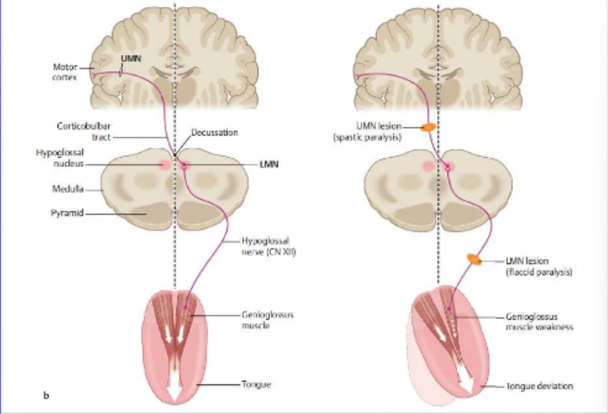
What is the pathway of the corticobulbar tract?
Origin: Motor cortex (face/head region).
Descends through genu of internal capsule → medial cerebral peduncle (crus cerebri).
Synapses on cranial nerve motor nuclei (V, VII, IX/X via nucleus ambiguus, XII) in midbrain, pons, and medulla.
Bilateral innervation except for lower face (CN VII) and part of CN XII, which are contralateral.
Controls voluntary movement of face, head, and neck muscles.
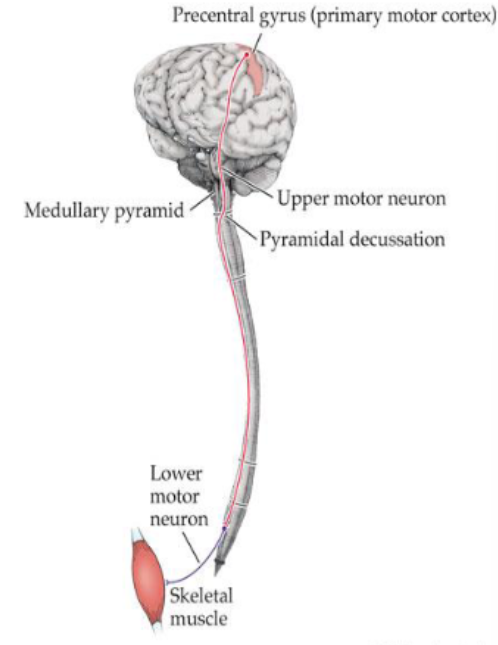
What is the corticospinal tract (CST)?
A collection of axons that carry movement-related information from the cerebral cortex to the spinal cord.
What is the lateral corticospinal tract?
Corticospinal axons that decussate in the lower medulla level
85% of lateral CST
What is the anterior corticospinal tract?
The uncrossed fibers of the corticospinal tract (15%).
What are upper motor neurons (UMNs)?
Motor neurons that start in the motor cortex of the brain and terminate within the medulla or within the spinal cord.
Damage to UMNs can result in exaggerated reflexes of muscles (spastic paralysis)

What are lower motor neurons (LMNs)?
Motor neurons that go from the spinal cord to a muscle.
The cell body is in the spinal cord, and its termination is in a skeletal muscle.
Loss of LMNs leads to weakness, twitching of muscle, and atrophy of muscles (flaccid paralysis).
What happens with damage to the direct corticospinal pathway?
Loss of fine motor control, skilled movements.
Hemiparesis (weakness).
Hemiplegia: Paralysis affecting only one side of the body, commonly caused by lesions of the corticospinal tract.
Corticospinal neurons are UMNs, so their death does NOT result in muscle atrophy.
What is Babinski's reflex?
Occurs when the great toe flexes toward the top of the foot and the other toes fan out after the sole of the foot has been firmly stroked.
Presence of a Babinski's reflex indicates damage to nerve pathways connecting the spinal cord and the brain (the corticospinal tract).
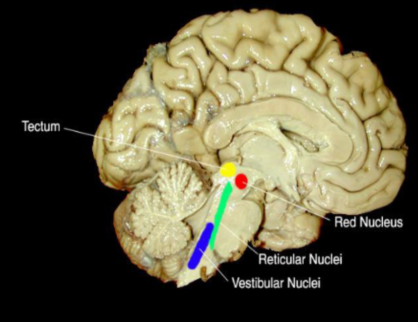
What are the indirect or extrapyramidal pathways?
UMN pathways that originate in the brainstem.
Axons do not run in the medullary pyramids.
Generally under direct control from the cortex.
Play an important supporting role in regulating posture and reflexes, and aid in voluntary movements.
Includes: Tectospinal Tract, Rubrospinal Tract, Reticulospinal Tracts, Vestibulospinal Tracts.

What happens with loss of cortical control (as in coma)?
Releases brain stem postures and leads to decorticate regidity or decerebrate state.
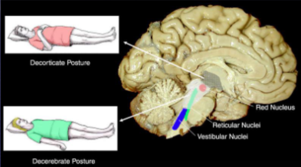
What is decorticate regidity?
Flexion of the elbows and wrists with extension of the legs and feet.
Causative lesion is located above the red nuclei and usually consists of diffuse cerebral damage.

What is the decerebrate state?
Extensor reflexes are exaggerated leading to rigid extension of the limbs and hyperreflexia and opisthotonus (spasm in which head, neck, and spine are arched backwards).
Usually caused by lesions in the brainstem region between the red nuclei and the vestibular nuclei.