Brain Anatomy II
1/58
Earn XP
Description and Tags
Brain Anatomy II quiz II from MRIQUIZ
Name | Mastery | Learn | Test | Matching | Spaced | Call with Kai |
|---|
No analytics yet
Send a link to your students to track their progress
59 Terms

letter A in image 136 is pointing to
anterior frontal vein

letter B in image 136 is pointing to
posterior frontal vein

Letter C in Image 136 is pointing to:
Parietal veins

Letter D in Image 136 is pointing to:
Superior sagittal sinus

Letter E in Image 136 is pointing to:
Torcular herophili

Letter F in Image 136 is pointing to:
Internal jugular vein

letter G in image 136 is pointing to:
right transverse sinus

Letter H in Image 136 is pointing to:
Superior sagittal sinus

Letter J in Image 136 is pointing to
right parietal veins

Letter K in Image 136 is pointing to:
vein of trolard

Letter L in Image 136 is pointing to:
left parietal veins

Letter M in Image 136 is pointing to:
Left sigmoid sinus

Letter N in Image 136 is pointing to:
Torcular herophili

Letter O in Image 136 is pointing to:
Superior sagittal sinus
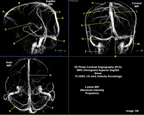
Letter P in Image 136 is pointing to:
Left transverse sinus

letter Q in image 136 is pointing to
right transverse sinus

Letter R in Image 136 is pointing to:
Basal vein/rosenthal vein

Letter S in Image 136 is pointing to:
Internal cerebral vein

Letter AA in Image 137 is pointing to:
cerebellar tonsils

Letter A in Image 137 is pointing to
internal carotid artery

Letter B in Image 137 is pointing to:
Caudate nucleus

Letter C in Image 137 is pointing to:
whtie matter

Letter D in Image 137 is pointing to
thalamus

Letter E in Image 137 is pointing to:
Gray matter

Letter F is Image 137 is pointing to:
straight sinus

Letter G in Image 137 is pointing to:
lateral ventricles
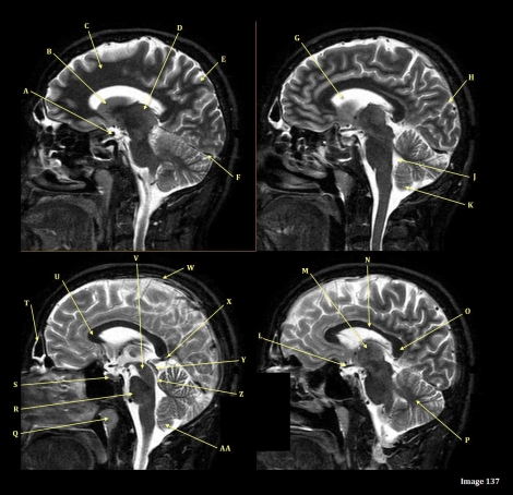
Letter H in Image 137 is pointing to:
Sulcus

Letter J in image 137 is pointing to
fourth ventricle

Letter K in Image 137 is pointing to:
Cerebellum

Letter L in image 137 is pointing to:
optic chiasm

Letter M in Image 137 is pointing to:
Thalamus

Letter N in Image 137 is pointing to what type of tissue?
White matter

Letter N in Image 137 is pointing to:
body of corpus callosum

letter O in image 137 is pointing to
splenium of corpus callosum

Letter P in Image 137 is pointing to:
cerebellum

Letter Q in image 137 is pointing to
uvula

Letter R in Image 137 is pointing to:
Pons

Letter S in Image 137 is pointing to:
pituitary gland

letter T in image 137 is pointing to:
frontal sinus

Letter U in Image 137 is pointing to:
Genu of corpus callosum

Letter V in Image 137 is pointing to:
Cerebral peduncle

Letter W in Image 137 is pointing to:
Superior sagittal sinus

Letter X in Image 137 is pointing to:
Vein of galen

Letter Y in Image 137 is pointing to:
Inferior colliculus of midbrain

Letter Z in Image 137 is pointing to:
Aqueduct of Sylvius

Letter A in Image 133 is pointing to:
VertebroBasilar junction (V-B)

Letter B in image 133 is pointing to:
left external carotid artery

Letter C in Image 133 is pointing to:
left common carotid bifurcation
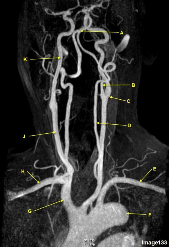
Letter D in Image 133 is pointing to:
left vertebral artery

Letter E in Image 133 is pointing to:
Left subclavian artery

Letter F in Image 133 is pointing to:
Thoracic aorta

Letter G in Image 133 is pointing to:
Brachiocephalic artery/innominate artery

Letter H in Image 133 is pointing to:
right subclavian artery

Letter J in Image 133 is pointing to:
right common carotid artery

Letter K in Image 133 is pointing to:
Right internal carotid artery

Image 54 is an MRI of the _________.
TMJ

Letter A in image 54 is pointing to:
mandibular condyle

Letter B in Image 54 is pointing to:
Articular disk
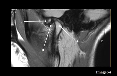
Letter C in Image 54 is pointing to:
Articular tubercle