Chapter 12 part 2
1/41
There's no tags or description
Looks like no tags are added yet.
Name | Mastery | Learn | Test | Matching | Spaced | Call with Kai |
|---|
No analytics yet
Send a link to your students to track their progress
42 Terms
Neuroglial cells protect the neurons and help them
function and outnumber neuron
CNS - 4 types
PNS - 2 types

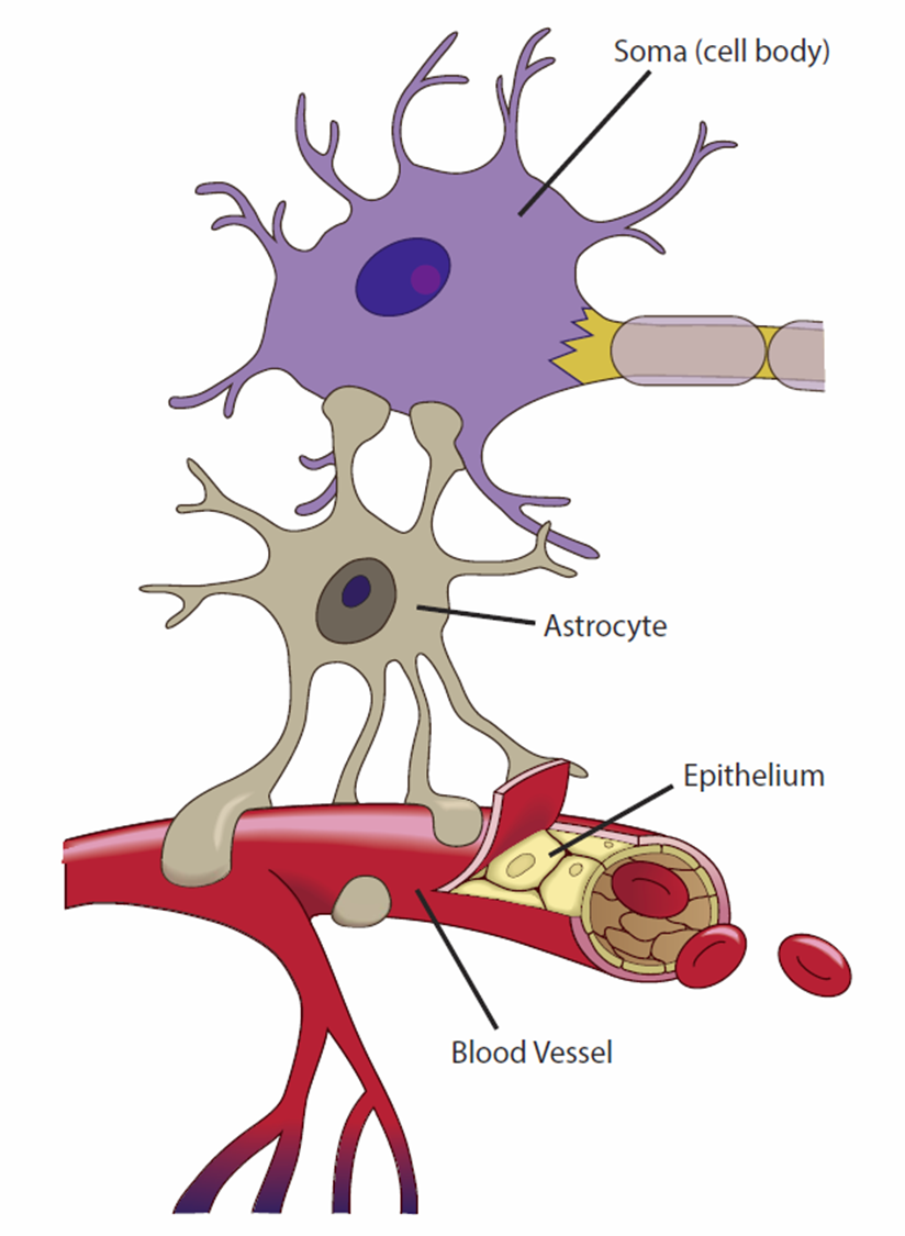
Astrocytes are the most abundant
glial cells in the CNS and constitute over 90% if the tissue in some areas of the brain

Astrocytes cover the entire brain surface and most
Non synaptic regions of the neurons in the gray matter

They are named for their many branched, somewhat starshaped and they have the most
diverse functions of any glia

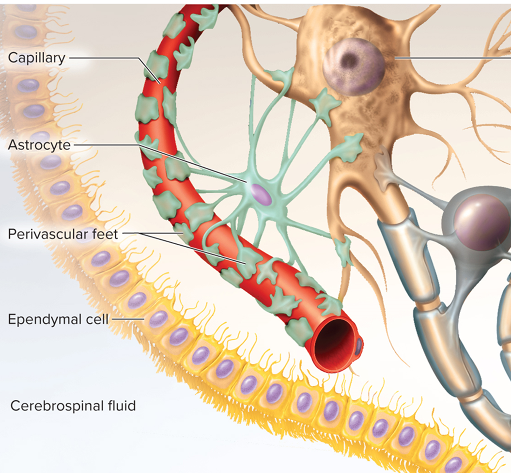
Astrocytes have extensions called perivascular feet, which contact the
blood capillaries and stimulate them to form a tight, protective seal called the blood-brain barrier

Astrocytes convert blood glucose to the lactate and supply this to the
neurons for nourishment

Astrocytes promote
synapse formation and fine-tune neural circuitry

When neurons are damaged, astrocytes form a harden scar tissue and eventually fill
space occupied by the neurons - this process is called Astrocytosis (or sclerosis)
Astrocytes regulate the composition of tissue fluid. When neurons transmit signals, they release
neurotransmitters and potassium ions
astrocytes absorb these and prevent them from reaching excessive levels in the tissue fluid

Astrocytes secrete nerve growth factors that
regulate nerve development
use lactate for energy
Astrocytes
gap junctions connect astrocytes
communication
slow Ca2+ pulses
chemical messengers
influence neuron functions
information processing
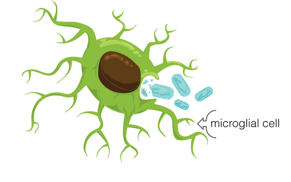
Microglia cells are small macrophages that develop from
white blood cells called monocytes

Microglia cells wander through the CNS, putting out fingerlike extensions to constantly probe the
tissue cellular debris or other problems
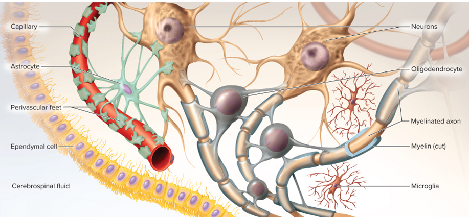
Microglial cells are thought to perform a complete checkup on the
brain tissue several times a day

Microglial cells become concentrated in areas damaged by
infection, trauma, or stroke

Pathologists look for clusters of microglia in brain tissue as a clue to sites of
injury or degeneration
microglia also aid in synaptic remodeling, changing the connections between neurons
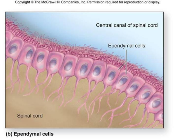
Ependymal cells produce cerebrospinal fluid (CSF), a liquid that
bathes the CNS and fills its internal cavities
line the internal cavities of brain and spinal cord (SC)

Ependymal cells have patches of cilia on their
apical surfaces that help to circulate the CSF
permeable barrier
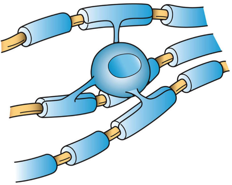
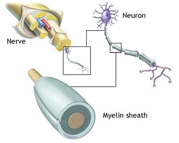
Oligodendrocytes have bulbous body with as many as 15 arms and each reaches out to an axon and
spirals around it like the electrical tape wrapped repeatedly around a wire

The wrapping in oligodendrocytes is called the
myelin sheath, insulates the axon from the extracellular fluid and speeds up signla conduction in the axon

PNS Neuroglial cells: Satellite cells surround the nerve cell bodies in
ganglia of the PNS, and provide insulation around the cell body and regulate the chemical environment of the neurons
Similar functions as astrocytes
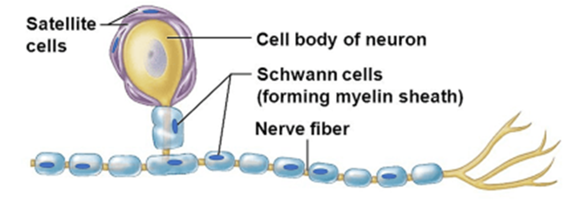
Schwann cells wind repeatedly around an axon and produces a
myelin sheath similar to the one produced in oligodendrocytes in the CNS

Schwann cells also assist in the
regeneration of damaged axons

Myelination: Grey matter
unmyelinated structures
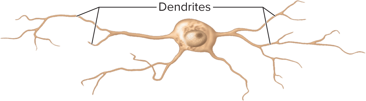
Cell bodies, dendrites
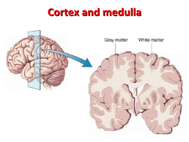
Myelination: white matter
myelinated axons
signals being sent
Myelin sheaths consist of the
plasma membranes of these glial cells, it is made of protein and lipids, but has some unique lipids
lipid rich (80%)

The myelin sheath is segmented, each gap between segments is called a
myelin sheath gap (node of Ranvier)

In the CNS, each oligodendrocyte reached out to myelinate several axons in its immediate vicinity. Since it is
anchored to multiple axons, it can’t migrate around any one of them like Schwann cell does

In the PNS, a Schwann cell spirals repeatedly around a single axon, laying down up to
100 compact layers of its own membrane with almost no cytoplasm between the membranes
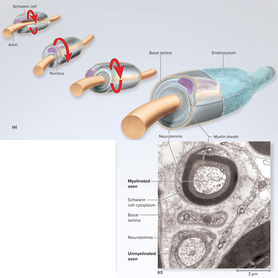
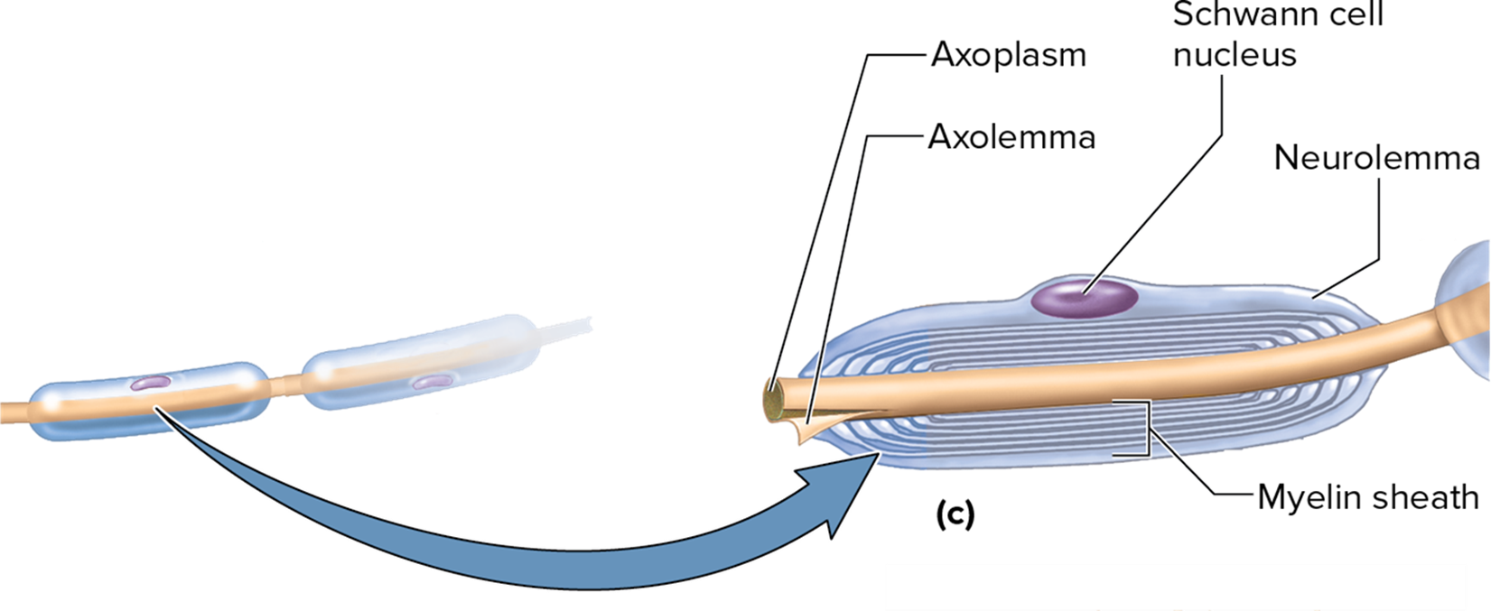
These layers constitute the myelin sheath, the Schwann cell spirals outward as it wraps the axons, finally ending with a
thick outermost coil called the neurolemma
Myelin sheath: PNS
No channel or carrier proteins
“Velcro” proteins, holds Schwann cells around the axon
Nodes of Ranvier
collateral branches
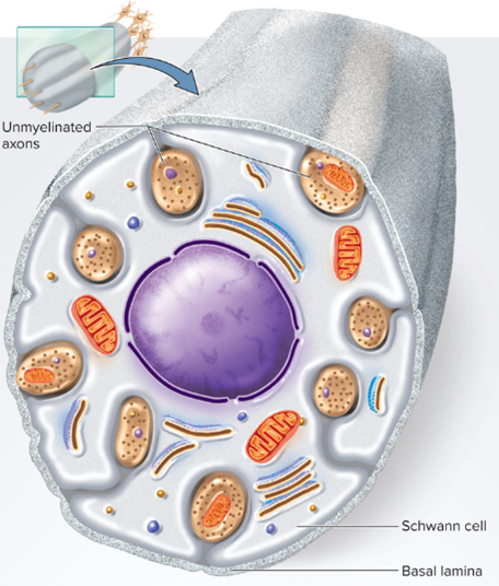
In the PNS even the unmyelinated axons are enveloped in Schwann cells, its membrane, doesn’t spiral repeatedly around the
axon as it does in myelin’s sheath, but folds once around each axon and may overlap itself along the edges
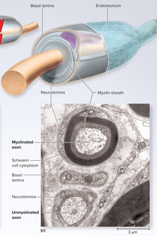
This wrapping is the neurolemma, several unmyelinated axons travel through individual
channels in the shared Schwann cell
a basal laminal surround the entire Schwann cell along with its axons
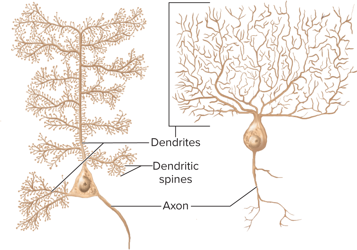
Structural Class: Multipolar neurons are those, like the preceding, that have
one axon and multiple dendrites
this is the most common type and includes most neurons of the brain and spina cords (CNS)
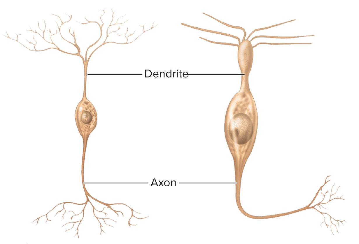
Structural class: Bipolar neurons have one axon and one
dendrite
examples include olfactory cell of the nose, neurons of the retina, and sensory neurons of ear

Structural Class: Unipolar Neurons have only a single
process leading away from the cell body
are represented by the neurons that carry signals to the spinal cord for such senses as touch and pain
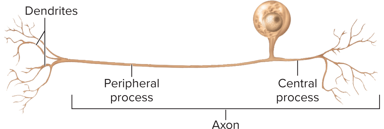
Unipolar neurons are a short distance away from the cell body, the process branches like a T into a
peripheral process and a cental process

In unipolar neurons the dendrites are considered to be only the short receptive ending. The rest of the process, both peripheral and central, is the
axon, distinguished by the presence of myelin and the ability to generate action potentials
Axon neurons have multiple
dendrites but no axon
communicate locally through their dendrites but don’t produce action potentials
in the retina they help visual processes
Functional classes: Sensory
Stimulus info —> CNS
Unipolar
Bipolar
Functional classes: Interneurons
integration
typically, multipolar
99% neurons
Functional classes: Motor
Signals to muscles and glands (effectors)
Multipolar