Basic Components of Living systems
1/38
There's no tags or description
Looks like no tags are added yet.
Name | Mastery | Learn | Test | Matching | Spaced |
|---|
No study sessions yet.
39 Terms
Define Magnification
the number of times larger an image is compared to the actual size of the object being viewed.
Define resolution
What is the resolving power of the unaided human eye?
the ability to see two objects as separate objects. The higher the resolution the more detail that can be seen.
Explain Cell Theory
both plant and animal tissue is composed of cells
cells are the b basic unit of all life
cells only develop from existing cells
Outline the development of cell theory
why was it not fully developed before the mid 19th century?
1665- cell first observed- Robert Hooke- thinly sliced cork- early light microscope - described compartments as cells- was dead plant tissue so he was looking at cell walls.
1674-1683- Anton van Leeuwenhoek- handcrafted microscopes- examined pond water samples- described bacteria and protoctists as animalcules(micro-organisms)- then observed other cells.
1832- evidence for the origin of new plant cells - new cells arise from within old cells.
1833- nucleus first observed- Robert Brown
1837-1838- the birth of universal cell theory -proposed that all plant tissues are composed of cells - Theodor Schwann - all living things are composed of cells and cell products.
1844 (55)- evidence for the origin of new animal cells - Robert Remak- observed cell division- Rudolf Virchow took his forgotten findings and published them a decade later.
1860- Louis Pasteur disproved the theory of spontaneous generation of cells by demonstrating that bacteria would only grow in a sterile nutrient broth after it had been exposed to the air.
Before the mid-19th century a lot of science was influenced by religion so many people were quite stuck in their beliefs and unwilling to accept new ideas, more importantly, they did not have the developed technology to provide clear evidence for their ideas.
what is the equation to calculate magnification?
magnification= image size/ actual size
Light microscope
maximum magnification
maximum resolution
purpose- what is it used to view?
how does it work?
advantages
disadvantages
x 2000
200nm
visualises whole cells and their larger components/tissues
contains 2 lens to magnify the images (objective lens and eyepiece lens). Specimen is illuminated by light from underneath it.
Specimen can be living or dead, vacuum is not required, natural colour of sample is seen, sample preparation does not usually lead to distortion, small, portable, inexpensive.
resolution is the limiting factor- if the image is blurred no more detail will be seen.
Scanning Electron microscope
maximum magnification
maximum resolution
purpose- what is it used to view?
how does it work?
advantages
disadvantages
x 500 000
0.2 nm
views smaller organelles and their internal details
a beam of electrons is transmitted through a specimen and focused to produce an image.
higher magnification and resolution
very expensive, needs a carefully controlled environment, only for dead specimens, complex sample preparation, vacuum is required, sample preparation often distorts material.
Transmission Electron microscope
maximum magnification
maximum resolution
purpose- what is it used to view?
how does it work?
advantages
disadvantages
x 100 000
3-10nm
views cell surfaces
a beam of electrons is sent across the surface of a specimen and the reflected electrons are collected.
3D Images
very expensive, needs a carefully controlled environment, only for dead specimens, complex sample preparation, vacuum is required, sample preparation often distorts material.
define mounting
the process of securing specimens on microscope slides, there are a few different common ways of doing so.
Dry Mount
description
example of what can be viewed using his technique
solid specimens are viewed whole or cut into very thin slices with a sharp blade (sectioning)- the specimen is placed on the centre of the slide and a cover slip is placed over the sample. sometimes a stain is used.
hair, pollen, dust, muscle tissues, plants, insect parts
Wet mount
description
example of what can be viewed using his technique
specimens are suspended in a liquid (such as water/immersion oil). The cover slip is applied at an angle. sometimes a stain is used.
aquatic samples
living organisms
Section Mount
description
a very thin cross-section of a specimen is cut using a microtome and placed on the slide for
squash slide
description
example of what can be viewed using his technique
prepare a wet mount then press down on the cover slip with a lens tissue/ another microscope slide
soft tissue
root tips- to view cell division
smear slide
description
example of what can be viewed using his technique
edge of a slide is sued to smear a sample
creates a thin even coating on another slide
cover slip placed over sample
blood cells
prepared mounts
- the specimen undergoes a complex process that can involve the following processes - fixing, dehydration, embedding, sectioning, staining, mounting
fixing
chemicals are used to preserve specimens in as near natural state as possible. Specimens can also be heated, micro-waved, or freeze-dried ( cryo-preservation)
staining
used to improve contrast as different structures will take up stains in different amounts. cytosol and other cell structures are often transparent.
dehydrating
water is removed - often using alcohol like ethanol
sectioning
specimen is dehydrated with alcohols and placed in a wax/resin mould to form a hard block. It is then sliced with a microtome.
embedded
specimen placed in a mould with wax or resin to form a hard block
which stain is used for animal cells- specifically cell nuclei and DNA ?
methylene blue
which stain is used for plant tissues or to identify starch?
iodine (in KI solution)
which stain colours nuclei of cells blue or dark purple?
h stain- hematoxylin
which stain colours proteins in the cytoplasm pink?
e stain - eosin
how would you prepare a sample for staining?
sample is first placed on a slide and allowed two air dry- using forceps
then it is passed through a flame to be heat-fixed
the specimen will be on the microscope slide and can take up stains.
how do positively and negatively charged dyes work?
they work due to the forces of either attraction or repulsion.
cell components in the cytosol and cytoplasm are negatively charged.
positively charged dyes are therefore attracted to the negatively charged materials and will stain the cell components, leaving the background unstained. e.g. crystal violet, or methylene blue dye
negatively charged yes are repelled by the cytosol and the dye stain the background, leaving cells unstained so that they stand out. e.g. nigrosin or Congo red dye
both techniques of staining allow for identification of different cells because they create contrast.
What is differential staining?
when multiple stains are used in order to distinguish between two types of organisms or cells or organelles.
explain the gram stain technique
used to separate bacteria into 2 groups.
gram-positive - susceptible to penicillin- inhibits the formation of cell walls
gram-negative- not susceptible to penicillin.
Crystal violet is first applied to a bacterial specimen on a slide, then iodine, which fixes the dye.
the slide is then washed with alcohol
Gram-positive bacteria will retain the crystal violet stain- appear blue or purple
gram-negative bacteria have thinner cell walls
they lose the stain
they are then stained with safranin dye - CALLED A COUNTERSTAIN.
these bacteria will then appear red.
explain the acid-fast technique
used to differentiate species of mycobacterium from other bacteria.
a lipid solvent is used to carry carbolfushsin dye into the cells.
cells then washed with a dilute acid-alcohol solution;
mycobacterium is not affected by wash and so retains stain.- bright red
other bacteria lose the stain and are exposed to a methylene blue stain, which I blue.
what must be carried out before applying a stain?
a risk assessment
as many of the stains are toxic or irritants.
what are rules for producing a good scientific drawing?
include a title
state magnification
use a sharp pencil for drawings and labels
use white, unlined paper
use as much of the paper as possible for the drawing
draw smooth, continuous lines
do not shade
draw clearly defined structures
ensure proportions are correct
label lines should not cross and should not have arrow heads
label lines should be parallel to the top of the page and drawn with a ruler.
conversions
nm-µm-mm-m
1000 nanometres= 1 micrometre
1000 micrometres = 1 millimetre
1000 millimetres= 1 metre
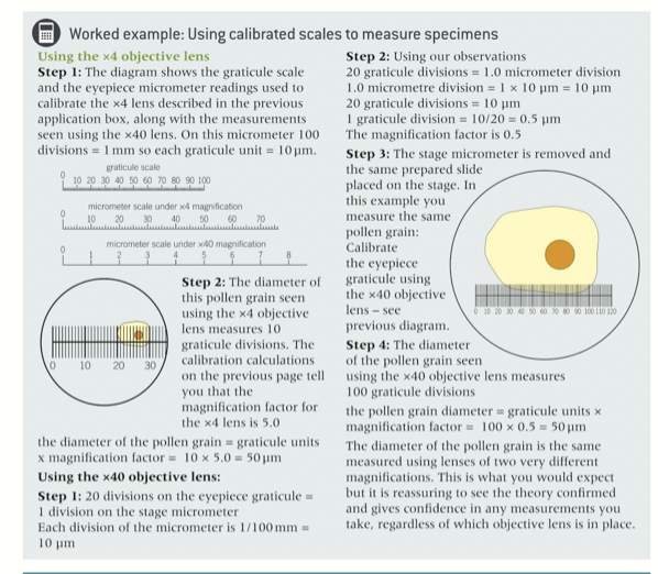
how to calibrate the eyepiece graticule with the x4 objective lens
put the stage micrometer in place and the eyepiece graticule in the eyepiece.
get the scale on the micrometer slide in clear focus.
align the micrometer scale with the scale in the eyepiece- take a reading from the two scales.
1 graticule division = number of micrometers / number of graticule divisions.
what is an eyepiece graticule used for?
- to measure objects with a light microscope, we use an eyepiece graticule
- the eyepiece graticule is a glass disc that is placed into the eyepiece
- you can measure the dimensions of a specimen by superimposing the eyepiece graticule over the specimen and measure the dimensions in eyepiece units (epu)
- scale is arbitrary
- image of specimen looks bigger at higher mag
- eyepiece scale has to be calibrated for each objective lens
what is a stage micrometer used for?
- to calibrate the eyepiece graticule
- this is a microscope slide with a scale etched into the centre circle region, like a tiny ruler
- stage micrometer is 1mm long (1000 um) and has 100 subdivisions, so one division is 0.01 um
What are cells? what are the different types? 2
the basic unit of all living things
prokaryotic and eukaryotic
prokaryotic
single-celled organisms with a simple structure of just a single undivided internal area called the cytoplasm.
eukaryotic
multicellular organisms- have a much more complicated internal structure - contain a membrane-bound nucleus and cytoplasm which contains many cellular components.
what is metabolism?
involves both the synthesis