Looks like no one added any tags here yet for you.
Dyspnea
Subjective sensation of uncomfortable breathing.
Orthopnea
Dyspnea while lying down.
Paroxysmal nocturnal dyspnea (PND)
Violent attacks of severe shortness of breath and coughing, generally occurring at night and awakening from sleep.
Chronic hypoxia
Long-standing low oxygen levels in the blood.
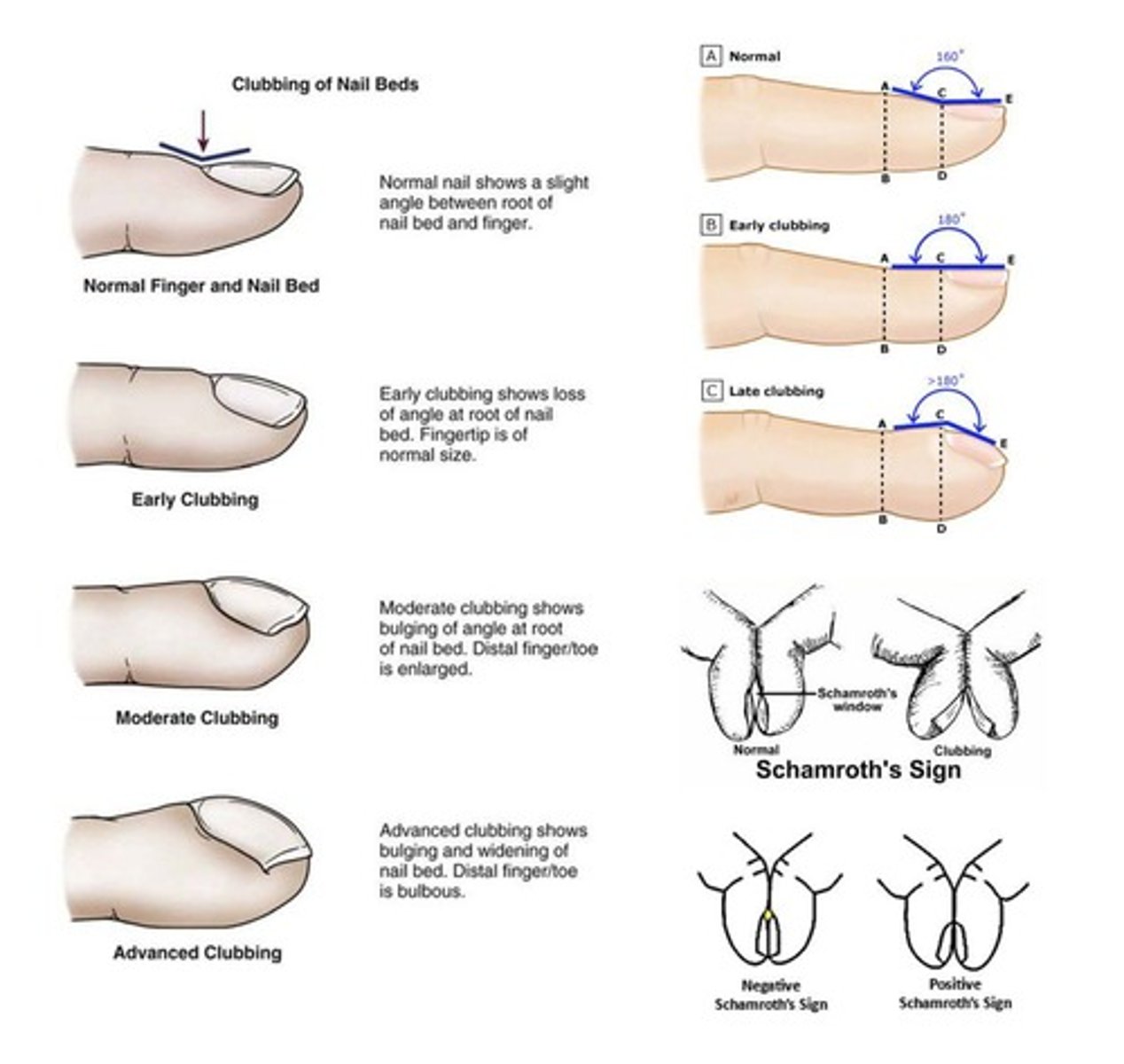
Clubbing
A physical sign characterized by the enlargement of the ends of fingers or toes.
Cyanosis
Patient appears blue; caused by low oxygen levels in the blood.
Cough
Protective reflex to clear the airway via an explosive expiration.
Productive cough
Cough with sputum.
Acute cough
Cough that resolves within 2 weeks.
Chronic cough
Cough that persists beyond 3 weeks.
Unproductive cough
Dry cough without mucus or other material.
Sputum
Mucus or other material that is coughed up from the lungs; can also be called phlegm.
Hemoptysis
Coughing up blood.
Pink (frothy) sputum
Associated with left-sided heart failure.
Tachypnea
Respiratory rate greater than 20 breaths per minute.
Bradypnea
Respiratory rate less than 20 breaths per minute.
Hypoventilation
Reduced breathing leading to high levels of CO2 in the blood (hypercapnia).
Hyperventilation
Increased breathing leading to low levels of CO2 in the blood (hypocapnia).
Wheezes
Sounds like a whistle.
Fine crackles
Sound like twirling your hair.
Coarse crackles
Sound like crumbling paper or velcro.
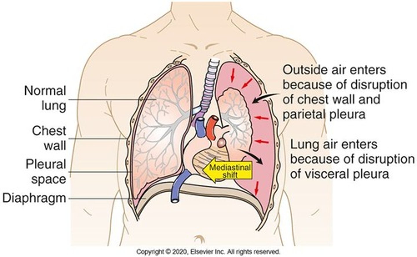
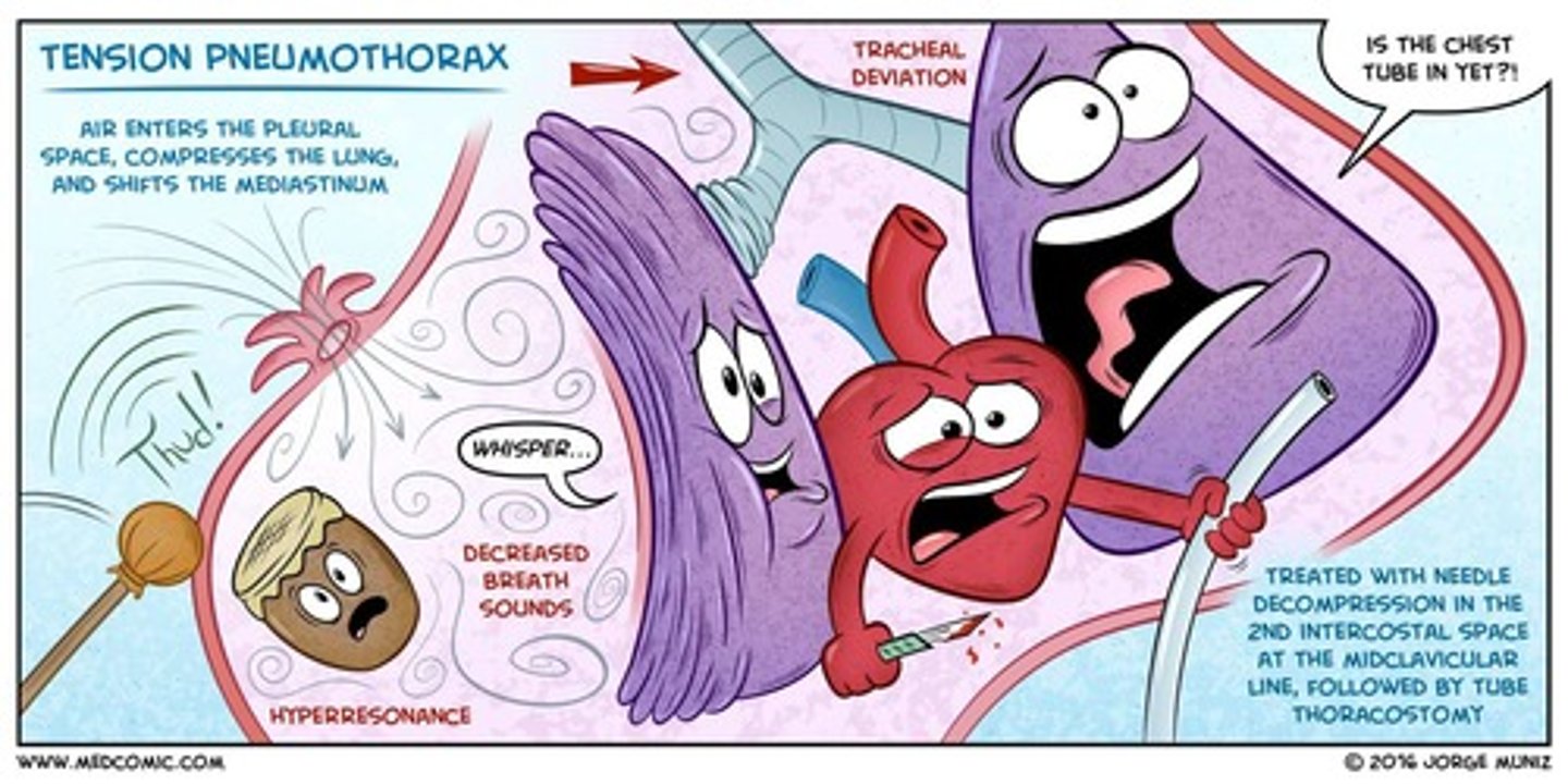
Pneumothorax
Accumulation of air in the pleural space.
Tension pneumothorax
Severe hypoxemia, tracheal deviation away from the affected lung, and hypotension.
Hypoxemia
Low levels of oxygen in the blood.
Pleurodesis
A procedure that sticks your lung to your chest wall to prevent fluid or air from building up.
Restrictive pulmonary disorder
Condition where lungs are restricted from fully expanding.
Obstructive pulmonary disorder
Condition where it is hard to exhale all the air in the lungs.
Acute respiratory distress syndrome (ARDS)
A life-threatening lung condition that causes severe difficulty breathing.
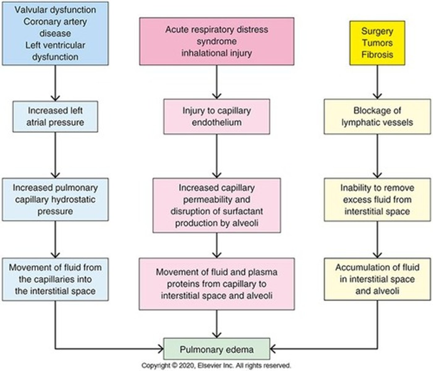
Pulmonary Edema
It occurs when fluid builds up in the lungs, preventing them from exchanging oxygen and carbon dioxide effectively.
Atelectasis
Collapse of the lung tissue (alveoli) with decreasing lung volume.
Atelectasis Symptoms
Alveoli become airless, inability of the lungs to expand, decrease gas exchange (perfusion) and hypoxia.
Types of Atelectasis
Absorption (resorptive or obstructive) atelectasis, compression, surfactant impairment (adhesive).
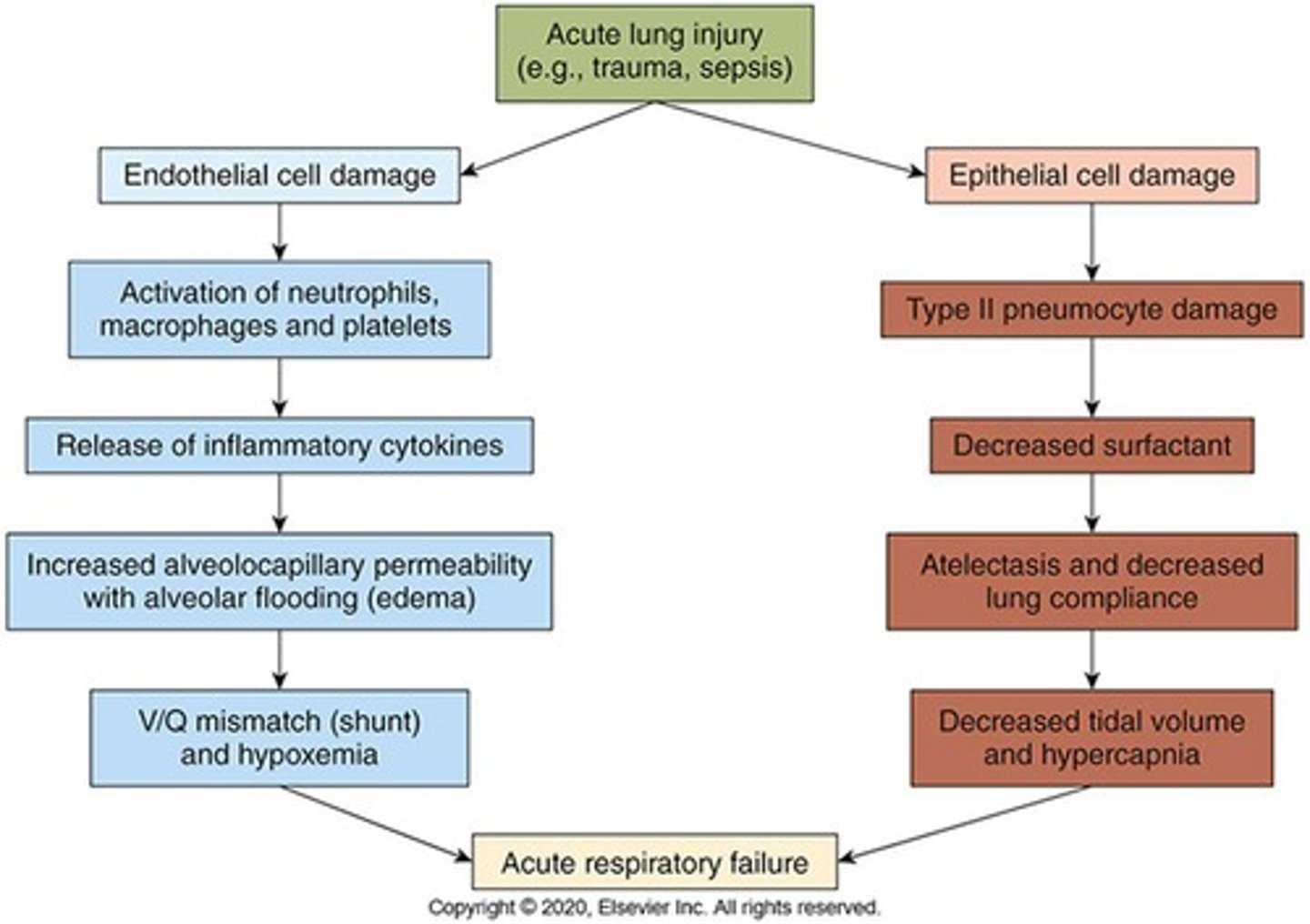
Acute Respiratory Distress Syndrome (ARDS)
Acute lung injury and inflammation of the alveolocapillary membrane; most severe form of acute lung injury.
ARDS Signs & Symptoms
Dyspnea & hypoxia, hyperventilation, decreased tissue perfusion, increased work of breathing, respiratory failure, decreased cardiac output, hypotension, death.
ARDS Treatment
Treat underlying cause and provide supportive therapy.
Obstructive Lung Diseases
Narrowing of airways leading to airway obstruction that is worse with expiration.
Obstructive Lung Diseases Symptoms
Required accessory muscles of expiration, increased work of breathing, ventilation-perfusion (V/Q) mismatch, decreased forced expiratory volume in one second (FEV1).
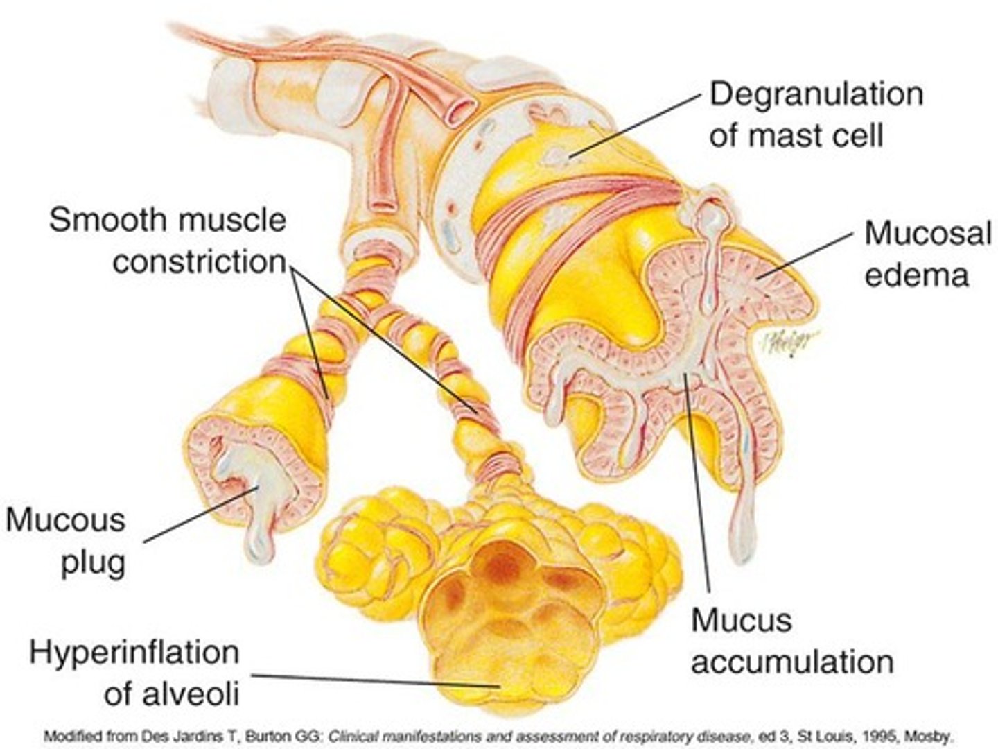
Asthma
Reversible airway obstruction characterized by airway inflammation and Type I hypersensitivity.
Asthma Prevalence
Prevalent chronic disease in childhood; multifactorial: genetic & environment.
Asthma Symptoms
Cough, marked dyspnea, wheezing.
Diagnosis of Asthma
Measures the levels of oxygen (O2), carbon dioxide (CO2), and pH (acidity) in the blood, and determines the percentage of hemoglobin that is saturated with oxygen.
Chest Imaging Techniques
Uses X-rays to create images of the chest organs and structure.
Chest Imaging Severity
Expiratory: mild to moderate; expiratory & inspiratory: severe.
Laboratory Assessments for Asthma
Laboratory assessments and studies are not routinely indicated for the diagnosis of asthma, but they may be used to exclude other diagnoses.
Arterial Blood Gas
A test that measures the levels of oxygen (O2), carbon dioxide (CO2), and pH (acidity) in the blood.
Pulse Oximetry
A test that determines the percentage of hemoglobin that is saturated with oxygen.
Computed Tomography (CT scan)
An imaging technique used to create detailed images of internal organs.
Magnetic Resonance Imaging (MRI)
An imaging technique that uses magnetic fields and radio waves to create detailed images of organs and tissues.
Nuclear Imaging
A technique that uses small amounts of radioactive material to diagnose and determine the severity of diseases.
Asthma Physical Signs
Rapid & labored breathing, nasal flaring, use of accessory muscles, exercise intolerance.
Corticosteroids
Anti-inflammatory medications used in asthma treatment.
Beta-agonists
Medications like albuterol that relax bronchial muscles to relieve asthma symptoms.
Anti-cholinergic
Ipratropium, a medication that helps open airways in asthma treatment.
Phosphodiesterase enzyme inhibitor
Theophylline (xanthine), a drug that relaxes the muscles of the airways.
Mast cell stabilizer
Cromolyn sodium, used to prevent asthma attacks.
Leukotriene receptor antagonist
Zafirlukast, a medication that blocks substances that cause asthma symptoms.
Monoclonal antibodies
Omalizumab, used for severe asthma treatment.
Combination drugs
Medications that combine a beta-agonist with a steroid for asthma management.
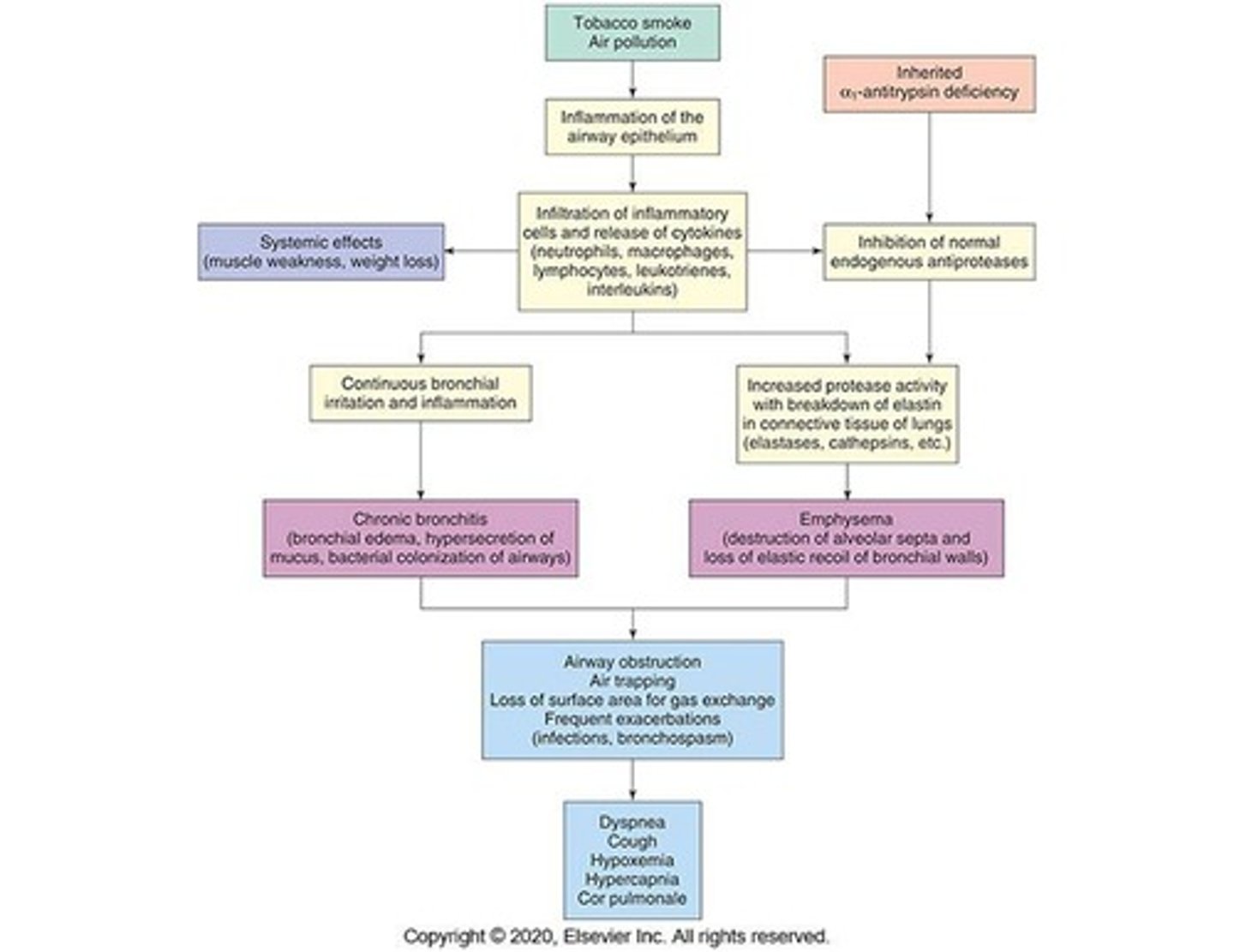
Chronic Obstructive Pulmonary Disease (COPD)
Characterized by persistent airflow limitation or obstruction.
Chronic bronchitis
Hypersecretion of mucus and chronic productive cough lasting at least 3 months for 2 consecutive years.
Emphysema
Abnormal permanent enlargement of the gas-exchange airways with destruction of alveolar walls.
Risk factors for COPD
Cigarette smoking, air pollution, and genetic factors.
Pathophysiology of Chronic Bronchitis
Includes inflamed mucosa, hypertrophy of mucous glands, and thickening of bronchial walls.
Clinical manifestations of Chronic Bronchitis
Cough, fever, and pain behind the sternum aggravated by coughing.
Treatment for Chronic Bronchitis
Includes smoking cessation, treatment of infection, and nutritional supplements.
Pathophysiology of Emphysema
Destruction of alveolar walls leading to loss of surface area for gas exchange.
Spirometry
Diagnostic test that evaluates lung function, with FEV1/FVC < 70% indicating COPD.
Signs & symptoms of COPD
Exertional dyspnea, increased shortness of breath, and development of barrel chest.
Diagnostic tests for COPD
Includes chest radiography, pulmonary function tests, and arterial blood gas analysis.
Treatment & Management of COPD
Focuses on improving functional status and preventing exacerbations.
Pneumonia
Any infection of the lung tissue of the lower respiratory tract.
Typical pneumonia
Characterized by high fever (> 101.5 degrees F) and productive cough.
Atypical pneumonia
Characterized by low-grade fever (< 100 degrees F) and non-productive cough.
Clinical Setting for Pneumonia
Involves assessing patients based on the setting in which they became sick.
Influenza
A viral infection that can cause respiratory illness.
Legionella
A genus of bacteria known to cause pneumonia.
Aspiration pneumonia
Pneumonia caused by inhaling foreign materials, often associated with alcohol use.
Atypical pneumonia
Pneumonia caused by organisms such as Mycoplasma and Chlamydia.
Respiratory syncytial virus
A virus that commonly affects children and can cause respiratory infections.
Para-influenza
A virus that commonly affects children and can lead to respiratory illness.
Influenza A & B
Types of influenza viruses that primarily affect older populations.
Adenovirus
A virus that can cause respiratory illness, especially in military barracks or close quarters.
Nosocomial pneumonia
Pneumonia acquired during hospital stays.
Gram (-) rods
Bacteria such as Klebsiella pneumoniae, E. coli, and Pseudomonas aeruginosa that can cause pneumonia.
S. aureus (MRSA)
Methicillin-resistant Staphylococcus aureus, a type of bacteria that can cause pneumonia.
Ventilator-associated pneumonia
Pneumonia that occurs in patients on mechanical ventilation, with the highest propensity for infection.
Pathogenesis of pneumonia
The process by which pneumonia develops, often involving compromised defense mechanisms.
Community-acquired pneumonia
Pneumonia acquired outside of a hospital setting, commonly bacterial in origin.
Exudative fluid
Fluid that fills alveolar air spaces in cases of pneumonia.
Diagnosis of pneumonia
Includes chest radiograph, gram stain of sputum, and blood cultures.
Treatment of pneumonia
Involves identifying the organism and selecting appropriate antibiotics.
Pulmonary Tuberculosis
An infectious disease caused by Mycobacterium tuberculosis.
Etiology of Tuberculosis
Caused by Mycobacterium tuberculosis, an acid-fast bacillus.
Epidemiology of Tuberculosis
Factors include living in crowded conditions, immunodeficiency, and chronic diseases.
Airborne droplet transmission
The method by which tuberculosis spreads from person to person.
Tubercle formation
Granulomatous lesions formed in response to tuberculosis infection.
Caseous necrosis
A cheese-like material that can form in tuberculosis infections.
Ghon tubercles
Fibrotic and calcified areas formed in the lungs as a result of tuberculosis.
Diagnostic tests for Tuberculosis
Includes tuberculin skin test, IGRA, and acid-fast sputum test.
Signs & symptoms of Tuberculosis
Include cough, night sweats, hemoptysis, and fatigue.