Anatomy - Scrotum, Thyroid, Misc.
1/91
Earn XP
Description and Tags
Name | Mastery | Learn | Test | Matching | Spaced | Call with Kai |
|---|
No analytics yet
Send a link to your students to track their progress
92 Terms
Aside from the pancreas, what other organ produces amylase?
A. salivary glands
B. adrenal glands
C. pituitary gland
D. liver
A. salivary glands
Amylase is a component of saliva
Calcitonin is produced by which of the following types of cells?
A. Parathyroid follicular
B. Thyroid follicular
C. Parathyroid parafollicular
D. Thyroid parafollicular
D. Thyroid parafollicular
Calcitonin is produced in the thyroid in the parafollicular cells. These cells are located in the tissues between the thyroid follicles.
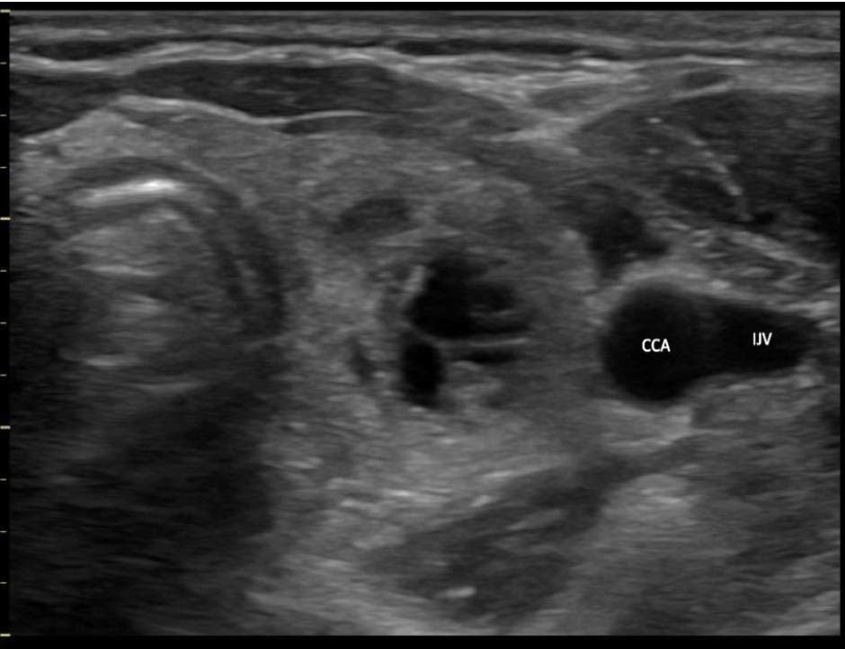
Which blood vessel is located lateral to the left lobe of the thyroid and demonstrates an anechoic ovoid shape in a transverse view of the mid thyroid?
A. superior vena cava
B. left internal jugular vein
C. left external carotid artery
D. left common carotid artery
B. left internal jugular vein
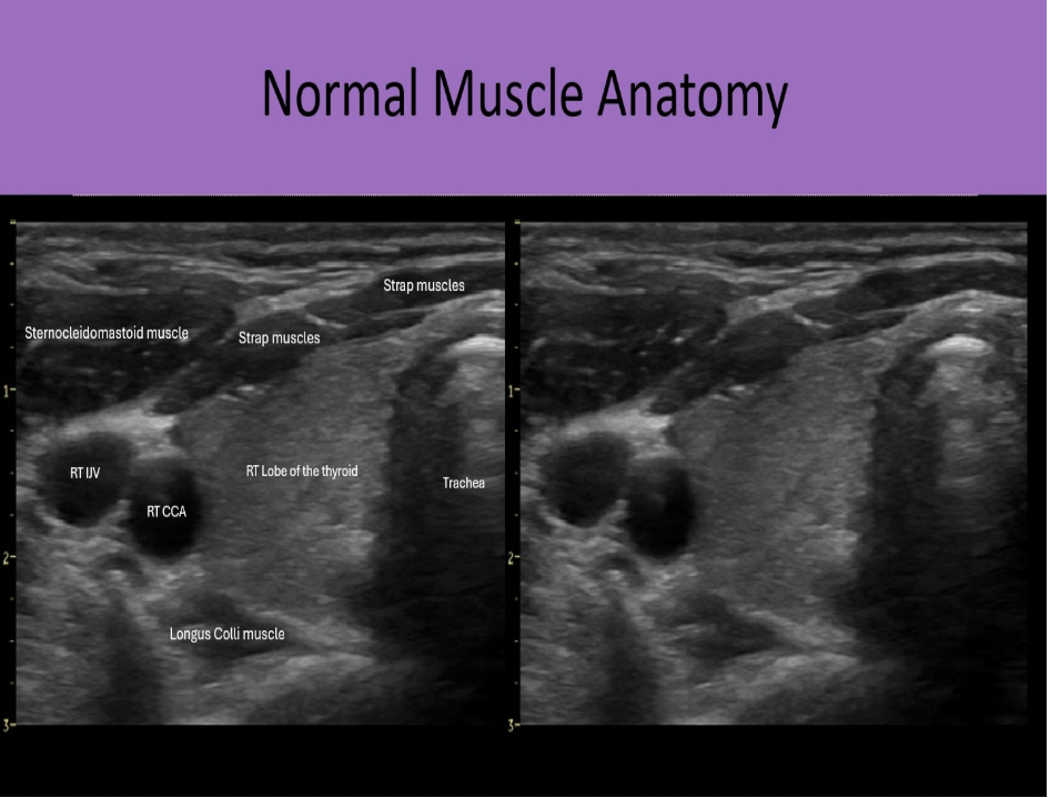
The internal jugular vein is located lateral to the left lobe of the thyroid and demonstrates an anechoic ovoid shape in a transverse view of the mid thyroid. The carotid artery is also lateral to the thyroid lobes but it should be a circular structure.
The image displays the left lobe of the thyroid with the circular CCA immediately lateral to the mid lobe and ovoid IJV lateral to the CCA.
The functional unit of the thyroid gland is called
A. Lobe
B. Nodule
C. Lobule
D. Follicle
D. Follicle
The follicles are tiny, cyst-like units of the thyroid gland that are lined with cuboidal epithelium and are filled with a colloid substance, about 30 to each lobule. There are numerous lobules in each lobe that are separated by webs of connective fascia.
The cardiac orifice is:
A. The opening in the diaphragm that allows the IVC to pass through
B. The opening by which the esophagus communicates with the stomach
C. The opening in the diaphragm that allows the aorta to pass through
D. The bare area of the liver that allows the hepatic veins to pass through
B. The opening by which the esophagus communicates with the stomach
The esophagus extends from the pharynx to the cardiac orifice of the stomach.
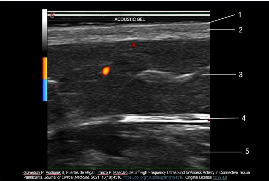
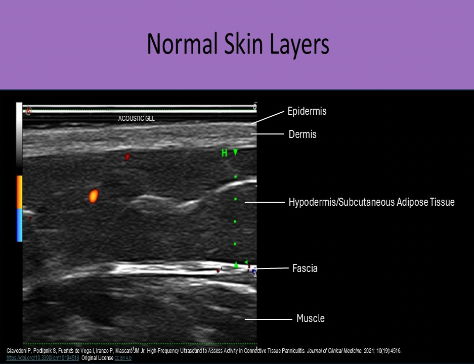
Which number indicates the dermis layer of the skin?
A. 3
B. 2
C. 1
D. 4
B. 2
The _______ is formed at the junction of the vas deferens and seminal vesicles.
A. ejaculatory duct
B. efferent ducts
C. rete testes
D. afferent ducts
A. ejaculatory duct
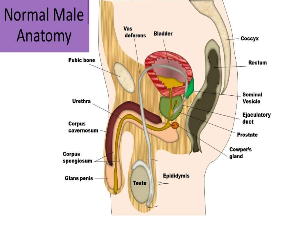
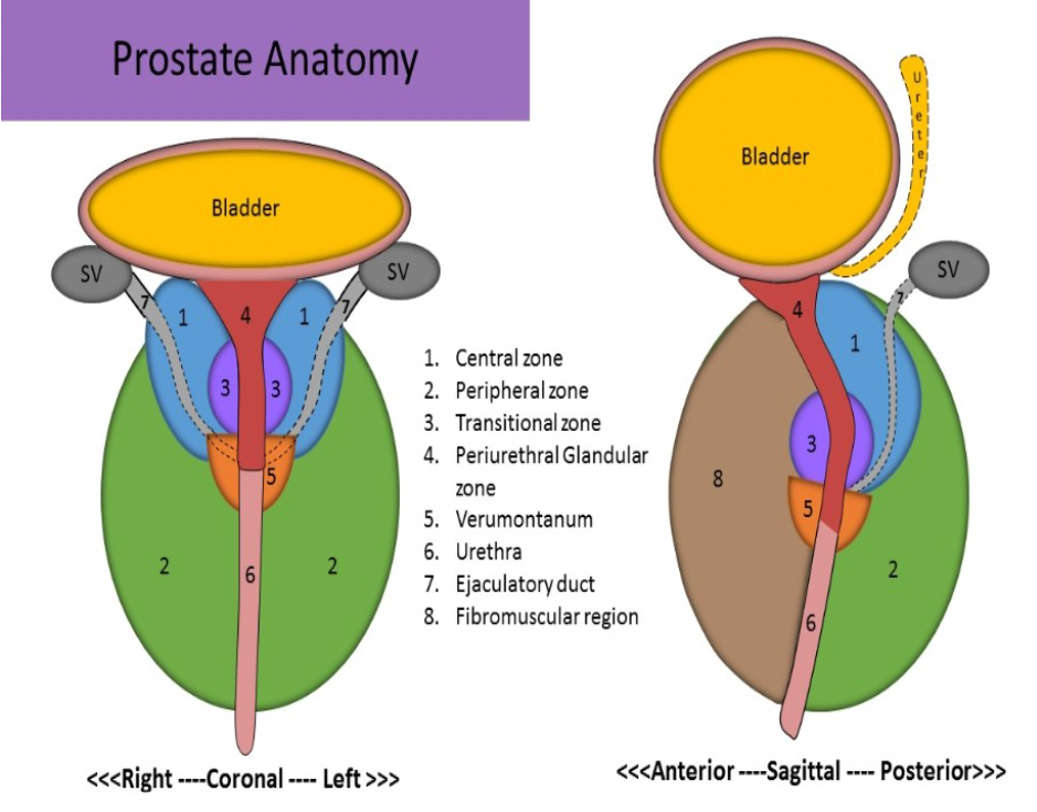
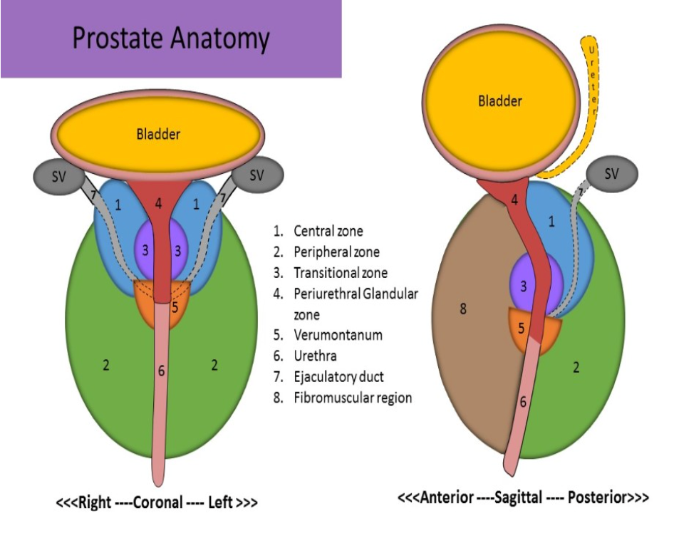
The _______ zone is the largest zone in the prostate.
A. peripheral
B. transitional
C. paraprostatic
D. central
A. peripheral
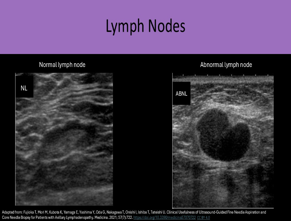
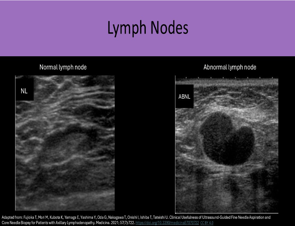
Which of the following is a characteristic of a normal lymph node?
A. posterior enhancement
B. round shape
C. thin hypochoic rim with hyperechoic central hilum
D. thick hypochoic rim with hypervascularity
C. thin hypochoic rim with hyperechoic central hilum
Normal lymph nodes are ovoid in shape. They do not demonstrate posterior enhancement. As lymph nodes enlarge with abnormal fluid accumulation, they appear more rounded and hypochoic/anechoic. The "plump" cortex causes decreased visualization of the central hilum. Color Doppler can be used to locate the hilum and main vascular supply. Posterior enhancement is present in structures posterior to abnormal nodes with significant fluid accumulation.
What blood vessels are demonstrated on the cross section of the penis?
A. cavernosal arteries
B. dorsal arteries
C. helicine arteries
D. urethral arteries
A. cavernosal arteries
The image demonstrates a cross section of the penis. The two corpus cavernosum and the corpus spongiosum are identified. The cavernosal artery courses through the corpus cavernosum.
Penile Vasculature: Internal iliac arteries give rise to the internal pudendal arteries, bulbourethral artery, and penile arteries. The penile artery branches into the dorsal and cavernosal arteries. The right and left cavernosal arteries enter the right and left CC, respectively. The helicine arteries are multiple tiny branches that penetrate the tissue. Superficial dorsal vein and deep dorsal vein provide the primary venous outflow routes
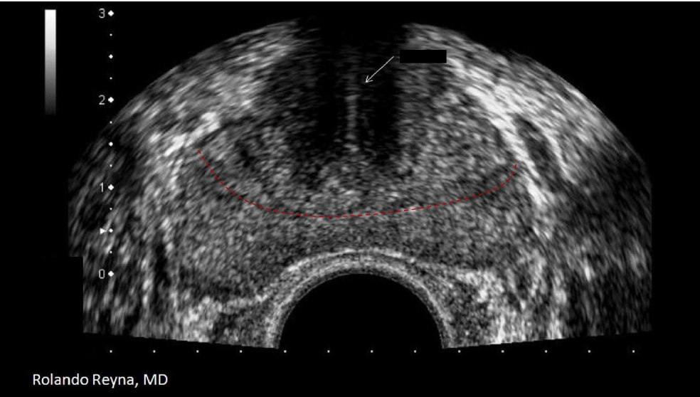
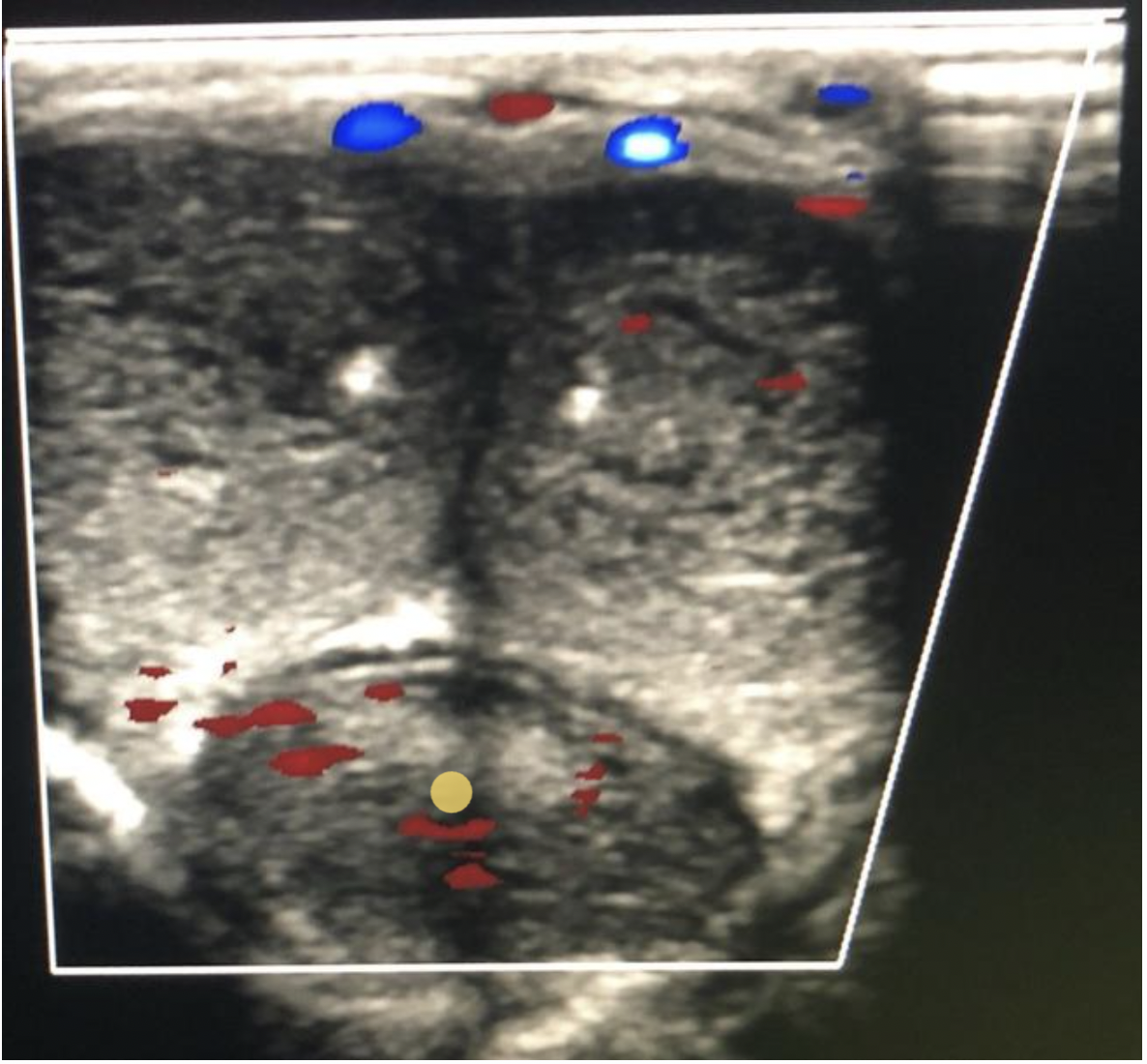
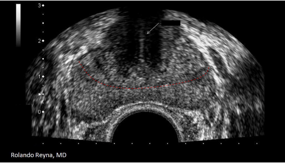
The red dotted line separates what two zones of the prostate?
A. peripheral and anterior fibromuscular
B. transitional and peripheral
C. verumontanum and central
D. central and transitional
B. transitional and peripheral
The white arrow indicates the urethra and the red dotted lines separate the transitional and peripheral zones.
The thyroid normally produces the most of which of the following hormones?
A. T3
B. T4
C. Thyrotropin
D. TSH
B. T4
The thyroid normally produces more T4 than T3
Pheochromocytoma, chromaffin cells and neurons in the central nervous system are responsible for the release of?
A. testosterone
B. aldosterone
C. sodium bicarbonate
D. epinephrine
D. epinephrine
Epinephrine and norepinephrine are produced in chromaffin cells of the adrenal glands and certain neurons in the CNS. Pheochromocytomas can also cause abnormal levels of epinephrine.
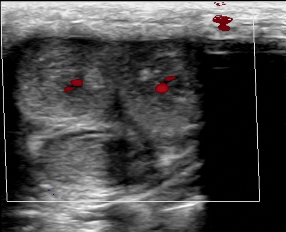
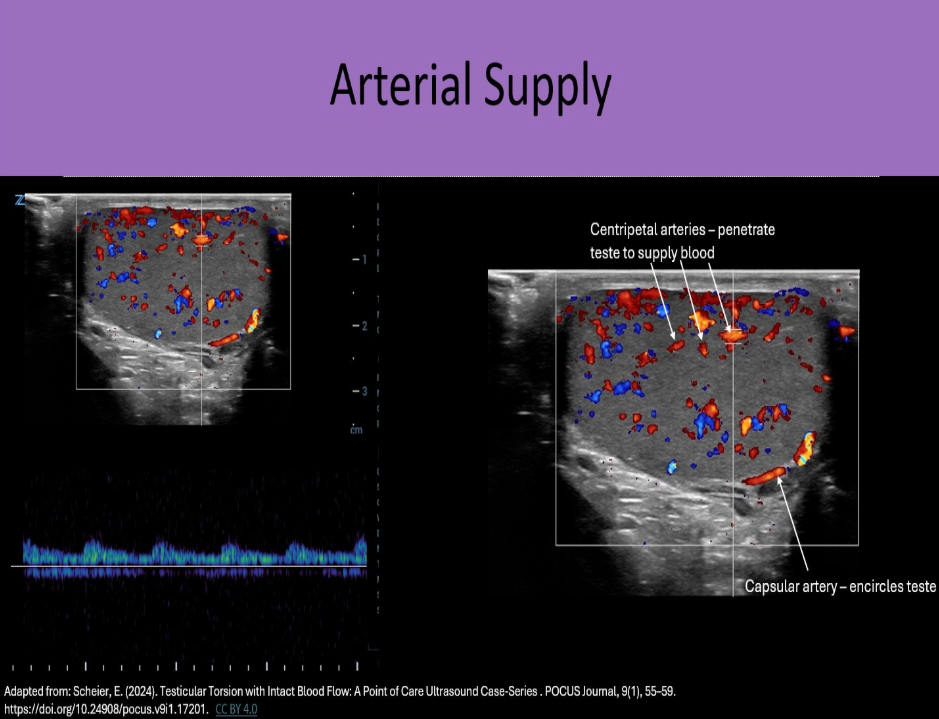
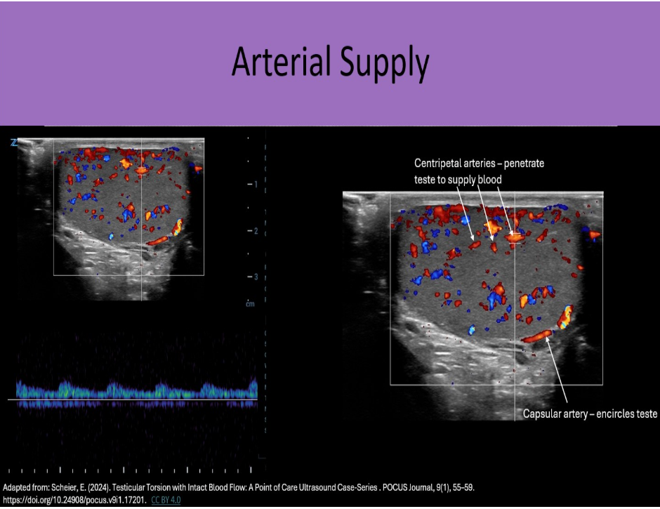
When evaluating intratesticular flow, which vessel is sampled by Doppler?
A. deferential
B. gonadal
C. cremasteric
D. centripetal
D. centripetal
Centripetal arteries course through the parenchyma to deliver oxygenated blood to the testicular tissues. These arteries are sampled with Doppler in a standard ultrasound evaluation.
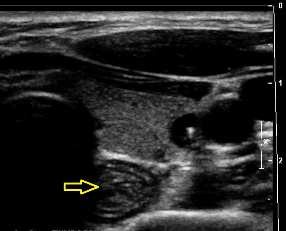
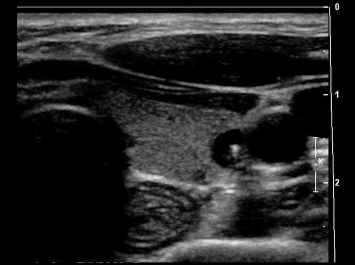
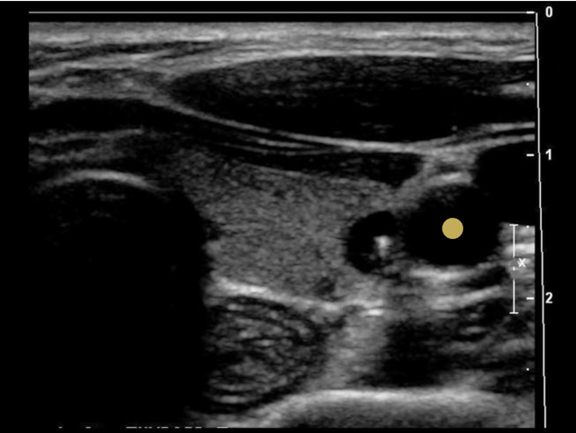
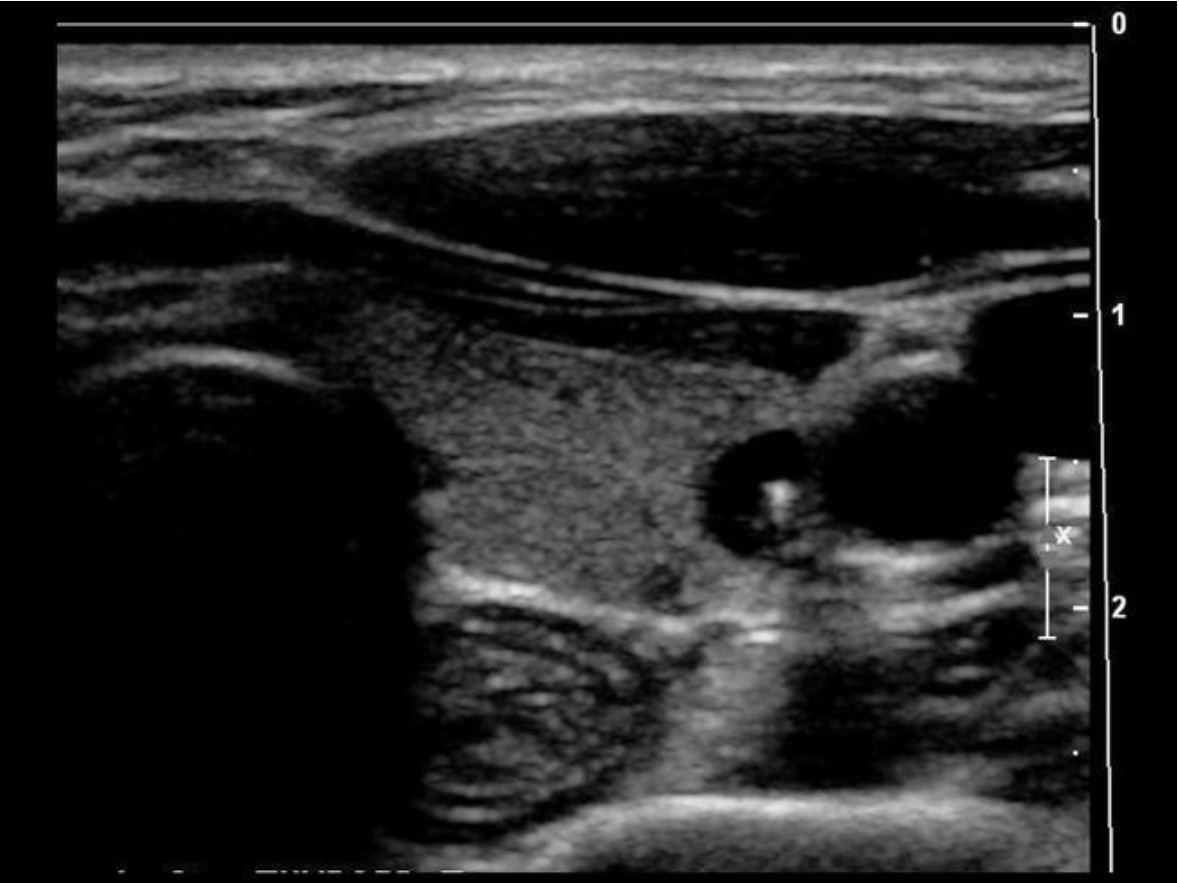
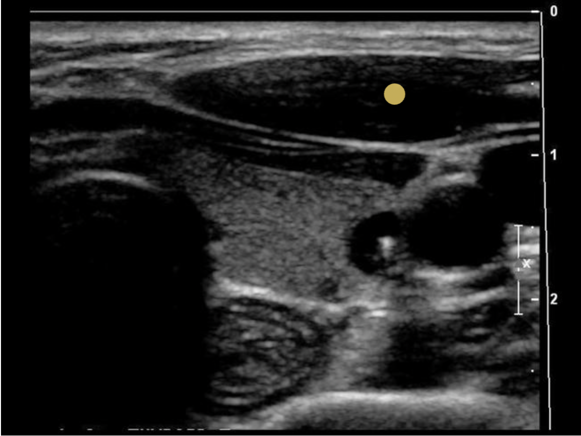
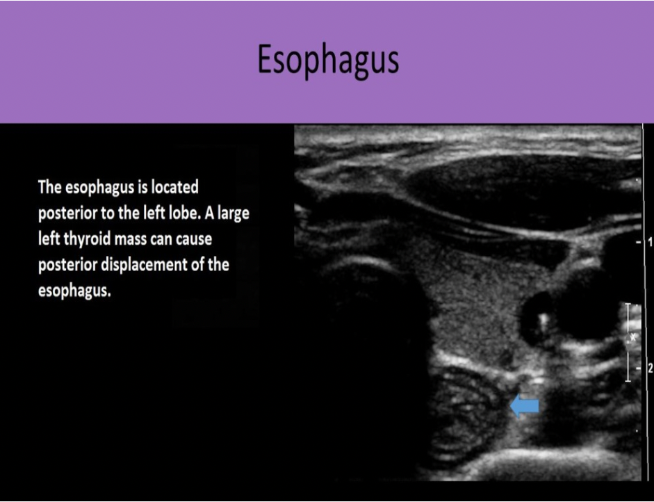
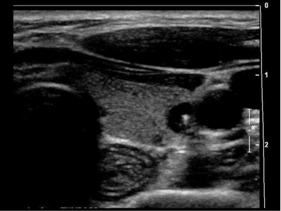
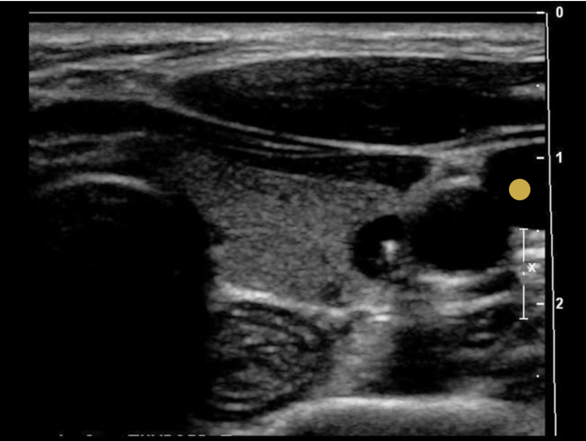
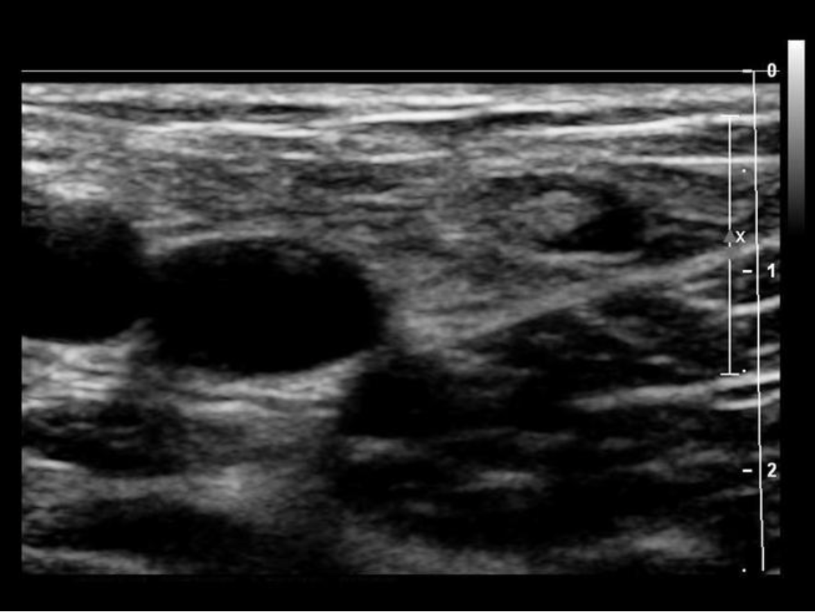
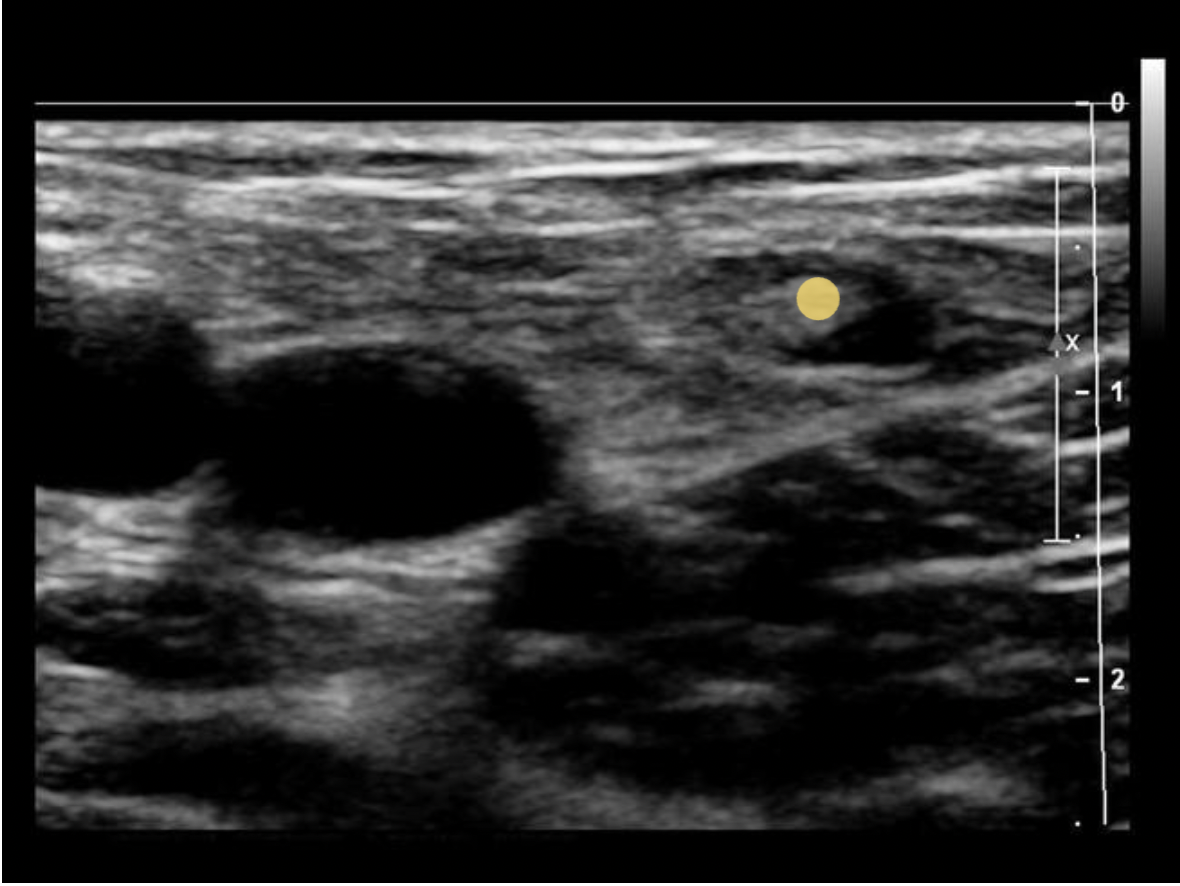
What is indicated by the yellow arrow?
A. parathyroid adenoma
B. longus colli muscle
C. esophagus
D. trachea
C. esophagus
The arrow indicates the esophagus. Note the rings of tissue seen with normal Gl tract structures.
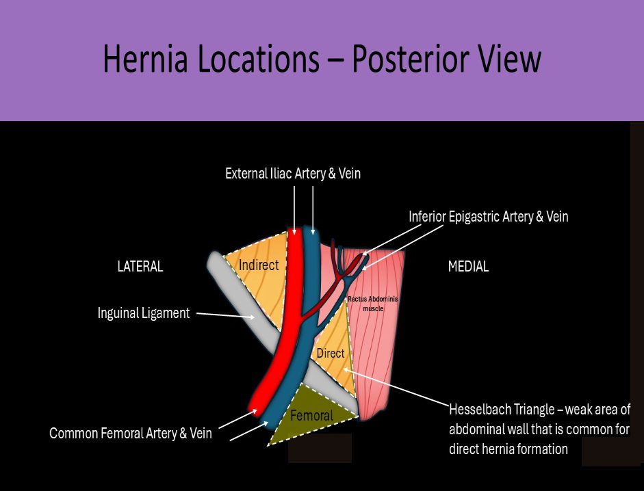
What abnormality involves the Hesselbach triangle?
A. direct inguinal hernia
B. spigelian hernia
C. femoral hernia
D. ventral hernia
A. direct inguinal hernia
The Hesselbach triangle is bordered inferiorly by the inguinal ligament, medially by the lateral aspect of the rectus abdominis muscle, and superolaterally by the Inferior Epigastric artery. Intra-abdominal structures move anteriorly through the aponeuroses at the Hesselbach triangle. The inguinal hernia sac lies posterior and medial to the spermatic cord in males, round ligament in females

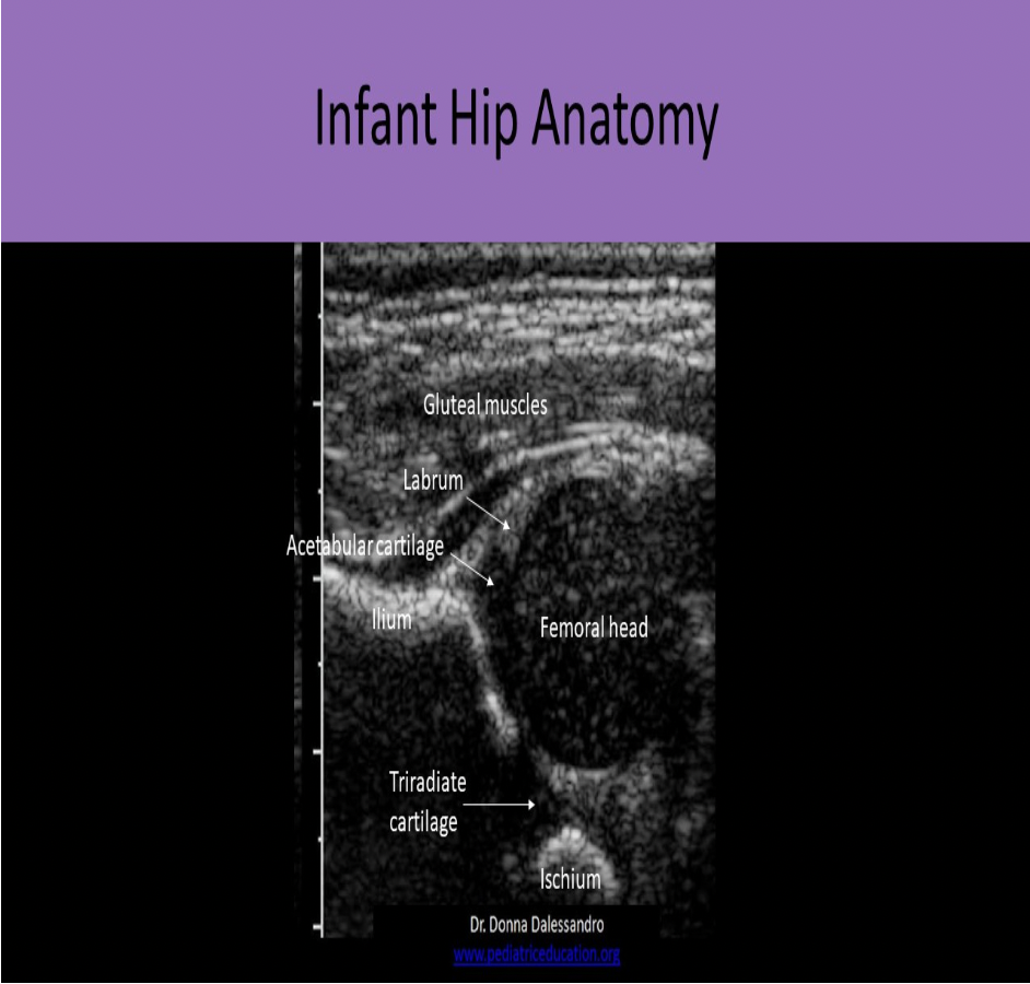
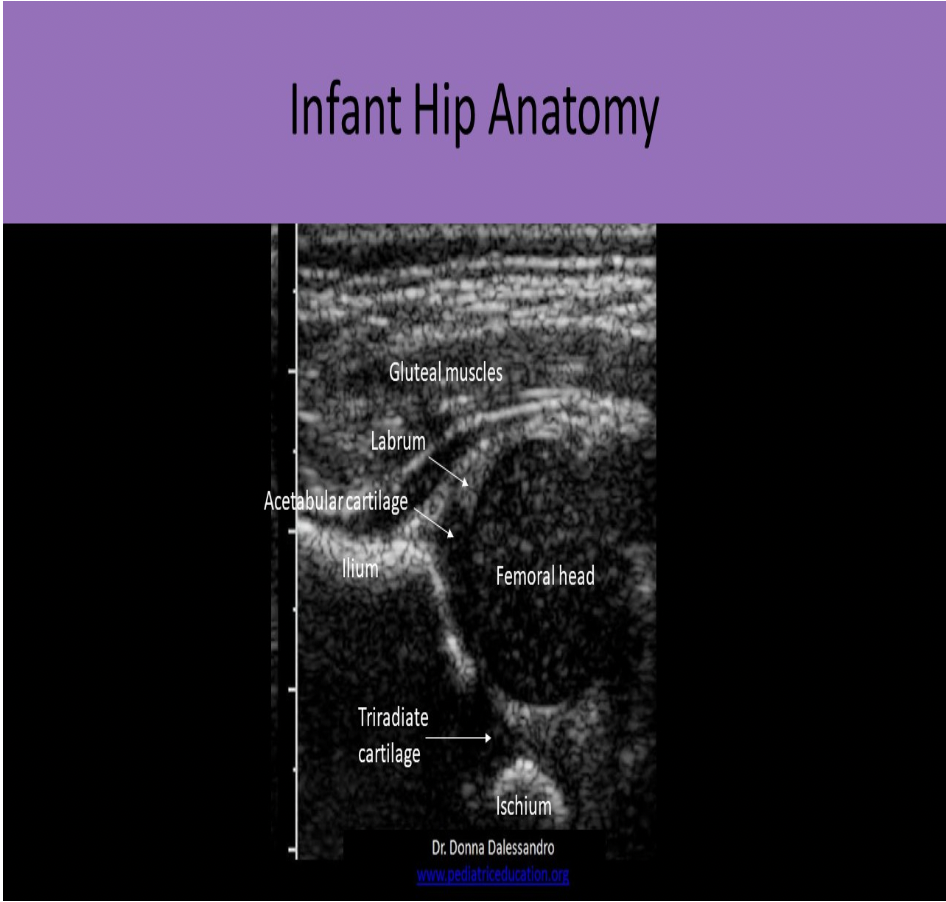
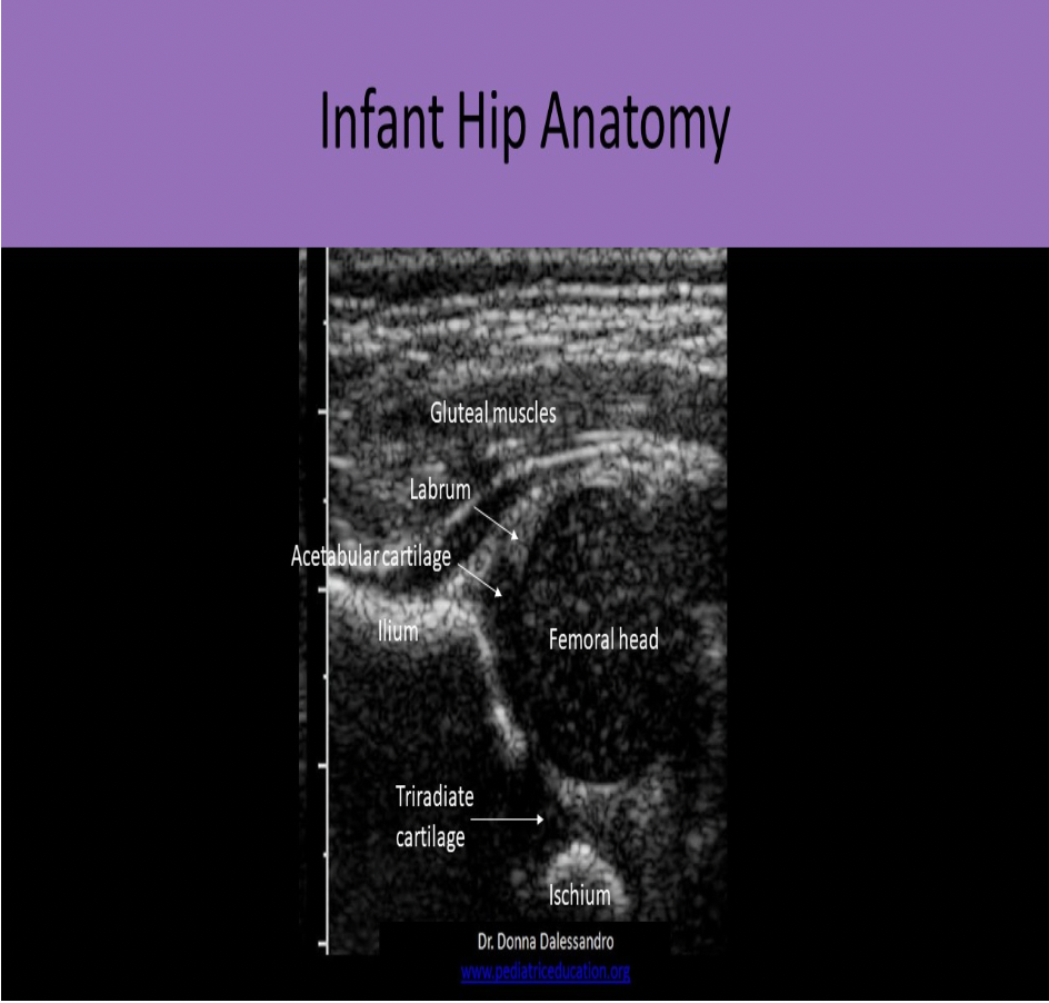
Locate the ilium
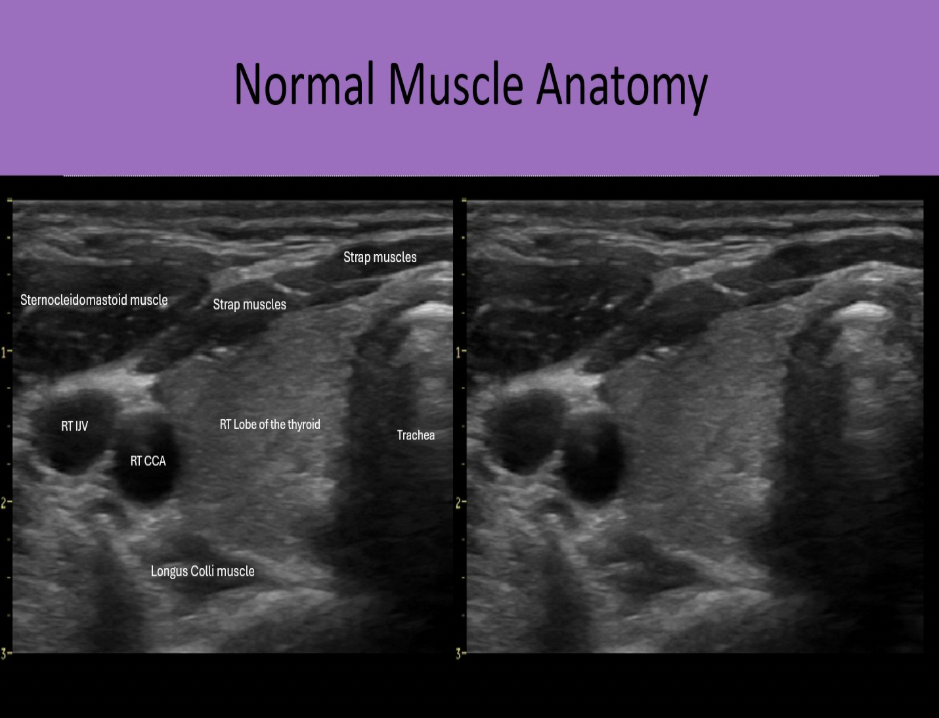
Which muscle group is located posterior to the thyroid gland?
A. strap muscles
B. tracheal muscles
C. longus colli muscle
D. sternocleidomastoid
C. longus colli muscle
Strap muscles are anteromedial to the gland. Sternocleidomastoid muscles are anterolateral to the gland. Longus Colli muscles are posterior to the thyroid lobes.
When evaluating the small bowel with ultrasound, which finding below is always considered abnormal?
A. Bull's eye appearance
B. 5 layers of gut wall
C. Intraluminal and extraluminal fluid
D. Color Doppler displayed with peristalsis
C. Intraluminal and extraluminal fluid
There are normally 5 layers of the gut wall identified on the US exam. In the cross-sectional view, the bowel segment will have a bull's eye appearance (in the normal or abnormal patient). Graded compression techniques are used to document compressibility expected from a normal segment. Color Doppler can be used to document peristalsis of a normally functioning segment. While fluid within the lumen is usually normal, any fluid in the walls or outside the segment (ascites) would be a sign of an abnormality.
The thyroid gland is considered normal in size if the:
A. AP measurement is <2cm
B. volume is <30mL
C. width measurement is <3cm
D. length measurement is < 4cm
A. AP measurement is <2cm
When the AP measurement is greater than 2cm, the gland is considered enlarged. Thyroid volume > 19mL indicates thyromegaly.
The fibromuscular stroma of the prostate can be referred to as the ______ zone.
A. anterior
B. central
C. peripheral
D. transitional
The fibromuscular stroma of the prostate is called the anterior zone.

Which of the following holds the psoas muscle?
A. anterior pararenal space
B. perirenal space
C. retrofascial space
D. Space of Retzius
C. retrofascial space
The retrofascial space holds the psoas and quadratus lumborum muscles.
The structure indicated by the arrow is composed of:
A. microvessels carrying venous blood
B. loops of spermatic cord
C. folds of tunica vaginalis
D. folds of tunica albuginea
D. folds of tunica albuginea
Folds of the tunica albuginea form sections within the testes that converge at a single location called the mediastinum testis (similar to a hilum). The rete testes is a series of channels within the mediastinum. Blood vessels and ductules enter/exit the testicle at the mediastinum.
The _______ is a connective sheath that is connected to the large intestine that provides structure and support, along with encasing/protecting blood vessels.
A. mesentery
B. mesocolon
C. greater omentum
D. lesser omentum
B. mesocolon
The mesocolon is a connective sheath that is connected to the large intestine that provides structure and support, along with encasing/protecting blood vessels.
The lesser omentum is a fibrous sheath that is connected to the superior aspect lesser curvature of the stomach that provides structure and support, along with encasing/protecting blood vessels.
The greater omentum is a fibrous sheath that is connected to the greater curvature of the stomach that provides structure and support, along with encasing/protecting blood vessels.
The mesentery is a connective sheath that is connected to the small intestine that provides structure and support, along with encasing/protecting blood vessels.
What structure is indicated by the arrow?
A. appendix teste
B. cremasteric ligament
C. centripetal ligament
D. mediastinum testis
D. mediastinum testis
Folds of the tunica albuginea form sections within the testes that converge at a single location called the mediastinum testis (similar to a hilum). The rete testes is a series of channels within the mediastinum. Blood vessels and ductules enter/exit the testicle at the mediastinum.
An increase in which of the following hormones causes increased calcium resorption in the small intestines?
A. thyroid stimulating hormone
B. calcitonin
C. T4
D. parathyroid stimulating hormone
D. parathyroid stimulating hormone
PTH and calcitonin control calcium absorption/excretion in the intestines and kidneys. Increased serum levels of calcium leads to hypercalciuria and hypercalcemia
Low calcium levels in blood: Increase in the release of PTH, Decrease in the release of calcitonin, Causes increased resorption of calcium in intestinal tract in order to increase the level of calcium in the blood, Kidneys also increase resorption of calcium during blood filtration. Increased resorption = reduced excretion
High calcium levels in the blood: Decrease in the release of PTH, Increase in the release of calcitonin, Causes increased excretion of calcium on the intestines to reduce level of calcium in the blood, Kidneys also increase excretion during blood filtration. Increased excretion = reduced resorption
Locate the common carotid artery
The common carotid artery is lateral to the thyroid gland. The internal jugular vein is slightly anterior and lateral to the common carotid artery.
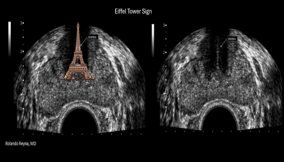
The Eiffel tower sign on ultrasound refers to:
A. duplex kidney
B. portal HTN
C. normal prostate
D. lymphadenopathy
C. normal prostate
Eiffel tower sign refers to shadowing created by dense tissues in the area of the urethra and verumontanum of the prostate.
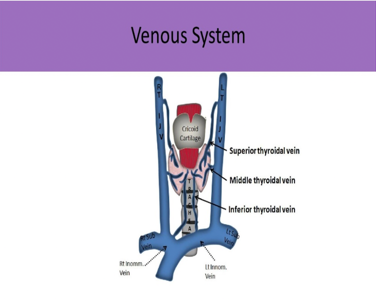
The internal jugular vein merges with what vein to form the brachiocephalic vein?
A. brachial vein
B. common carotid vein
C. innominate vein
D. subclavian vein
D. subclavian vein
The internal jugular vein merges with the subclavian vein to form the brachiocephalic vein. The brachiocephalic vein is also referred to as the innominate vein.
Which scrotal artery supplies the epididymis?
A. centripetal
B. deferential
C. cremasteric
D. capsular
B. deferential
Capsular artery courses along the testicle periphery and produces branches called centripetal arteries which course through the parenchyma. The deferential artery supplies vas deferens and epididymis with blood. The cremasteric artery supplies scrotal sac with blood.
Which of the following structures is not included in the spermatic cord?
A. Gonadal artery
B. Cremasteric artery
C. Centripetal artery
D. Vas deferens
C. Centripetal artery
Centripetal arteries are small branches of the capsular artery surrounding the testicle. These branches penetrate the testicular tissue.
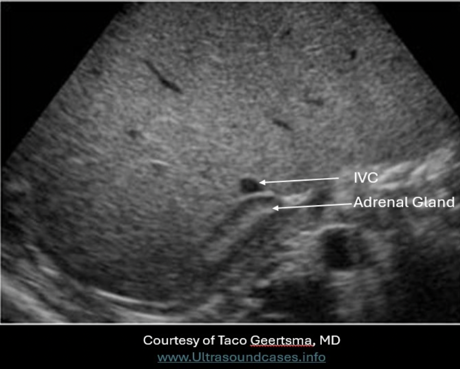
Which of the following is true regarding the anatomic position of the right adrenal gland?
A. posterior to the IVC
B. lateral to the right lobe of liver
C. lateral to the left diaphragm crura
D. anterior to the aorta and SMA
A. posterior to the IVC
Right Adrenal Gland - medial to right lobe of liver; posterior to IVC; superomedial to the kidney; lateral to the right crura
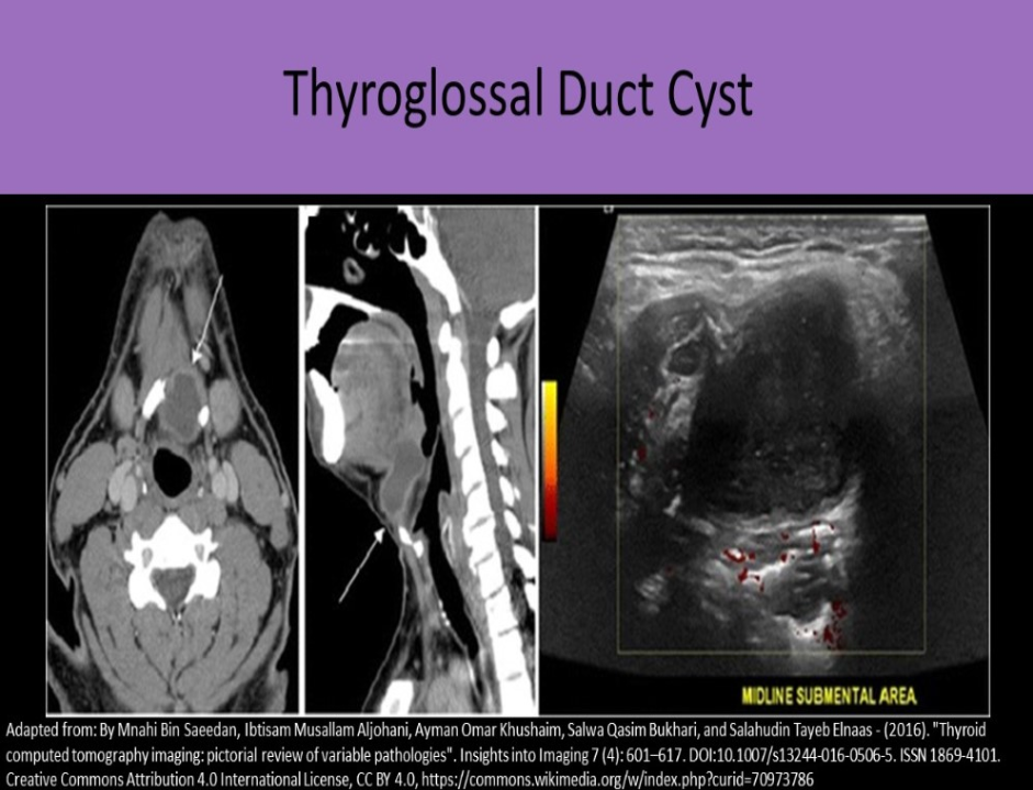
A patient presents with a palpable round lump on the midline anterior neck about 2cm below the jaw. The structure appears anechoic with thin, smooth walls on ultrasound. Which of the following is the most likely explanation for the findings?
A. thyroglossal duct cyst
B. pharyngeal pouch cyst
C. ectopic thyroid cyst
D. nasopharyngeal duct cyst
A. thyroglossal duct cyst
Thyroglossal duct connects the thyroid to the pharynx and regresses after thyroid gland reaches its normal position in the neck. The duct can fail to regress completely leaving a cystic structure behind. The cyst usually forms high in neck, near midline. It is the most common congenital neck cyst.
Sonographically, the cyst appears as an anechoic structure anterior to the pharynx and superior to thyroid gland that has all qualities of simple cyst.
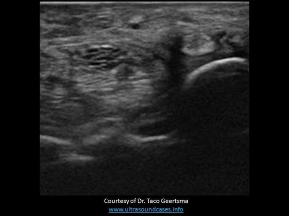
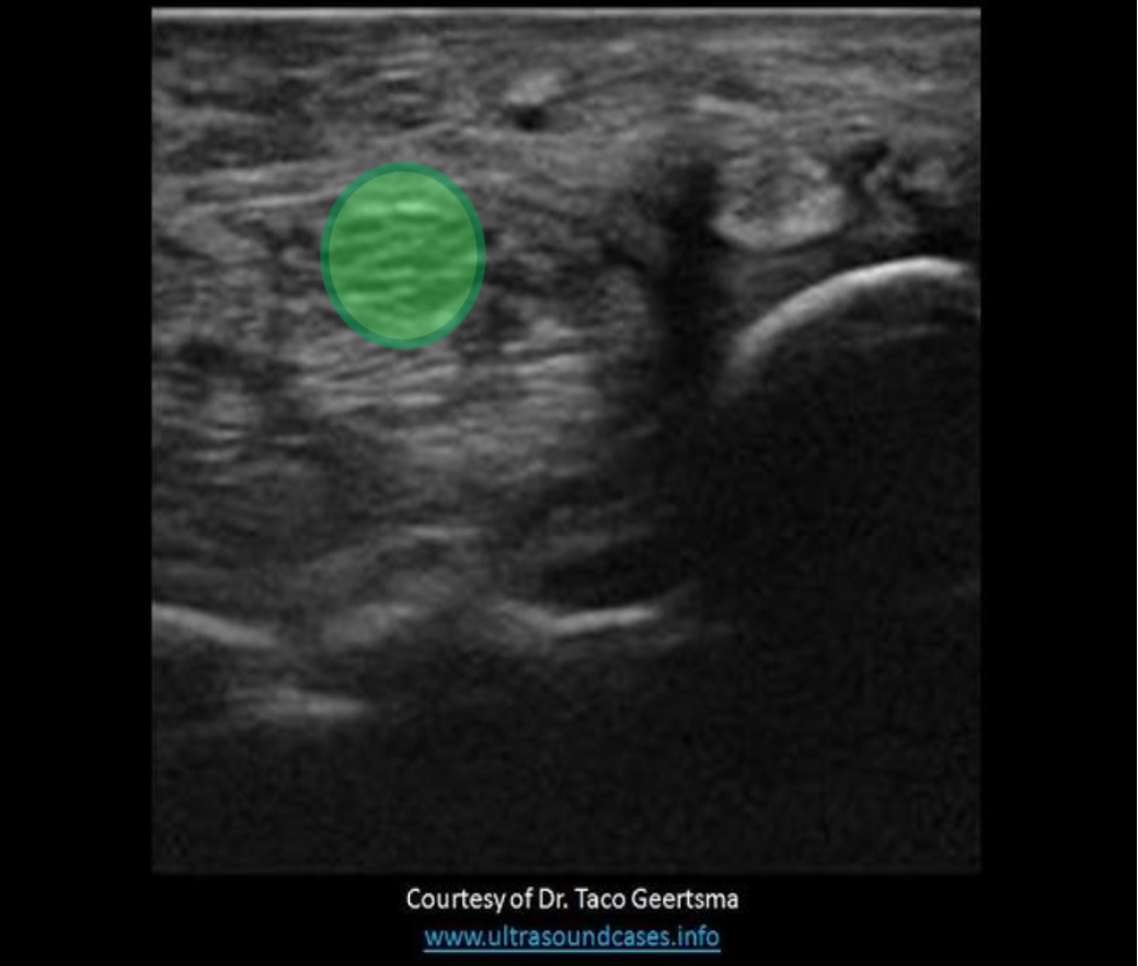
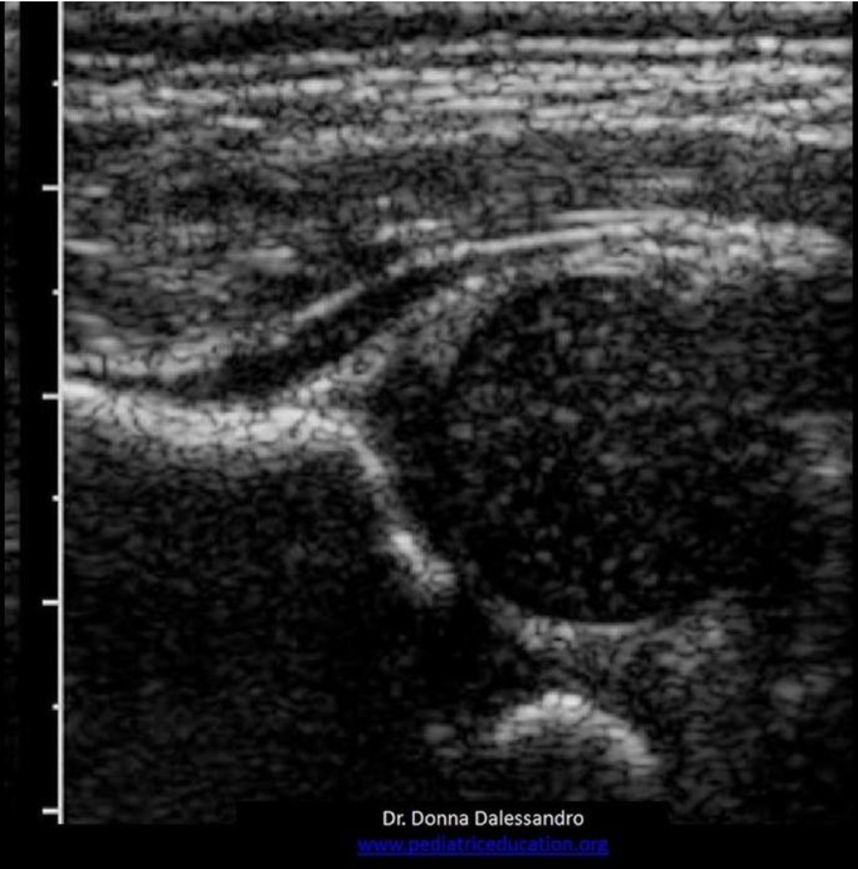
The image demonstrates a transverse view of the wrist. Find the center of the median nerve
The median nerve is a hypochoic striated ovoid structure in the anterior wrist.
This nerve is compressed/injured in carpal tunnel syndrome.
The _______ is a connective sheath that is connected to the small intestine that provides structure and support, along with encasing/protecting blood vessels.
A. mesentery
B. greater omentum
C. mesocolon
D. lesser omentum
A. mesentery
The lesser omentum is a fibrous sheath that is connected to the superior aspect lesser curvature of the stomach that provides structure and support, along with encasing/protecting blood vessels.
The greater omentum is a fibrous sheath that is connected to the greater curvature of the stomach that provides structure and support, along with encasing/protecting blood vessels.
The mesentery is a connective sheath that is connected to the small intestine that provides structure and support, along with encasing/protecting blood vessels.
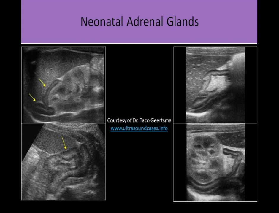
On an ultrasound exam, the normal adrenal cortex appears:
A. as an anechoic ring around the echogenic medulla
B. isoechoic to the medulla
C. as a hypochoic ring around the echogenic medulla
D. as a hyperechoic ring around the hypochoic medulla
C. as a hypochoic ring around the echogenic medulla
While scanning the thyroid, you identify a 0.6cm ovoid structure outside the thyroid, lateral to the left carotid artery. The structure has a thin hypoechoic rim surrounding a hyperechoic center. These findings are most suggestive of:
A. thyroglossal duct cyst
B. parathyroid carcinoma
C. normal esophagus
D. normal lymph node
D. normal lymph node
Normal lymph nodes are ovoid in shape. They do not demonstrate posterior enhancement. As lymph nodes enlarge with abnormal fluid accumulation, they appear more rounded and hypoechoic/anechoic. The "plump" cortex causes decreased visualization of the central hilum. Color Doppler can be used to locate the hilum and main vascular supply. Posterior enhancement is present in structures posterior to abnormal nodes with significant fluid accumulation.
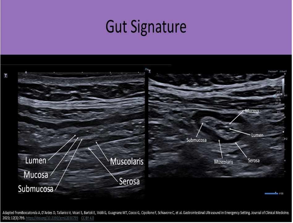
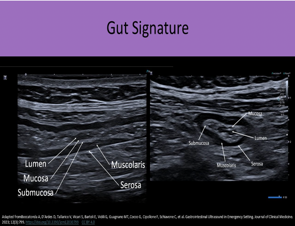
How many layers are identified in the normal sonographic "gut signature"?
A. 3
B. 5
C. 6
D. 8
B. 5
Serosa, Muscularis, Submucosa, Deep Mucosa, Mucosa/Lumen
Locate the triradiate cartilage
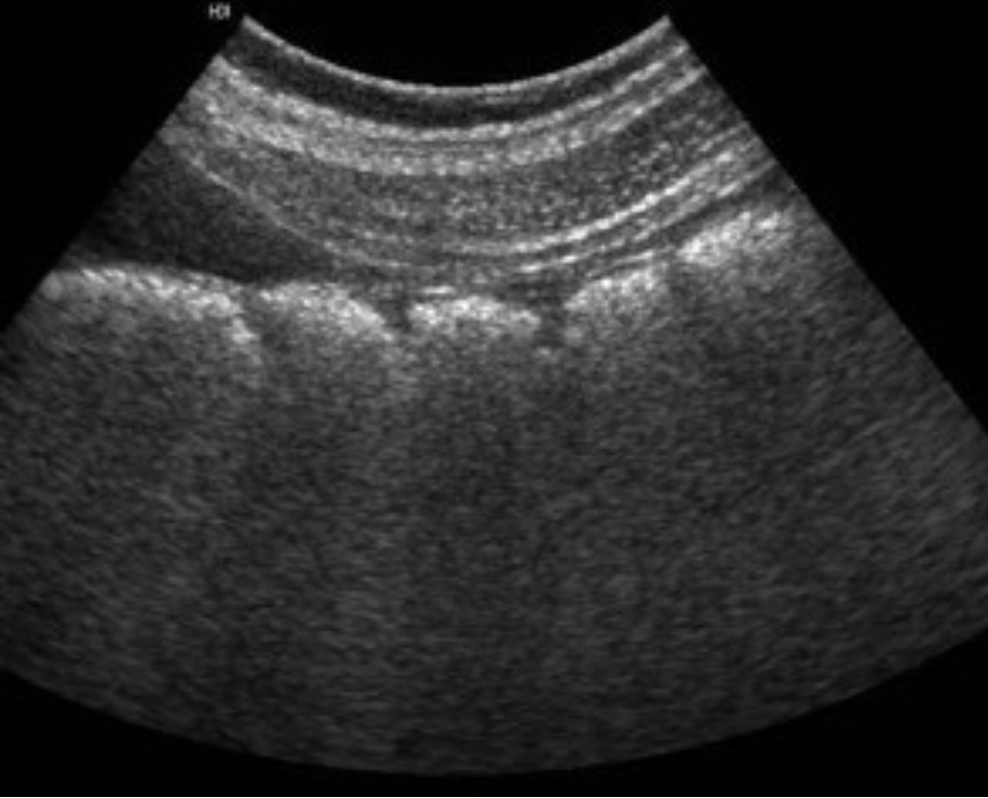
What portion of the Gl tract is demonstrated on the image?
A. duodenum
B. stomach
C. esophagus
D. colon
D. colon
Note the wall undulations called haustra
Which of the following vessels can be used to help locate the left adrenal gland?
A. celiac axis
B. right renal vein
C. splenic vein
D. distal SMA
C. splenic vein
The splenic artery and vein course between the tail of the pancreas and the left adrenal gland. Identifying these vessels can aid in identifying the left adrenal gland.
Which blood vessel is located lateral to the right lobe of the thyroid and demonstrates an anechoic circular shape in a transverse view of the mid thyroid?
A. superior vena cava
B. right internal jugular vein
C. right common carotid artery
D. left common carotid artery
C. right common carotid artery
The right common carotid artery is located lateral to the right lobe of the thyroid and demonstrates an anechoic circular shape in a transverse view of the mid thyroid.
The image displays the left lobe of the thyroid with the circular CCA immediately lateral to the mid lobe and ovoid IJV lateral to the CCA.

Normal wall thickness of distended small bowel is:
A. <1 cm
B. <3mm
C. <7mm
D. <3cm
B. <3mm
Normal wall thickness in distended bowel < 3mm. Normal wall thickness in non-distended bowel < 5mm.
Find the sternocleidomastoid muscle
The sternocleidomastoid muscles are anterolateral to the thyroid gland
Which structure is visualized posterior and medial to the left lobe of the thyroid gland?
A. Left longus coli muscle
B. Left omohyoid muscle
C. Esophagus
D. Left sternocleidomastoid muscle
C. Esophagus
The strap muscles and the omohyoid muscle, one of the strap muscles, are anterior to the gland. The sternocleidomastoid muscles are anterior and lateral.
The longus colli muscles are on either side of the spine, posterior to the thyroid lobes. The esophagus is posterior and medial to the left thyroid lobe.
In infants and children, the superior vena cava, aorta, and ______ are well visualized using the thymus as an acoustic window
A. pulmonary artery
B. renal arteries
C. distal carotid arteries
D. inferior vena cava
A. pulmonary artery
Find the mediastinum testis
The mediastinum testis is an infolding of the tunica albuginea that enters the posterior testicle. Blood vessels and tubules enter and exit the testicle through the mediastinum. testis. and it can be considered the hilum of the testicle.
The Achilles' tendon is normally less than _____ in diameter.
A. 7mm
B. 10mm
C. 4mm
D. 13mm
A. 7mm
The Achilles' tendon is normally 5-7 mm in diameter
When evaluating a patient with testicular pain:
A. The US system settings should be established while scanning the affected side and remain unchanged when scanning the unaffected side
B. The testicle on the affected side should be scanned first
C. The US system settings should be established while scanning the unaffected side and remain unchanged when scanning the affected side
D. The epididymis on the affected side should be scanned first
C. The US system settings should be established while scanning the unaffected side and remain unchanged when scanning the affected side
When a patient presents with acute scrotal pain,begin the scrotal ultrasound by imaging the unaffected side
The 2D image should be optimized using the normal testicle and then maintain those settings when evaluating the affected side
Use the dual screen function for comparison views for echotexture and perfusion
Scan the affected side with the same settings to look for differences in echotexture and vascularity
Comparison views should also be taken with equipment settings optimized for the UNAFFECTED side
Which of the following connects the subphrenic space with Morison pouch?
A. foramen of Morgagni
B. epiploic foramen
C. foramen magnum
D. foramen of Luschka
B. epiploic foramen
The posterior right subhepatic space communicates with the lesser sac via the epiploic foramen, also called the foramen of Winslow.
Which of the following is true regarding the anatomic position of the left adrenal gland?
A. anterior to the pancreas tail
B. lateral to the left diaphragm crura
C. anterior to the IVC
D. superolateral to the left kidney
B. lateral to the left diaphragm crura
Left Adrenal Gland - medial to the spleen; posterior to pancreas tail and stomach; lateral to the aorta and left diaphragm crura
The cremasteric artery and deferential artery supply blood to the:
A. stomach
B. thyroid
C. adrenal glands
D. testicles
D. testicles
The cremasteric artery branches from the external iliac artery. The deferential artery branches from the internal iliac artery. The gonadal arteries originate from the anterior aorta at or below the level of the renal artery origin. All of these arteries supply the testicles with blood.
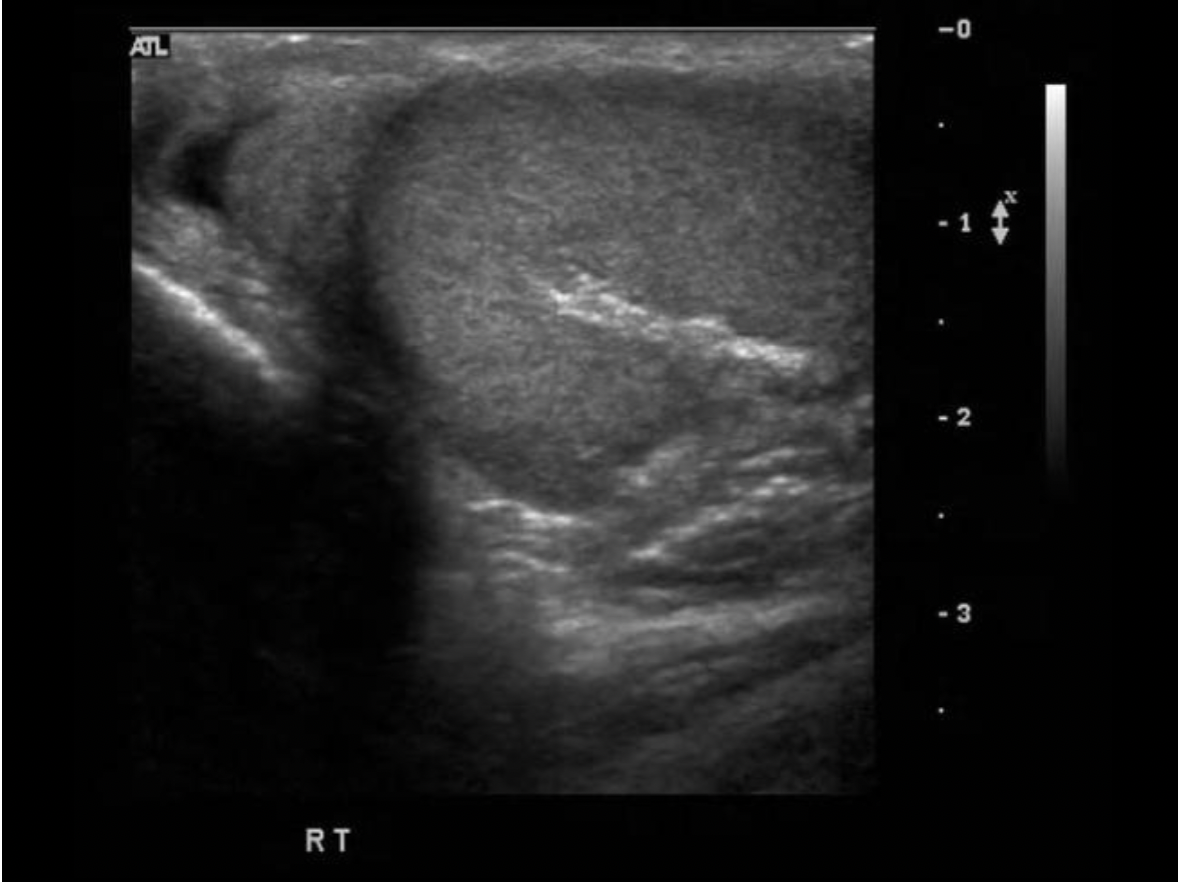
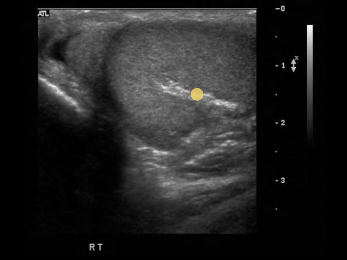
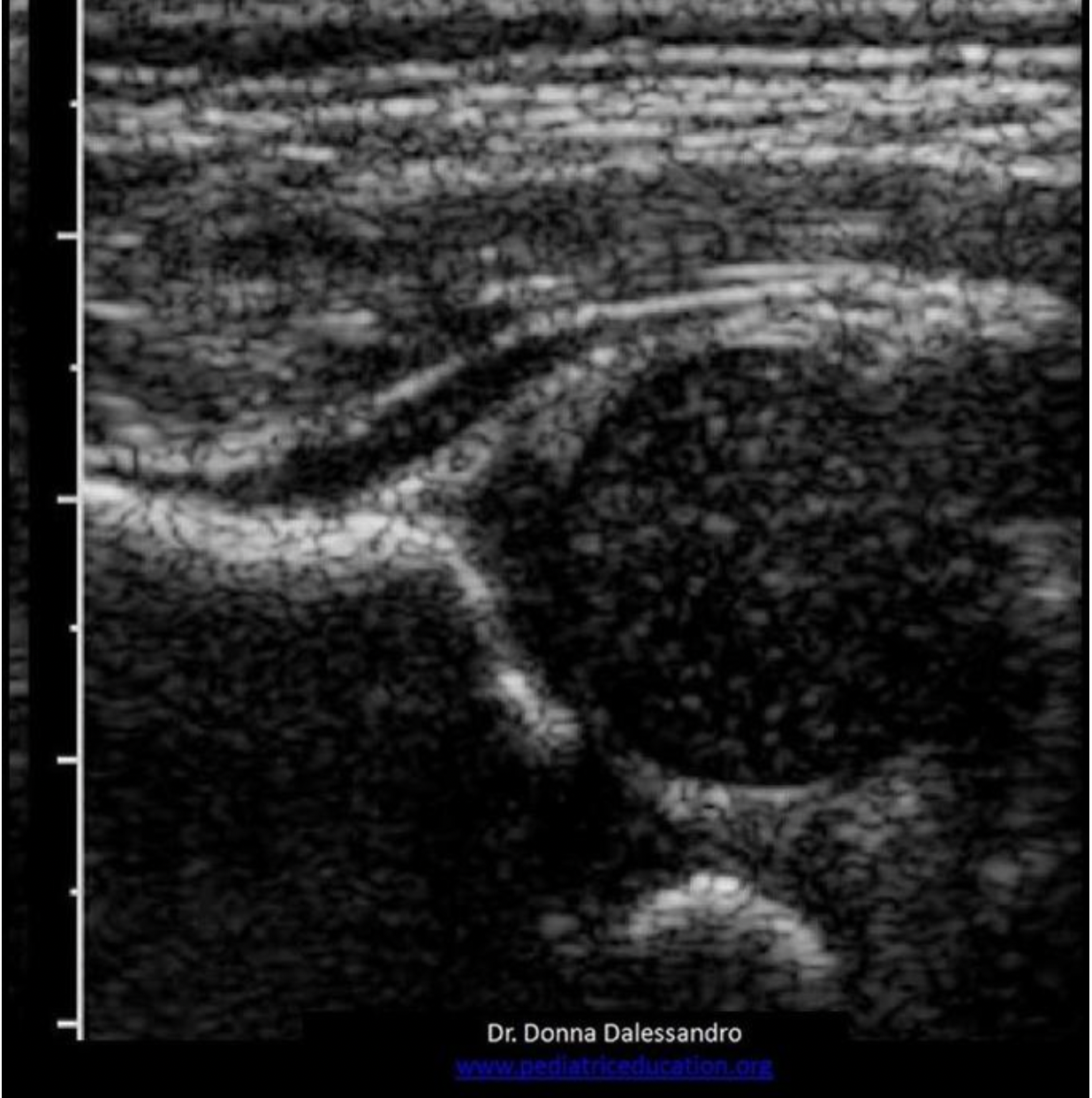
Which of the following correctly describes the findings on the image?
A. There is loculated hydrocele present.
B. There is an extratesticular mass present.
C. There is an appendix teste present
D. There is a scrotal pearl present.
C. There is an appendix teste present
The appendix teste is a small ovoid structure usually located between the superior pole of the testicle and epididymal head. It is normally the same echogenicity of the epididymis.
The paracolic gutters:
A. are divided by the linea alba
B. trap fluid in the pelvic area
C. trap fluid in the lateral abdomen
D. allow fluid to move between the abdomen and pelvis
D. allow fluid to move between the abdomen and pelvis
The paracolic gutters allow fluid to move between the abdomen and pelvis.
Fluid in the subhepatic spaces can move through the paracolic gutters to reach the pelvis.
Which two gut layers are normally hypoechoic on the ultrasound image?
A. Submucosa, muscularis propia
B. Deep mucosa, muscularis propia
C. Serosa, muscularis propia
D. Serosa, superficial mucosa
B. Deep mucosa, muscularis propia
The Gut Signature (Sonographically):
Superficial Mucosa - epithelial lining (and lumen); echogenic
Deep Mucosa-- Consists of loose connective tissue and muscularis mucosa;
hypoechoic
Submucosa - echogenic
Muscularis propia-- inner circular fibers and outer longitudinal fibers;
hypoechoic
Serosa or Adventitia - echogenic
Which of the following normally secretes moderate amounts of mucus?
A. pancreas
B. bronchial alveoli
C. duodenum
D. seminal vesicles
C. duodenum
The duodenum secretes mucus to protect the small bowel from gastric acid. The bronchial alveoli are located in the lungs. Lung infections cause abnormal mucus secretion.
Where is the Dartos muscle and fascia?
A. in the abdominal wall
B. in the scrotal sac
C. in the cystic duct
D. in the urinary bladder
B. in the scrotal sac
The dartos muscle is a layer of tissue found in the penile shaft and scrotum. It is a layer of smooth muscle located just under the skin of the scrotum. It helps form the median raphe and regulate scrotal temperature.
A top normal thyroid isthmus measurement would be:
A. 4cm
B. 6mm
C. 4mm
D. 6cm
B. 6mm
The normal epididymis is located:
A. posterolateral to the testicle
B. posteromedial to the testicle
C. anterolateral to the testicle
D. anteromedial to the testicle
A. posterolateral to the testicle
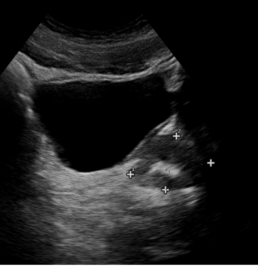
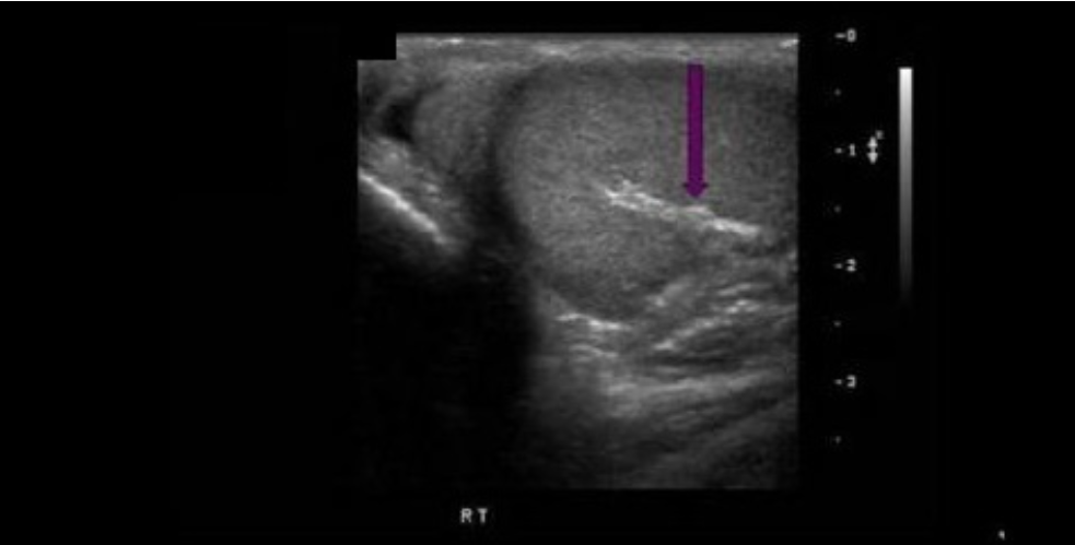
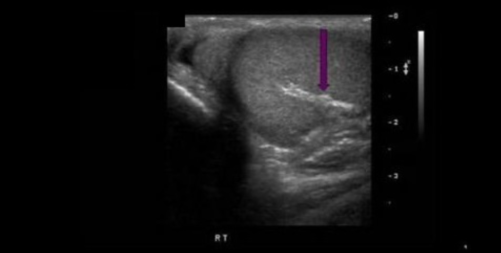
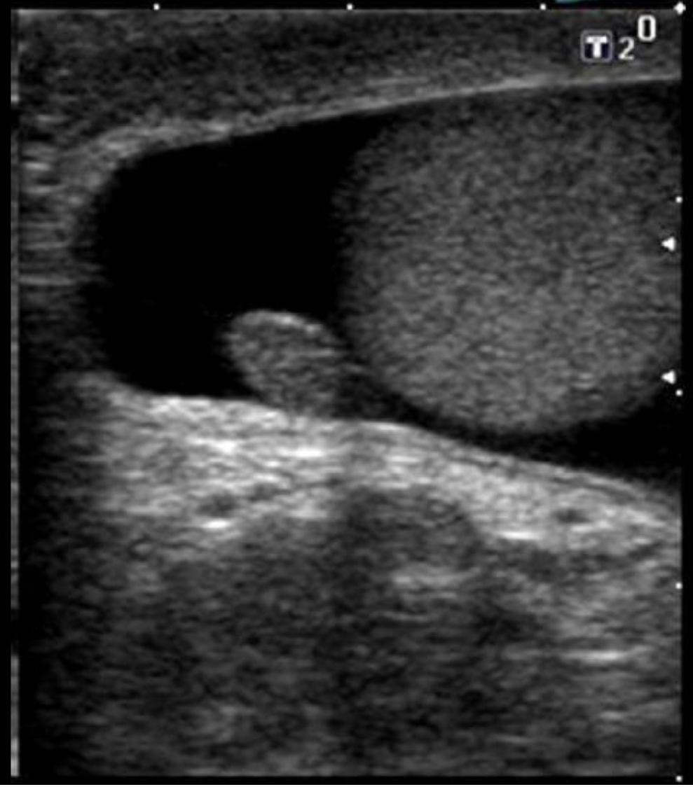
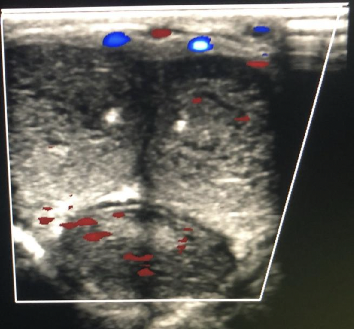
The findings on the image are most suggestive of:
A. Enlarged seminal vesicle
B. Normal seminal vesicle
C. Normal sized prostate with echogenic appearance of the central sinus related to the hypoechoic outer cortex
D. Normal sized prostate with a corpora amylacea.
D. Normal sized prostate with a corpora amylacea.
The normal prostate usually demonstrates a somewhat homogeneous, hypochoic appearance. The image demonstrates a hyperechoic focus in the center of the gland (calcification). Corpora amylace refers to benign calcification formation in the prostate gland.
Find the internal jugular vein
The common carotid artery is lateral to the thyroid gland. The internal jugular vein is slightly anterior and lateral to the common carotid artery.
Which scrotal arteries penetrate the testicular parenchyma to supply oxygenated blood?
A. centripetal
B. capsular
C. cremasteric
D. deferential
A. centripetal
Capsular artery courses along the testicle periphery and produces branches called centripetal arteries which course through the parenchyma. The deferential artery supplies vas deferens and epididymis with blood. The cremasteric artery supplies scrotal sac with blood.

The white arrow on the image indicates:
A. air tract from biopsy
B. central zone
C. urinary catheter
D. urethra
D. urethra
The white arrow indicates the urethra and the red dotted lines separate the transitional and peripheral zones.
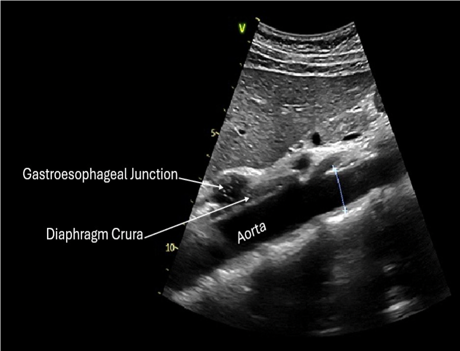
The gastroesophageal junction is best seen in the ______ plane, anterior to the aorta and posterior to the left lobe of the liver.
A. longitudinal
B. radial
C. coronal
D. transverse
A. longitudinal
Which of the following is synthesized by the adrenal medulla?
A. Cortisol
B. Epinephrine
C. Aldosterone
D. Androgens
B. Epinephrine
The adrenal cortex synthesizes cortisol, androgens, estrogen, progesterone and aldosterone. The adrenal medulla synthesizes catecholamines (epinephrine, norepinephrine).
Which muscle group is located anterolateral to the lobes of the thyroid gland?
A. strap muscles
B. tracheal muscles
C. sternocleidomastoid
D. longus colli muscle
C. sternocleidomastoid
Strap muscles are anteromedial to the gland. Sternocleidomastoid muscles are anterolateral to the gland. Longus Colli muscles are posterior to the thyroid lobes.
Ascites can be identified in all of the following spaces/areas, except:
A. Morison pouch
B. Paracolic gutters
C. Between the coronary ligaments within the bare area of the liver
D. Subhepatic space below the right lobe
C. Between the coronary ligaments within the bare area of the liver
Because the liver is in direct contact with the diaphragm in the bare area, fluid cannot accumulate there.
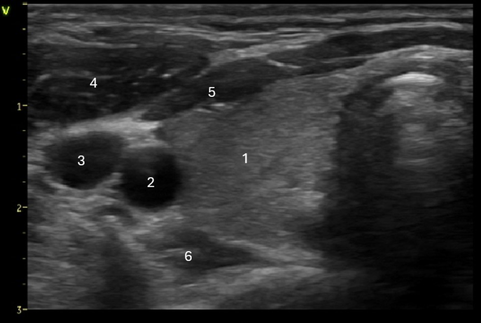
Which of the following structures is labeled #6?
A. Longus Colli muscle
B. Sternocleidomastoid muscle
C. Strap muscle
D. Esophagus
A. Longus Colli muscle
Right lobe of the thyroid
Right carotid artery
Right internal jugular vein
Sternocleidomastoid muscle
Strap muscles
Longus colli muscle
Which of the following scrotal structures carries the seminal fluid from the rete testis to the epididymis?
A. vas deferens
B. efferent ducts
C. mediastinum testis
D. seminiferous tubules
B. efferent ducts
The seminiferous tubules funnel the sperm toward the rete testis. The efferent ductules carry the sperm from the rete testis to the epididymis.
Which of the following is not a layer of the scrotal sac?
A. Dartos muscle
B. Epithelial/Skin
C. Tunica Vaginalis
D. Detrusor muscle
D. Detrusor muscle
The detrusor muscle is located in the urinary bladder.
In pediatric patients, the volume of what structure is calculated using the Lambert or Ellipsoid formula?
A. testicles
B. spleen
C. liver
D. kidneys
A. testicles
In pediatric patients, testicular volumes can be calculated using:
Lambert formula = length X width X height X 0.71
Ellipsoid formula = length X width X height X 0.52
Which scrotal artery encircles the testicular periphery?
A. cremasteric
B. capsular
C. deferential
D. centripetal
B. capsular
Capsular artery courses along the testicle periphery and produces branches called centripetal arteries which course through the parenchyma. The deferential artery supplies vas deferens and epididymis with blood. The cremasteric artery supplies scrotal sac with blood.
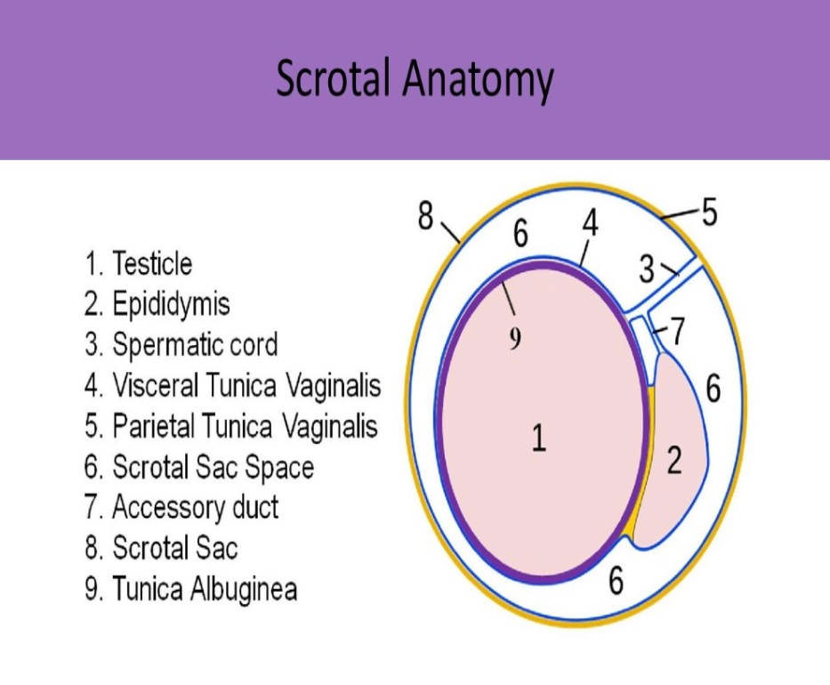
Which of the following lines the inside of the scrotal sac?
A. tunica albuginea
B. visceral tunica vaginalis
C. rete testis
D. parietal tunica vaginalis
D. parietal tunica vaginalis
The parietal tunica vaginalis lines the inside of the scrotal sac. The visceral layer is the inner layer covering the testes, epididymis and lower part of the spermatic cord
Which muscle group is located anteromedial to the lobes of the thyroid gland?
A. longus colli muscle
B. tracheal muscles
C. strap muscles
D. sternocleidomastoid
C. strap muscles
Strap muscles are anteromedial to the gland. Sternocleidomastoid muscles are anterolateral to the gland. Longus Colli muscles are posterior to the thyroid lobes.

The ______ gland is located anterior to the ear and is
drained by the ______
A. submandibular, Stensen duct
B. submandibular, Wharton duct
C. parotid, Stensen duct
D. sublingual, Wharton duct
C. parotid, Stensen duct
Parotid gland - located anterior to the ear, drained by Stensen duct
Submandibular gland - located under the mandible, drained by Wharton duct
Sublingual glands - located under the tongue
Locate the labrum

The inguinal ligament extends:
A. from the anterior superior iliac spine to the pubic tubercle
B. from the tip of the coccyx to the pubic tubercle
C. from the distal right oblique muscle to the distal left oblique muscle
D. from the linea alba to the anterior superior iliac spine
A. from the anterior superior iliac spine to the pubic tubercle
The inguinal ligament extends from the anterior superior iliac spine to the pubic tubercle. The inguinal canal runs between the external oblique and the transversalis fascia, superficial to the inguinal ligament. A weakness in the pelvic floor allows pelvic contents to drop through the canal into the groin/scrotum.
Increased serum levels of _______ will reduce urine output and increase blood volume in the body.
A. thyroxine
B. alkaline phosphatase
C. aldosterone
D. bilirubin
C. aldosterone
Aldosterone is secreted by the adrenal cortex and increases the rate of sodium resorption and potassium excretion in the kidneys. It adjusts urine output to increase or decrease blood volume. Higher sodium levels will increase water retention by reducing urine output. This will increase blood volume and blood pressure. Elevated aldosterone levels = increased blood volume and pressure. Reduced aldosterone levels = decreased blood volume and pressure.
The ______ gland is located under the mandible and is drained by the _______
A. submandibular, Stensen duct
B. submandibular, Wharton duct
C. sublingual, Wharton duct
D. parotid, Stensen duct
B. submandibular, Wharton duct
Parotid gland - located anterior to the ear, drained by Stensen duct
Submandibular gland - located under the mandible, drained by Wharton duct
Sublingual glands - located under the tongue
Which of the following retroperitoneal spaces contains the adrenal gland?
A. retrofascial space
B. perirenal space
C. anterior pararenal space
D. Glisson capsule
B. perirenal space
Perirenal space holds kidney, perirenal fat, proximal ureter, adrenal gland.
Renal infection can lead to fluid, abscess or gas within the space which will distort renal fascia.
Normal retroperitoneal lymph nodes usually measure less than ___ in length.
A. 0.5cm
B. 1cm
C. 1.5cm
D. 2cm
B. 1cm
The right common carotid artery is a branch of which artery?
A. right subclavian artery
B. right vertebral artery
C. innominate artery
D. aortic arch
C. innominate artery
There is only one innominate artery. It is the first branch of the aortic arch and quickly divides into the right common carotid artery and right subclavian artery.
The left common carotid artery is the second branch of the aortic arch, followed by the left subclavian artery.
What is the term for a cone-like mass of normal thyroid tissue protruding superiorly from isthmus?
A. Thyroglossal lobe
B. Early Graves' disease
C. Pyramidal lobe
D. Hyperthyroidism
C. Pyramidal lobe

Locate the ischium
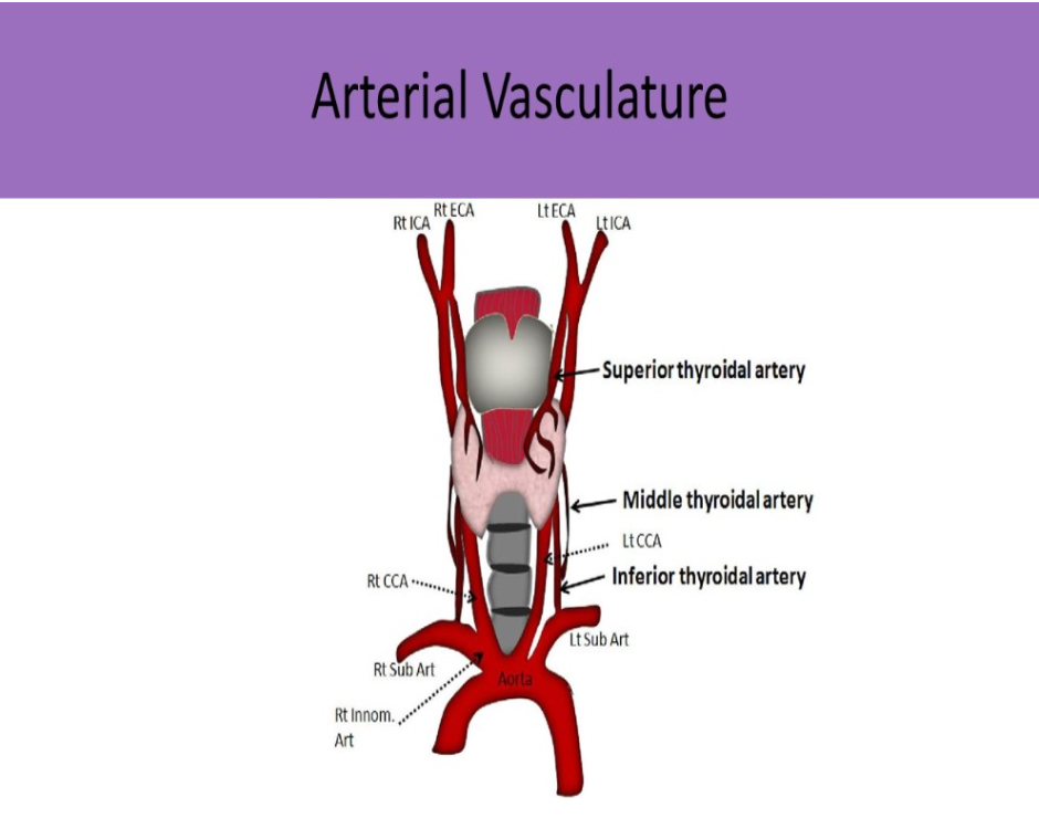
The superior thyroidal artery is a branch of the _______
A. vertebral
B. internal carotid artery
C. innominate
D. external carotid artery
D. external carotid artery

What is the primary function of lymph nodes?
A. filter lymphatic fluid and protect the body from infection
B. release enzymes that fight infection in the body
C. filter blood and protect the body from infection
D. release endorphins that aid in blood filtration
A. filter lymphatic fluid and protect the body from infection
The lymphatic system carries lymph throughout the body to assist in fighting infection and inflammation. Lymph nodes filter foreign substances out of the lymphatic fluid. They produce phagocytes that are carried through the lymph system to destroy bacteria, viruses in the body. They also control the amount of lymphatic fluid in the tissues of the body.

Find the longus colli muscle
The longus colli muscles are posterior to the thyroid gland
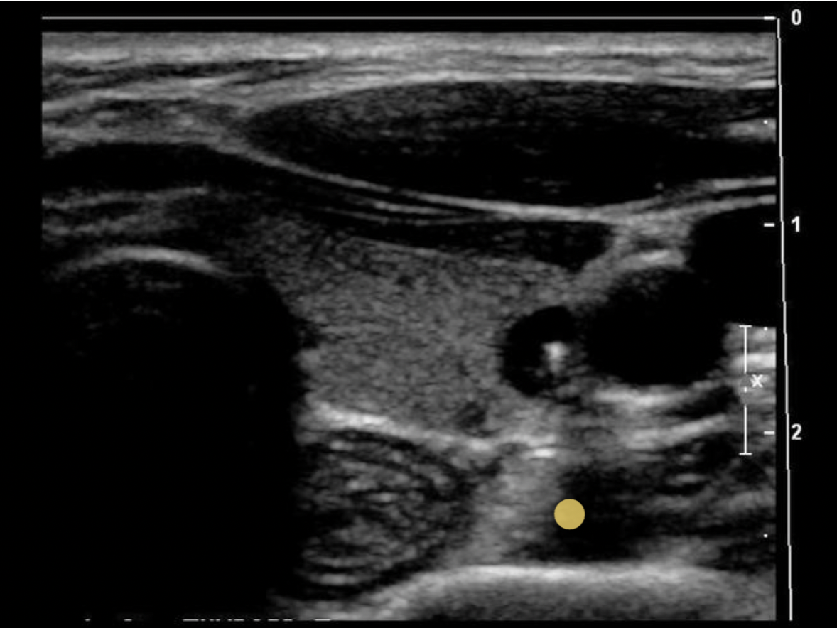
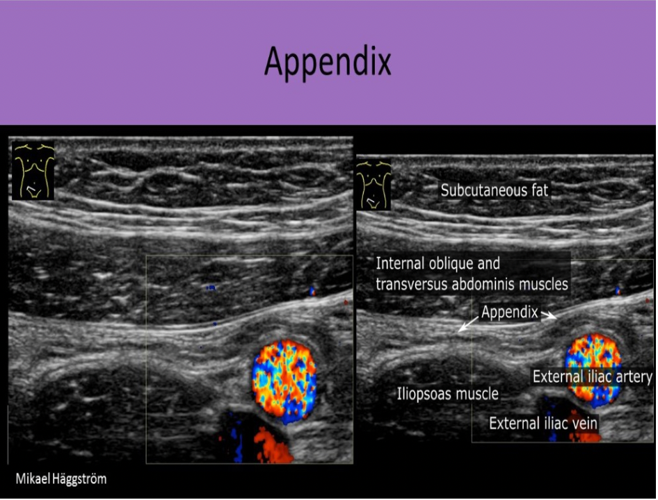
The appendix is normally identified:
A. posterior to the terminal ileum and anterior to the iliac vessels
B. anterior to the terminal ileum and lateral to the iliac vessels
C. originating from the sigmoid colon at the level of the inguinal ligament
D. only in patients less then 150lbs
A. posterior to the terminal ileum and anterior to the iliac vessels
The appendix is also referred to as the vermiform appendix. It originates from the cecum in most patients and can be identified posterior to the terminal ileum and anterior to the iliac vessels.
The _______ is a part of the prostate gland that is mostly composed of smooth muscle cells and makes up approximately 1/3 of the gland.
A. fibromuscular region
B. transitional zone
C. peripheral zone
D. central zone
A. fibromuscular region
The prostate gland contains four glandular zones and a fibromuscular region.
The peripheral zone is the largest glandular segment. Most prostate cancer occurs in the peripheral zone and is also the most common site of chronic prostatitis.
The transitional zone, the site of benign prostatic hyperplasia (BPH), surrounds the urethra.
The central zone holds most of the remaining glandular tissue.
The glandular tissue that surrounds the proximal prostatic urethra is called the periurethral glandular zone.
The anterior fibromuscular region is mostly smooth muscle and occupies about one-third of the prostate.
The testicles normally descend into the scrotum:
A. 7 days after birth
B. by 1 yr old
C. in utero
D. at the onset of puberty
C. in utero
The testicles normally descend into the scrotum in utero, between 26 and 34 weeks gestation. If one or both fail to descend, cryptorchidism is present.
Find the normal lymph node
A normal lymph node is oval in shape and demonstrates a thin hypoechoic rim of tissue around an echogenic hilum. Abnormal lymph nodes demonstrate a more rounded shape, thickened cortex that can appear anechoic. and there is loss of differentiation of the hilum.
Find the area where the urethra would normally be found
The urethra courses through the center of the corpus spongiosum on the ventral side of the penis.