A&P of Domestic Animals-Test 1
1/48
There's no tags or description
Looks like no tags are added yet.
Name | Mastery | Learn | Test | Matching | Spaced | Call with Kai |
|---|
No analytics yet
Send a link to your students to track their progress
49 Terms
What is the difference between anatomy and physiology? Provide examples
Anatomy is the structure of the organs
Physiology is how the organs function
Ex: Anatomy- the design of the engine of a car
Physiology- how the car functions from the engine
What are the four levels of organization in the body? What is the smallest level capable of life?
Cells, Tissues, Organ, Organ System
Cells
What do cells need to live? How do they obtain what they need?
Water, Oxygen, Nutrients, Living Environment, and Waste Removal.
Through the bloodstream
What are the four basic tissues in the animal body and what are their functions? Describe a body organ containing numerous types of tissue and explain how those tissues work within the organ.
Epithelial- protect the body, absorb nutrients, secrete, filter, and excrete waste
Muscle- Allows for movement throughout the body; support
Connective- binding & supporting organs; transporting nutrients
Nervous- controls and regulates the body
Heart- epithelial protects outside of the heart
Muscle makes the heart pump
Connective allows the heart to move blood throughout body
Nervous tells the heart to beat
Correctly classify tissues in the body into one of the four basic tissue types.
Epithelial- epidermis of skin, lining of digestive tract
Connective- Blood, bone, cartilage
Muscle- skeletal, cardiac, and smooth muscle
Nervous- neurons, spinal cord, and nerves
Apply directions terms, including: Left and Right, Cranial and Caudal, Rostral, Dorsal and Ventral, Proximal and Distal, Medial and Lateral, Superficial and Deep, Palmar and Plantar
Left/ Right- Animal’s left/ right
Cranial- toward the animal’s head
Caudal- toward the animal’s rump/tail
Rostral- (use on the head) toward nose/ front of skull
Dorsal- toward top surface of standing animal
Ventral- Toward belly (ground) of standing animal
Medial- Toward median (middle) plane of body
Lateral- away from median (middle) plane of body
Superficial- toward surface of the body
Deep- toward center of the body
Proximal (on extremities only)- toward body, closer to body
Distal ( on extremities only)- away from body, further from body
Palmar- below carpus on FRONT LEG
Plantar- below tarsus on REAR LEG
How are directional terms used to describe the front and back surfaces of the limbs? *Remember that different terminology is used above / below the carpus/tarsus.
Cranial- front limb surface above carpus/tarsus
Caudal- back limb surface above carpus/tarsus
Dorsal- front/top surface of limb below carpus/ tarsus
Palmar- back surface below carpus/ tarsus on front limb
Plantar- back surface below carpus/ tarsus on hind limb
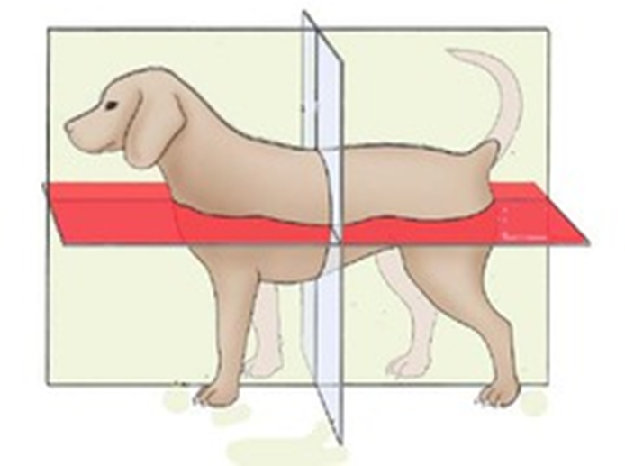
Describe, apply, and sketch planes of reference, including: sagittal, median, transverse, and dorsal
Transverse- gray plane; divides body into head/ rump parts (hula hoop plane)
Sagittal- like medial, but not exactly centered
Medial- Yellow plane; equal right and left parts
Dorsal- Red Plane; dorsal and ventral parts of body
What is the general plan of the animal body? What body cavities are present and what do they contain? What membranes line these cavities and cover the organs within them?
Two main body cavities
Dorsal- (Cranial- brain and Spinal- spinal cord)
Ventral- (Thoracic- lungs/heart and Abdominal- other organs)
Thoracic lined by thin membrane called pleura;viceral pleura covers OUTER surface of organs; Parietal Pleura- lines INSIDE of cavity
Abdominal- peritoneum; visceral- INNER surface; parietal- OUTER
Explain how body systems are essential to the functioning of the animal body. What are the basic functions of each system?
They each contribute to the body functioning perfectly, some can be removed with no damage.
Digestive- how food/nutrients get absorbed
reproductive- creates human life/ produces hormones
Urinary- how waste is excreted
Endocrine- regulates processes throughout the body
Respiratory- oxygen exchange
Integumentary- protects outside of body
Skeletal- provides support for body, protects organs
Muscular- facilitates body movement
Cardiovascular- carries essential nutrients throughout the body
Immune- first line of defense against invaders
Lymphatic- returns fluids to the bloodstream
Nervous- controls the entire body and how it functions
Define health. What is disease?
The absence of disease; disrupts the normal function of the body that may cause a disfunction
Define homeostasis? Explain what is involved in the animal body maintaining homeostasis
An organisms stable internal environment; nervous system, endocrine system
Describe the functions of epithelial tissue. How do these functions vary depending upon where the epithelial tissue is located in the animal body?
It protects, filters, absorbs, manufactures secretions and excretion, and provides sensory input.
Single layer- GI tract absorption
Stratified- outermost layer of skin
Glandular- glands of the body
What are general characteristics of epi tissue?
Avascular, Innervated, contains basement membrane (connects epi to connective tissue)
What is keratin? Why is it important when it comes to epithelial cells?
A tough, waterproof protein; prevents bacterial penetration
What is the difference between simple and stratified epithelium? What is the benefit/downside of each type of epithelial tissue? Where would we find simple and stratified epithelial tissue in the animal body? Why?
Simple contains only one type of cell shape, stratified contains many
Simple offers little protection, but can absorb; stratified is tougher, but nutrients cannot absorb as fast
Simple- Lines alveoli in lungs; stratified- outer layer of skin
Compare and contrast keratinized and non-keratinized stratified squamous epithelium. Where is each found in the animal body and why?
Keratinized- outer layer of skin, outside of body/ dead
Non-keratinized- mouth, protects tissues underneath
What is unique about transitional epithelium and where is it found in the animal body?
It allows area to stretch and then return back to normal size
bladder, ureters, urethra
Determine if a gland is endocrine or exocrine. Give an explanation of the differences between these types of glands.
Endocrine- secretes hormones directly into bloodstream
Exocrine- secrete substances through ducts onto internal or external body surface
Understand and combine basic medical terminology prefixes and suffixes from the notes (for example: cyte, osteo, etc.)
Chondro-cartilage
Osteo-bone
Leuko- white
Adipo- fat tissue
-cyte-cell
-blast-immature
What are the functions of connective tissue?
Supports, protects, and connects tissues and organs
What are the 3 components of CT? Which are living and which are non-living? Which components make up the extracellular matrix?
Specialized Cells-Living, Protein Fibers- nonliving, and Ground Substance- nonliving. All types of cells
Compare and contrast the following types of CT: areolar CT, adipose tissue, dense regular CT
Compare-All three are types of connective tissue, all contain cells
Contrast- Areolar is loose and airy, adipose contains fat cells, dense regular is very dense and tightly packed
Describe specialized CT (cartilage, bone, blood) in the animal body. What unique features do these CT’s have?
Cartilage- cushions, protects, and supports
Bone- stores calcium, supports body
Blood- stores electrolytes and transports many substances and nutrients
Describe cartilage and how it stands out from other types of CT. How is it different? Why is this difference important?
Flexible and strong connective tissue, but it is noninnervated and avascular. Because it acts as a shock absorber and cushion for different areas of the body
Where are the various types of cartilage found in the animal body?
Hyaline- in joints, nose, growth plates, rib cage (most common)
Elastic- ear pinnae, voice box
Fibrocartilage-intervertebral disks, pubic symphysis
What are the components of blood?
plasma, red blood cells, white blood cells, and platelets
What does the term hematopoietic mean? Where is hematopoietic tissue found within bone?
Development of blood and blood cells
Bone marrow
What are the unique features of bone as a CT? Describe various bone cells and their functions.
Hardest, most rigid type of CT, living and changing tissue
Osteocytes-mature bone cells
Osteoblasts-help bones grow and develop
Osteoclasts- form new bones and add growth to existing ones
What is osteoarthritis? Is it painful? What types of connective tissue are involved? Explain.
Bone and joint inflammation in the body. It is painful. Cartilage wears away and innervated bone rubs against each other, causing pain
Compare and contrast the 3 basic types of muscle in the animal body. Where is each type of muscle located?
Skeletal- found in limbs; striated, multinucleated, voluntary
Cardiac- found in heart; striated, 1 nucleus; involuntary
Smooth- found in viscera; not striated, 1 nucleus; involuntary
What is the function of nervous tissue? What is the primary cell type in nervous tissue?
transmit sensory information from body to brain and brain out to body; neurons- nerve cells
What makes neurons high maintenance cells?
high requirement for oxygen, cannot reproduce, need support (glial cells)
What are glial cells?
support cells for neurons, protect neurons
Use appropriate medical terminology for the integument (derm, pilus, tricho, hypo)
Derm-skin
Plius- relating to hair
tricho- relating to hair
Hypo- below
What are the functions of the integumentary system? Why are these functions important for the body?
Protection, preventing desiccation (drying out)
Sensation
Temperature Regulation
Production of Vitamin D
Secretion and Excretion via glands
What are the characteristics of each layer of the integument? Vascular or avascular, innervated, etc. How are these features important to the functioning of the area?
Epidermis- avascular, but innervated. Allow for bleeding not all the time
Dermis- dense, irregular, vascular and innervated
Hypodermis- deepest layer, insulator and shock absorber. allows skin to move freely over bone
Describe the layers of the epidermis – where are living, dividing cells found? What happens to cells as they move towards the surface of the integument?
Deepest- dividing, living epi cells
Outermost layers made of keratinized, dead epithelial cells that are shed constantly
Describe the special features of the integument, including pigmentation, paw pads, planum nasale, ergots and chestnuts, and cutaneous pouches of sheep
pigmentation- melanocytes produce pigment; color
paw pads-forms the lower, weightbearing part of the foot
planum nasale- hairless pigment of muzzle
ergots and chestnuts- extra toes or pads from ancestors
cutaneous pouches- waxy substance that gives off a distinct color
Describe and diagram the parts of hair. What layer of the integument is hair anchored in? What are the functions of hair?
Hypodermis
Protects, insulator, and sensory organ
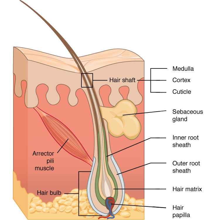
What is the function of the arrector pili muscle? Is it a voluntary or involuntary muscle? How does it contribute to the fight or flight response?
to contract and cause hair to stand up. Adrenaline can cause this from fear
Compare and contrast the various types of hair: primary, secondary, tactile
Primary- long, stiff straight; outermost layer
Secondary- short, thin, soft; soft undercoat, close to skin
Tactical- thicker and longer than primary; whiskers or sinus hairs
Compare and contrast the types of glands found in the skin. What are the functions of each? Are the glands endocrine or exocrine? Why?
sebaceous and sweat glands, both exocrine
Describe claws and dew claws.
Claws- hard outer covering of distal digits
Dewclaws- extra toe on dog, first digit
Explain a feline declaw surgery and understand the anatomy involved.
Amputation of entire third phalanx bone and claw. Claw grows directly from P3 so failure could result in regrowth of claw
What is an ungulate? Which species are ungulates?
a hoofed, four-legged mammal that typically walks on the tips of its toes.
How many digits (toes) do ruminants bear weight on? How many digits do horses (equines) bear weight on?
2;1
Explain the anatomy of the equine hoof. How to all of the components work together? What are the features of each area of the hoof? Which areas are insensitive to pain? Which are sensitive to pain?
outer part- hoof wall- toe, quarters, heel- not innervated or vascular
inner- sole, frog- non-innervated
deepest- corium, secures hoof wall and sole to underlying P3 bone- innervated and vascular
What happens when a horse has laminitis? Identify the areas of anatomy that are involved and describe the physiology.
Inflammation of the laminae of the corium
Swelling can cause separation of P3 bone and hoof wall. P3 rotates downward due to weight of horse