Unit 4: The Nervous System & Sense Organs
1/212
There's no tags or description
Looks like no tags are added yet.
Name | Mastery | Learn | Test | Matching | Spaced | Call with Kai |
|---|
No analytics yet
Send a link to your students to track their progress
213 Terms
nervous system
brain, spinal cord, and nerves
coordinate the body’s systems by receiving and sending information through electrical signals; maintains homeostasis
functions of the nervous system
sensory input: receives internal and external bodily information through sensory receptors
integration: processes and interprets sensory input to determine where information is sent
motor output: activates effector organs to respond to signals and produce a response
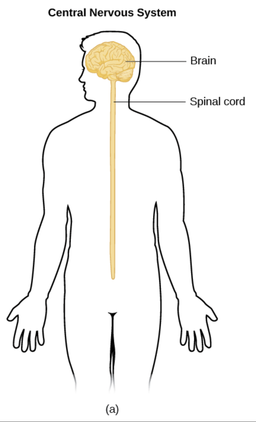
central nervous system (CNS)
brain and spinal cord; body’s command center for processing information
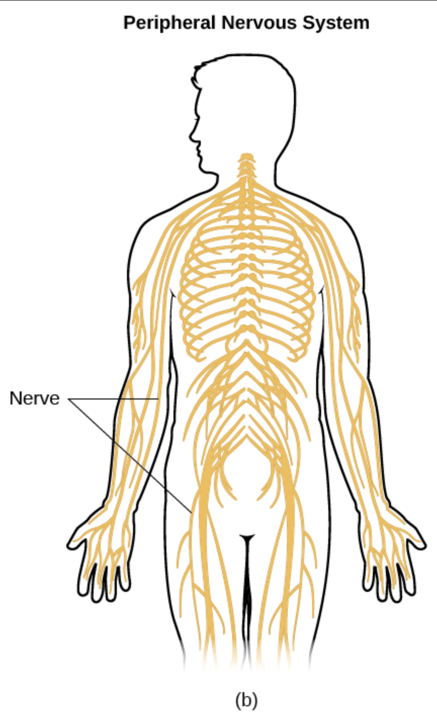
peripheral nervous system (PNS)
31 pairs of spinal nerves, 12 pairs of cranial nerves
nerves that branch out from the CNS and connect to other body parts to relay sensory input and motor commands
somatic nervous system (SNS)
division of the PNS; facilitates voluntary movement of skeletal muscles, transmits sensory information to the CNS
autonomic nervous system (ANS)
division of the PNS; regulates involuntary physiological processes
parasympathetic nervous system
division of the ANS; network of nerves that relaxes the body after periods of stress and danger (“rest and digest”)
lowers heart rate, boosts metabolism, calms the body
sympathetic nervous system
divison of the ANS; network of nerves that stimulate and prepare the body for stress, danger, or exercise (“fight or flight”)
increases heart rate, dilates pupils, widens airways, redirects blood flow to muscles
parasympathetic and sympathetic divisions act
antagonistically, one is excitatory, other inhibits
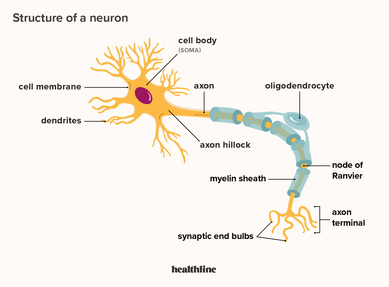
neurons
nerve cells that transmit information; fundamental unit of the brain and nervous system
cell body (soma)
contains the nucleus and other cell organelles
dendrites
short, branched extensions of a neuron, receives information and transmits to the cell body
axons
single long fibers of a neuron, conducts information away from the neuron
myelin
fatty insulating sheath surrounding axons; increases neural signal conduction speed
produced by oligodendrocytes or Schwann cells
nodes of ranvier
gaps in the myelin sheath along the axon
neurofibrils
fibers and structural proteins within the axons of neurons, provide structural support to the neuron
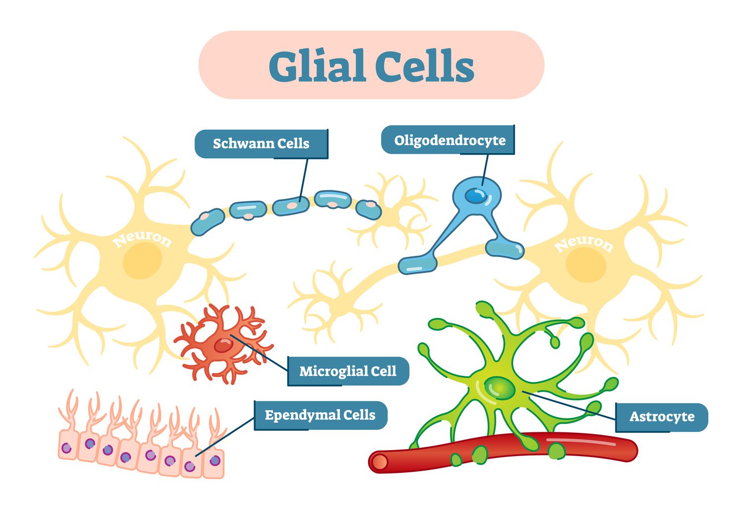
neuroglial cells
non-neuronal cells in the CNS and PNS that outnumber neurons and provide support, protection, and maintenance of neurons
microglial cells
immune function (neuroprotection), digest debris, kills bacteria, synaptic pruning
oligodendrocytes
make myelin sheath around neurons in the CNS
schwann cells
make myelin sheath around neurons in the PNS
ependymal cells
forms membranes around simple epithelium tissue, filters blood to make and regulate cerebrospinal fluid (CSF)
astrocytes
physically connect blood vessels to neurons to regulate energy and oxygen supply to neurons; most abundant glial cell in the CNS
white matter
myelinated axon that transmits signals rapidly, appears white due to the fat component of myelin sheath
grey matter
unmyelinated axon that primarily processes information
lesions
evidence of nerve cell damage in the brain or spinal cord
symptoms of lesions
varies depending on the location of the lesion;
spinal cord lesions may cause motor problems
back of brain lesions may cause balance problems
reflex arc
the shorter neural pathway that bypasses the brain to mediate an involuntary and rapid response to stimuli, provides protection and facilitates survival
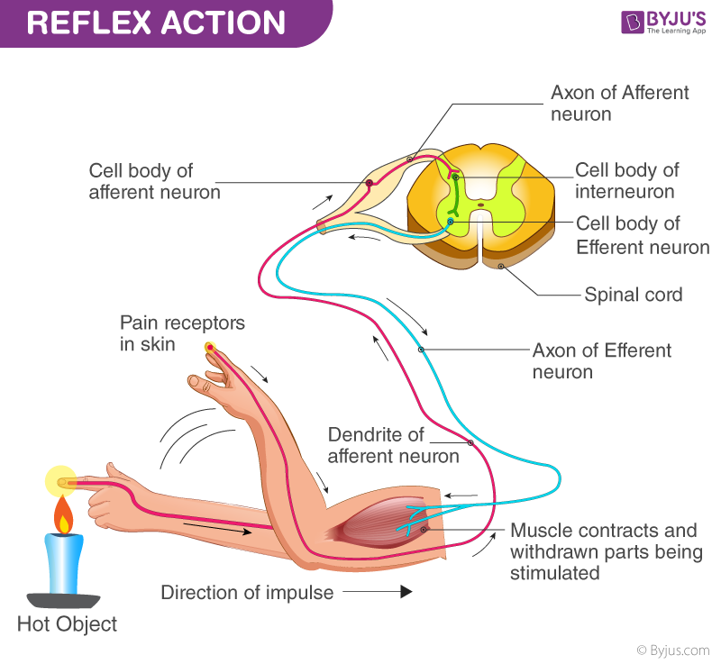
components of a reflex arc
stimulus, receptor, sensory neuron, CNS (spinal cord), interneuron, motor neuron, effector pathway, response
brain function in a reflex arc
message gets sent to the brain later, can choose to override the initial response or act further
simple reflex
reflex arc designed for protection or homeostasis without requiring conscious brain involvement
conditioned reflex
learned physiological, motor, or emotional responses to a certain stimulus
sensory (afferent) neurons
transmit impulses from receptors to the CNS to perceive stimuli
motor (efferent) neurons
transmit impulses from the CNS to muscles and glands to enable action
relay neurons (interneurons)
connect sensory neurons to motor neurons to transmit signals between neurons to facilitate quick communication such as in reflex arcs
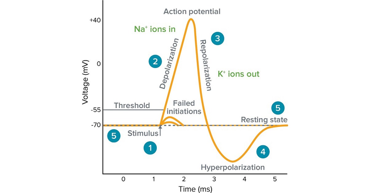
action potential (nerve impulse)
a rapid electrical signal traveling along a neuron’s membrane to enable communication throughout the nervous system
action potentials originate from
cell body
direction of action potential
dendrites receive signal, signal passes down axon, crosses to the dendrites of a neighboring neuron
resting membrane potential
the difference in electrical charge (voltage) between the inside and outside of a cell membrane when the cell is at rest and not actively signaling, typically -70 mV
steps of an action potential
resting state (1), threshold, depolarization, repolarization, hyperpolarization, resting state (2)
resting state (1)
stable negative electrical charge of a neuron’s interior (-90 to -70 mV relative to outside), NA+ and K+ channels are closed, neuron is ready but not actively sending a message
threshold
critical membrane potential needed for an action potential to fire (-55 mV), stimulus causes ligand-gated NA+ channels to open, some NA+ begins to diffuse into the cell from outside the cell
depolarization
membrane potential rises from -55 mV to 30 mV, when threshold of -55 mV is met NA+ channels open and causes rapid diffusion of positive NA+ ions into the cell, action potential is elicited
repolarization
membrane potential decreases from 30 mV to a negative value after depolarization peak, caused by NA+ channels closing after depolarization peak, K+ channels open to allow K+ ions to diffuse out of the cell to restore resting membrane potential
hyperpolarization
membrane potential becomes more negative than the resting potential (less than -70 mV) as K+ ions diffuse out of cell, K+ channels close, cell resets, action potentials are temporarily inhibited during refractory period
resting state (2)
sodium-potassium pump uses ATP to pump 3 NA+ out of the cell and 2 K+ into the cell, re-establishes the original ionic gradient, returns the neuron to its initial resting state
summarized steps of an action potential
neuron membrane maintains resting potential
threshold stimulus is received
sodium channels open
sodium ions diffuse inward, depolarizing the membrane
potassium channels open
potassium ions diffuse outward, repolarizing the membrane
action potential causes a local bioelectric current that stimulates the membrane
wave of action potentials travel the length of the axon as a nerve impulse
speed of nerve impulses
speed is proportional to the size of the axon, greater diameters produce a faster impulse
myelinated axon conduction
myelinated axons conduct impulses faster than an unmyelinated one; most neurons are myelinated, but not all are
damaged myelin sheath
slows down, disrupts, fails to get the nerve impulse to their destination
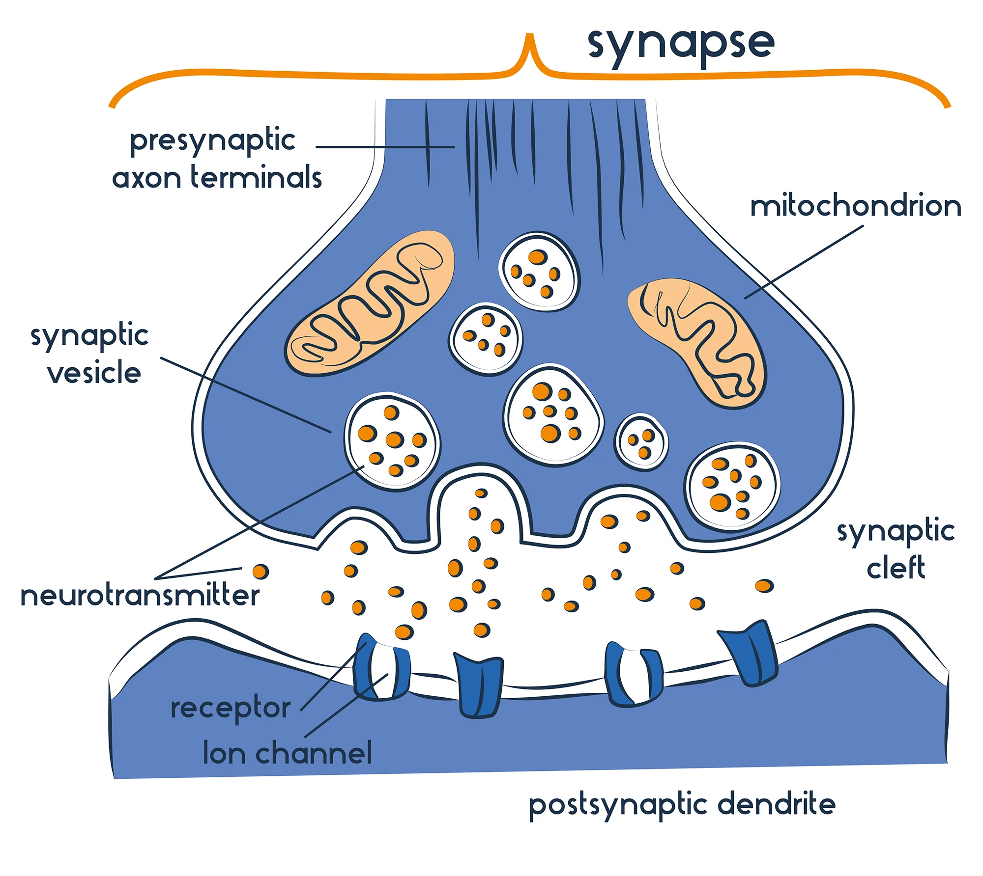
synapse
junction between two neurons where signal transmission occurs, converts electrical action potentials into neurotransmitters to pass information
synaptic transmission
the process at synapses when a chemical signal is released across the synaptic cleft in order to communicate
mitochondria
maintains high-energy synaptic transmission
vesicle
stores, transports, and releases neurotransmitters into the synaptic cleft
receptor
specialized proteins that detect neurotransmitters and convert the chemical signals into an electric response
calcium channel
translates electrical signals into chemical messages
transporter (reuptake area)
end neural signals by absorbing neurotransmitters from the synaptic cleft
nerve pathway
nerve impulse travels from neuron to neuron
neurotransmitters
chemical messengers that transmit signals across a synapse between neurons, muscles, or gland cells
role of neurotransmitters
released at the gap of a synapse to signal to the next neuron to complete the signal, land on receptors on the dendrite receive the chemical message
excitatory neurotransmitters
increase membrane permeability, increases the chance for threshold to be achieved to fire an action potential
inhibitory neurotransmitters
decrease membrane permeability, decrease chance for threshold to be achieved to fire an action potential
reuptake
process in which neurotransmitters are returned to the original cell
what happens if the reuptake transporter is blocked?
leaves the neurotransmitter to remain in the synaptic cleft and continues to stimulate receptors
reuptake inhibitors
drugs that increase the amount of neurotransmitter levels by blocking their reabsorption into the presynaptic neuron
selective serotonin reuptake inhibitors (SSRIs)
antidepressants that increase serotonin levels in the brain by blocking its reuptake (reabsorption) into neurons, leaves serotonin active for longer
acetylcholine
stimulates muscle contraction
dopamine
mood, happiness
serotonin
sleepiness, mood
endorphins
pain reduction, mood
agonists
molecule that has the same effect on the neuron as the original neurotransmitter, it mimics the molecule
antagonist
molecule that blocks the effects of a neurotransmitter
neurological disorder
a disorder affecting the brain, spinal cord, and nerves; disrupts the nervous system
multiple sclerosis (MS)
autoimmune disorder when the immune system mistakenly attacks the myelin sheath in the CNS nerve fibers
causes of MS
autoimmune disorder when the immune system mistakenly attacks the myelin sheath in the CNS nerve fibers
risk factors of MS
genetics, viral infections
signs and symptoms of MS
muscle weakness, numbness, tingling in limbs, fatigue, difficulty with coordination and balance, muscle stiffness, cognitive difficulties
difficulty in diagnosing MS
myelin can be attacked at various locations within the CNS which affects different muscles; varies from person to person
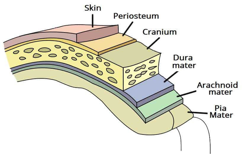
meninges
three layers of tissue membrane between bone and soft tissue
dura mater, arachnoid mater, pia mater
layers of brain
skin, periosteum, bone, dura mater, arachnoid, pia mater
cerebrospinal fluid (CSF)
a clear liquid that circulates the brain and spinal cord, protects and cushions the brain by acting as a shock absorber
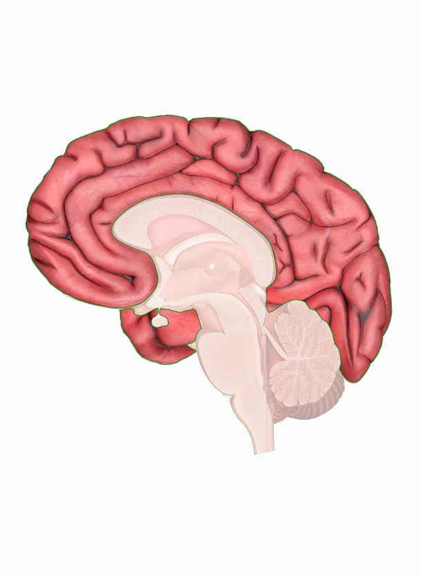
cerebrum
largest, uppermost part of the brain, consists of left and right hemispheres and outer cerebral cortex
function: higher mental functions, problem solving
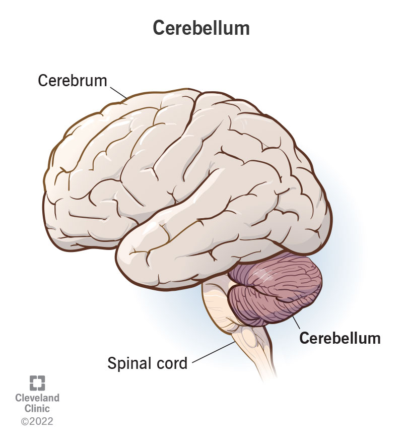
cerebellum
hindbrain structure at the back of the skull
function: coordination of voluntary movements, motor learning, and balance and posture
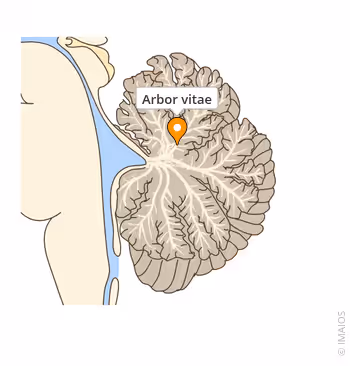
arbor vitae
branching, tree-like white matter within the core of the cerebellum
function: brings sensory and motor information to and from the cerebellum
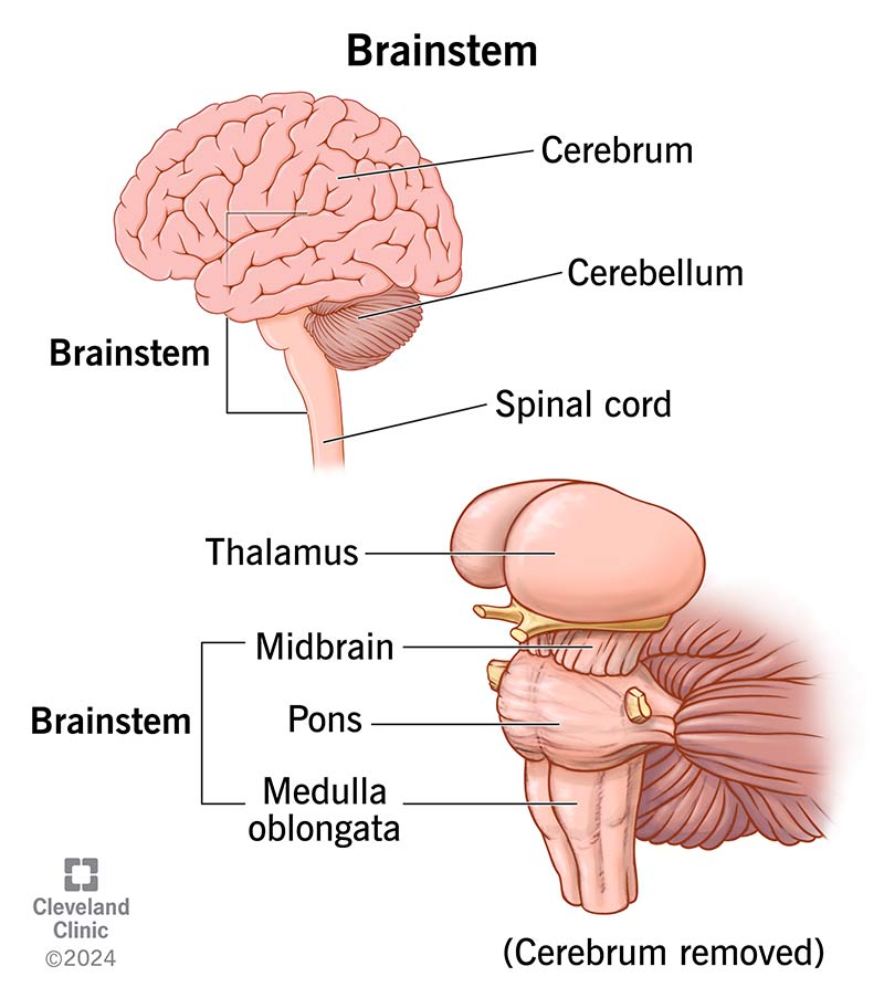
brain stem
connects the brain and the spinal cord
function: carries signals that regulate autonomic bodily functions (breathing, heart rate, blood pressure, digestion)
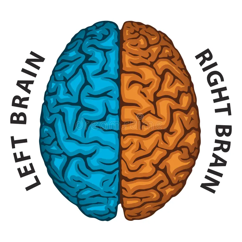
cerebral hemispheres
left and right sides of the brain
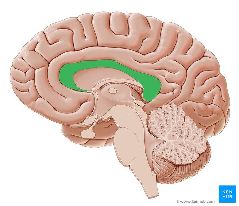
corpus callosum
bundle of nerve fibers connecting the left and right cerebral hemispheres
brain convolutions
the folds (gyri) and grooves (sulci) on the surface of the cerebral cortex, increases the brain’s surface area to allow more neurons and neural connections
fissures
deep groove
sulcus
shallow groove
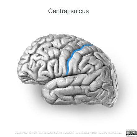
central sulcus
divides frontal and parietal lobes
gyrus
raised ridges or bumps
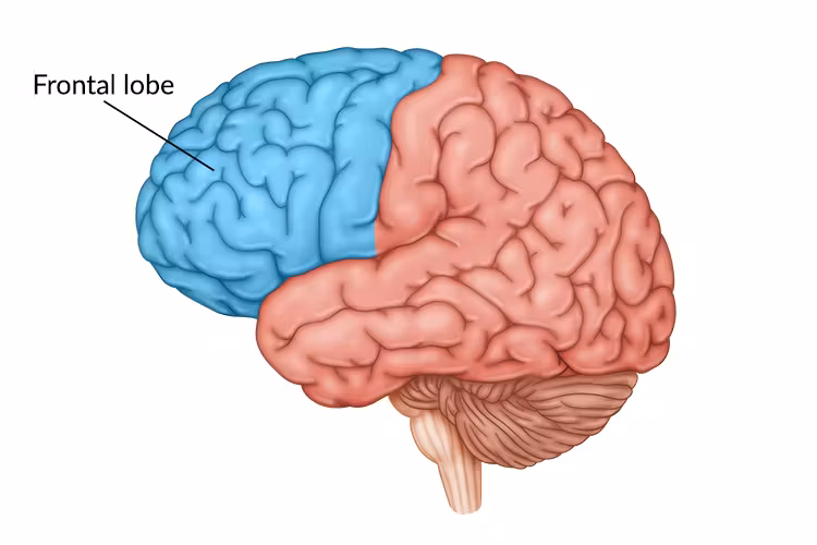
frontal lobe
executive functions, high-level cognitive functions, motor control, decision-making, personality, social behavior
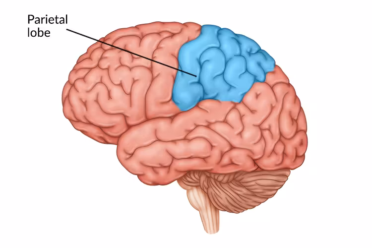
parietal lobe
perception, spatial awareness, navigation, sense-making, math
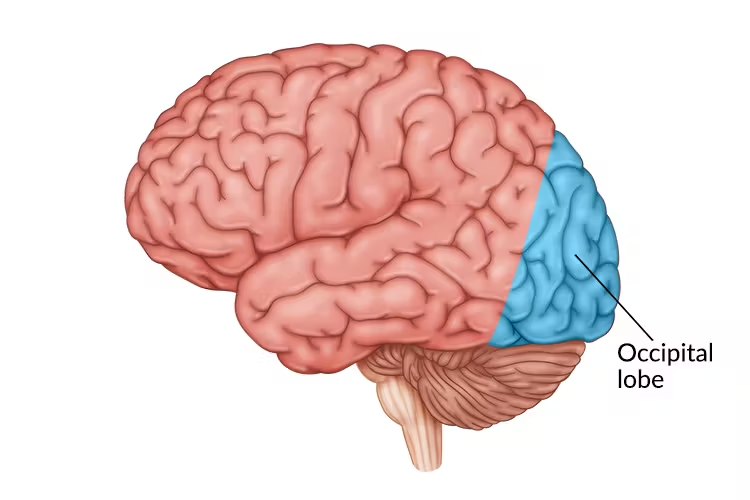
occipital lobe
processing, integrating, and interpreting visual stimuli
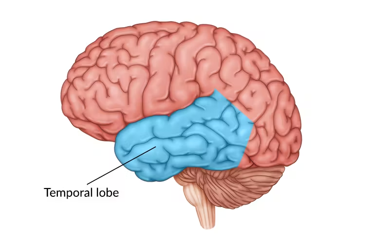
temporal lobe
higher level visual processing, primary auditory cortex, language processing, long-term memory, identifying visual objects and faces
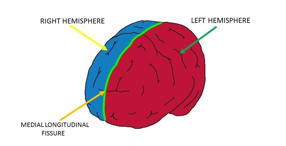
longitudinal fissure
separates right and left cerebral hemispheres, allows each hemisphere to have specialized functions
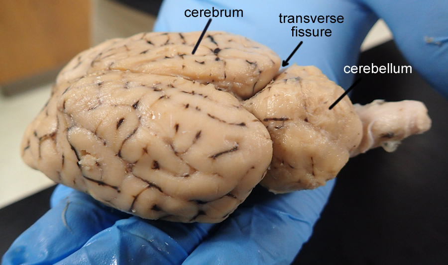
transverse fissure
separates cerebrum and cerebellum
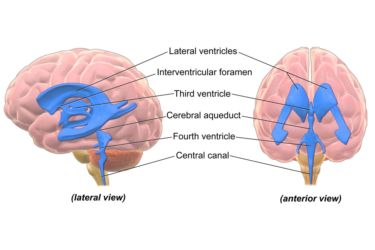
ventricles of the brain
network of four connected fluid-filled cavities that produce, circulate, and store cerebrospinal fluid (CSF), cushion and protect the brain
names of ventricles of the brain
lateral ventricles (2), 3rd ventricle, 4th ventricle
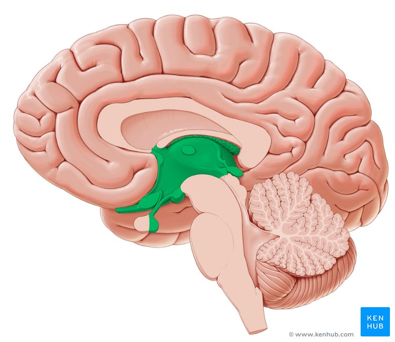
diencephalon
between the cerebrum and the brainstem, consists of the thalamus and hypothalamus
function: relaying sensory and motor signals, maintaining homeostasis, managing emotions and autonomic activities (hormone release)