StemUp: AQA A level Biology 3.2.1 Cell structure
1/58
There's no tags or description
Looks like no tags are added yet.
Name | Mastery | Learn | Test | Matching | Spaced | Call with Kai |
|---|
No analytics yet
Send a link to your students to track their progress
59 Terms
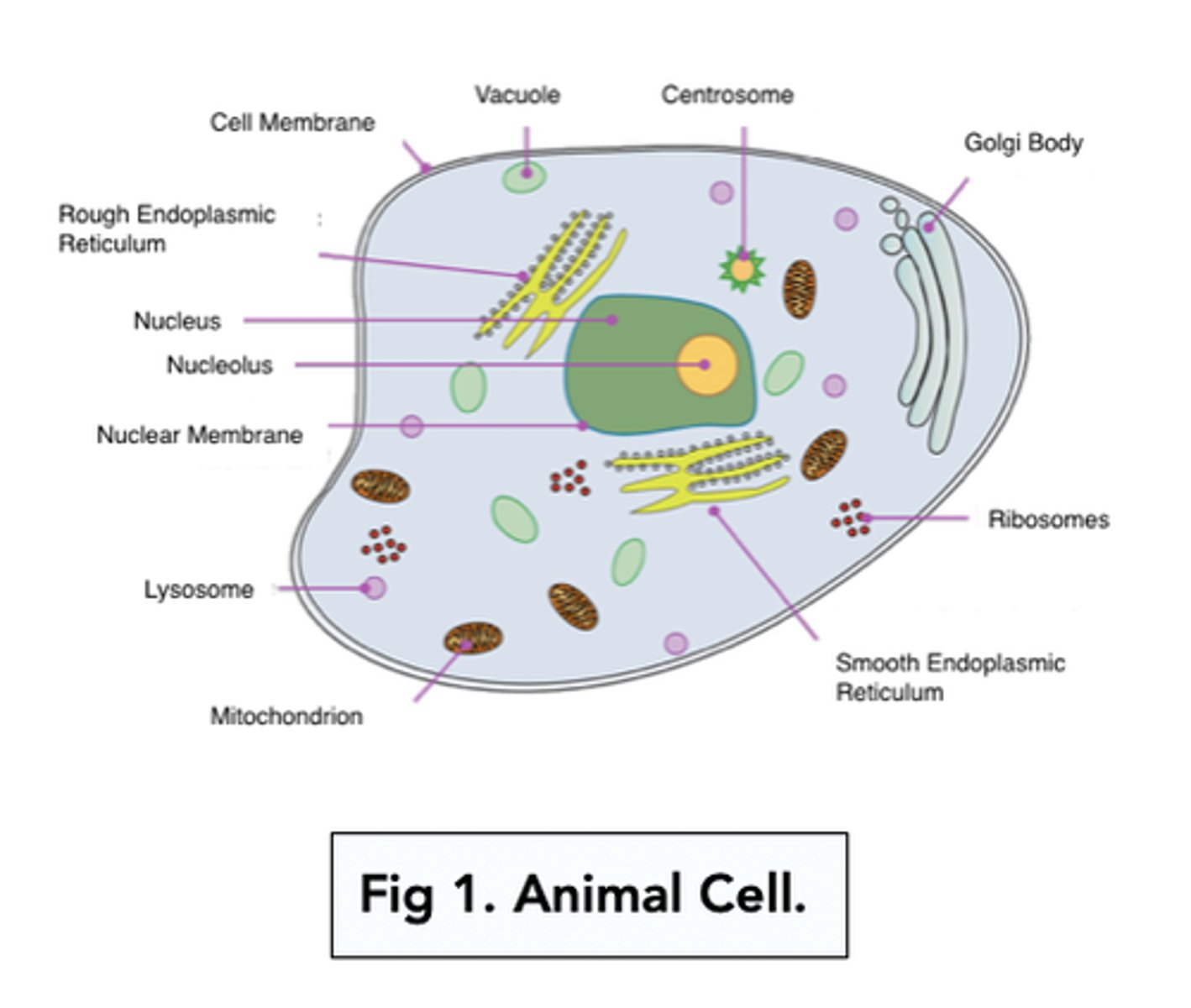
Draw and label the structure of a typical animal cell (7)
Draw and label the structure of a typical plant cell (7)
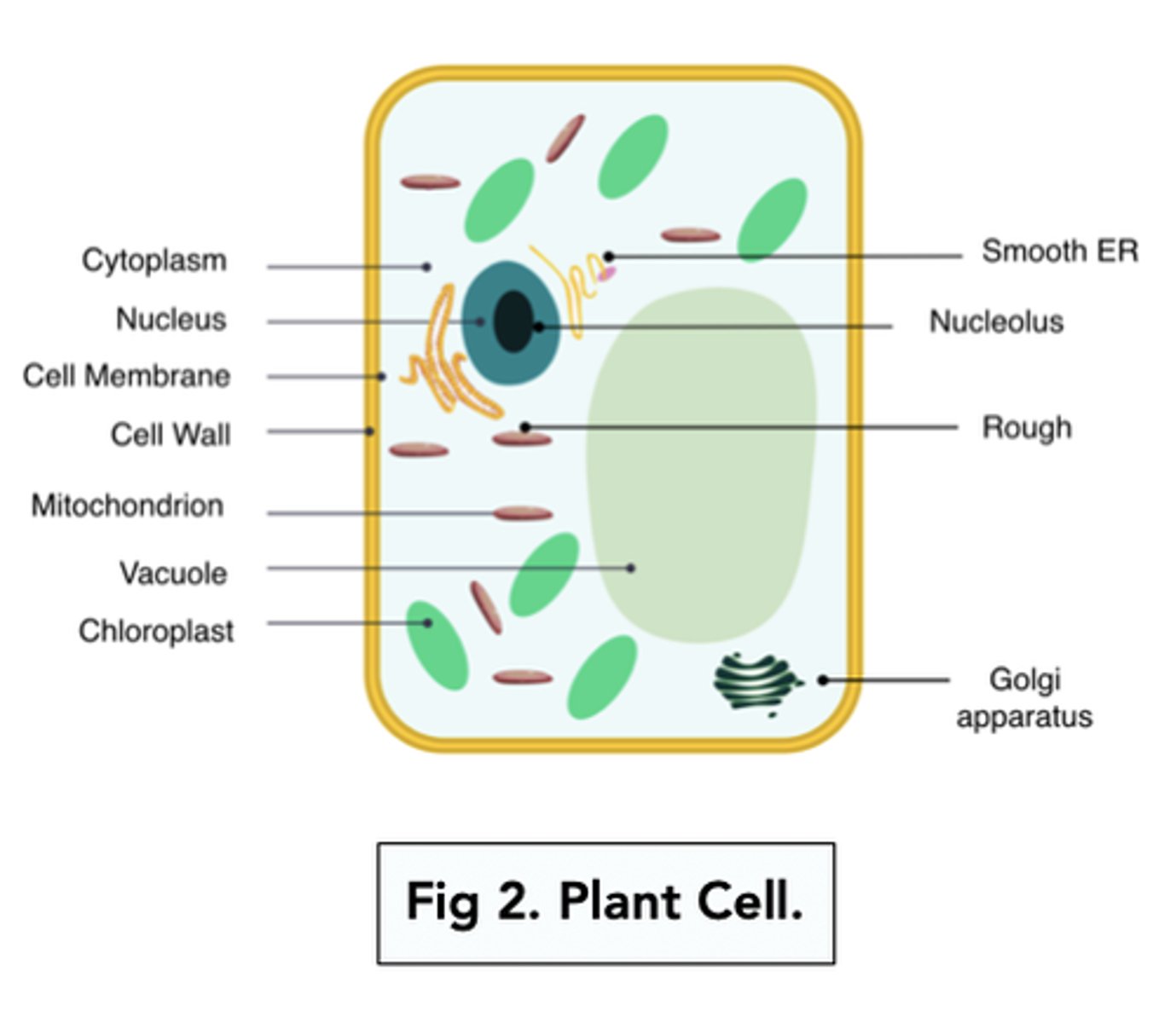
What organelles are only present in plant cells? (3)
- Cellulose cell wall
- Chloroplasts
- Vacuole
What is the main difference between fungal cells and plant cells? (2)
- Fungal cell walls are made of chitin, instead of cellulose
- Fungal cells don't have chloroplasts
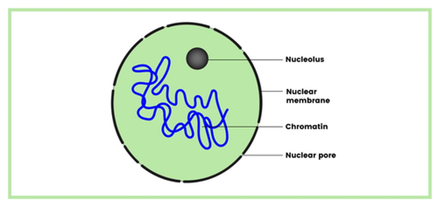
Draw and label the structure of a nucleus (4)
- Nucleolus
- Nuclear membrane
- Chromatin
- Nuclear pore
What does the nucleus contain within its structure to accommodate for its function? (3)
- Histones (protein)
- Linear DNA
- One or more RNA nucleoli
What is the function of the nucleus? (2)
- Controls protein synthesis
- So controls the development and function of the cell
What components make up the structure of ribosomes? (2)
- Proteins
- Ribosomal RNA
Draw the structure of the rough endoplasmic reticulum (1)
It looks a lot like a maze with ribosomes on its surface
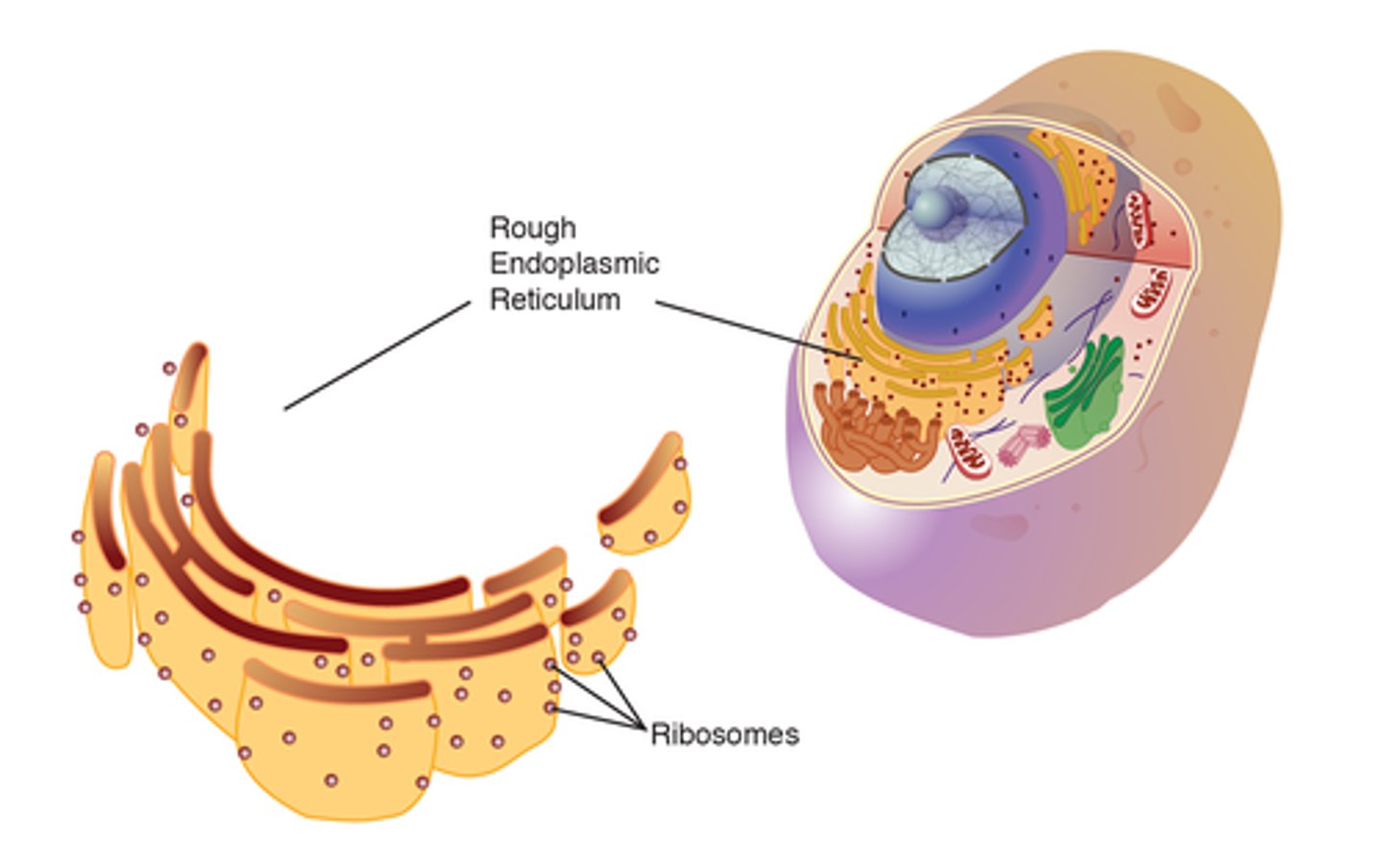
Describe the function of the rough endoplasmic reticulum (2)
- Has ribosomes on its surface that produce secretory proteins (proteins that are released out of the cell)
- These secretory proteins are sent to Golgi Apparatus for modification and packaging
What is the difference in structure between the RER & SER? (1)
RER contains ribosomes on its surface, whereas, SER lacks ribosomes
What is the basic function of the SER? (1)
Involved in the production and transport of carbohydrates and lipids
NOTE: This has barely came up in exam papers, interpret it how you like :)
Draw the structure of Golgi apparatus (1)
It looks a lot like a wifi symbol
NOTE: you won't need to label each component of the Golgi apparatus at all, but in an exam but you may be asked to recognise it from a micrograph
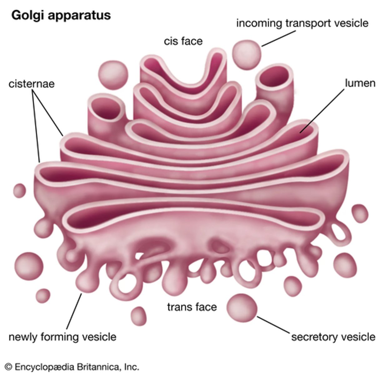
Describe the several functions of the Golgi apparatus (3)
- Adds carbohydrates to proteins received from the RER to form glycoproteins
- Packages proteins/glycoproteins into Golgi vesicles (sacks) for secretion
- Produces lysosomes - a type of Golgi vesicle that releases lysozymes (hydrolytic enzymes)
What are the roles of the organelles that are involved in the production, transport and release of proteins from eukaryotic cells? (5)
1. DNA in the nucleus codes for these proteins
2. Ribosomes in the RER produce these proteins via protein synthesis
3. Mitochondria produces the ATP that is required for protein synthesis
4. Golgi apparatus packages and modifies these proteins into vesicles
5. Vesicles fuse with the cell membrane and release proteins outside of cell
Describe the functions of lysosomes (3)
- Digests material that is taken in by phagocytosis
- Non-functioning organelles within the cell are engulfed and digested by lysosomes
- Releases enzymes outside of the cell
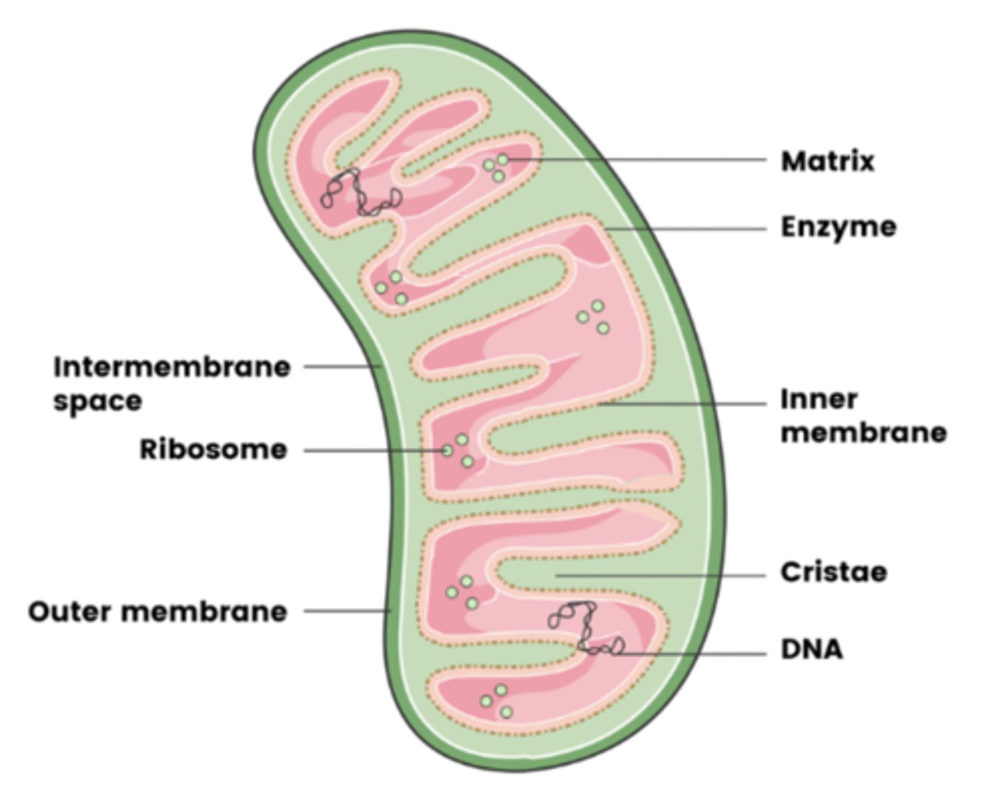
Draw and label the structure of the mitochondria (8)
- Matrix
- Enzymes
- Inner membrane
- Cristae
- (Circular) DNA
- Inter-membrane space
- Ribosomes
- Outer membrane
What is the basic function of the mitochondria? (2)
- Involved in aerobic respiration
- Which produces ATP
Why may some cells have lots of mitochondria and could you give an example of a cell? (2)
- To provide energy to cells that require a large amount of ATP
- E.g. muscle cells
What is the basic function of the cell wall? (1)
Provides support, strength and shape to the cell
What is the basic function of the chloroplasts? (2)
- Contains chlorophyll
- Which absorbs light energy for photosynthesis
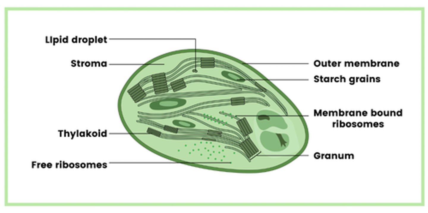
Draw and label the structure of chloroplasts (8)
- Lipid droplets
- Stroma
- Thylakoids
- Free ribosomes
- Circular DNA
- Starch grains
- Membrane bound chromosomes
- Granum
What is the granum that is found in chloroplasts? (1)
Stacked thylakoids
How do granum join together to form grana (plural for granum)? (1)
Link together by thin pieces of membrane - lamellae
What is the basic function of the large vacuole? (1)
Contains soluble sugars, salts and pigments
Draw and label the structure of a typical prokaryotic cell (8)
- Cell wall (made of murein - a glycoprotein)
- Cell surface membrane
- Free circular DNA molecule in cytoplasm
- Ribosomes (70s ribosomes)
- Cytoplasm
- Capsule surrounding the cell wall (in some)
- One or more plasmids (in some)
- One or more flagella (in some)
NOTE: you only need to know the structures we have outlined, you do not need to know any of the functions (expect of course the ones that we have made separate flashcards for)
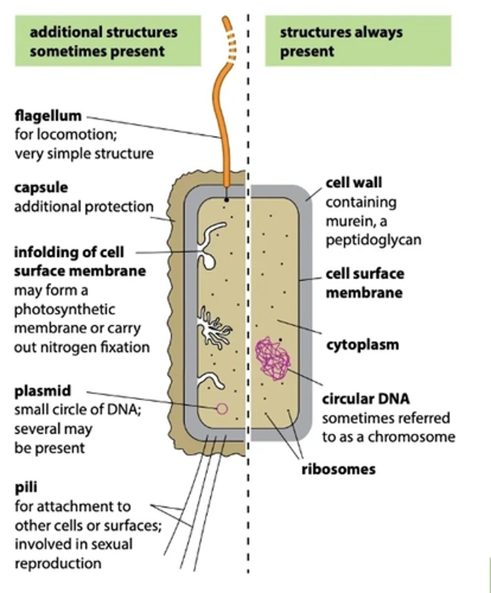
What structures are present in all prokaryotic cells? (5)
- Cell wall (made of murein - a glycoprotein)
- Cell surface membrane
- Free circular DNA molecule in cytoplasm
- Ribosomes (70s ribosomes)
- Cytoplasm
What structures are only present in some prokaryotic cells? (3)
- Capsule surrounding the cell wall
- One or more plasmids
- One or more flagella
Describe the DNA of prokaryotic cells (4)
- No nucleus is present
- DNA is free in the cytoplasm
- Circular DNA
- Not attached to any histone proteins
Describe the properties of plasmids found in prokaryotic cells (3)
- Contain antibiotic resistance genes (amongst others)
- Plasmids are able to be passed between prokaryotes
- Some prokaryotic cells have several plasmids, others have no plasmids
Compare prokaryotic cells and eukaryotic cells (5)
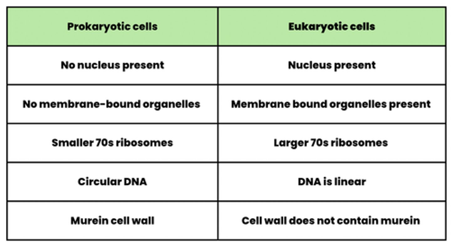
What is the difference in the type of ribosome in bacterial and human cells? (2)
Bacterial cells have 70S ribosomes whereas, human cells have 80S ribosomes
What are the two key properties of viruses? (2)
- Acellular
- Non-living (as they contain no organelles)
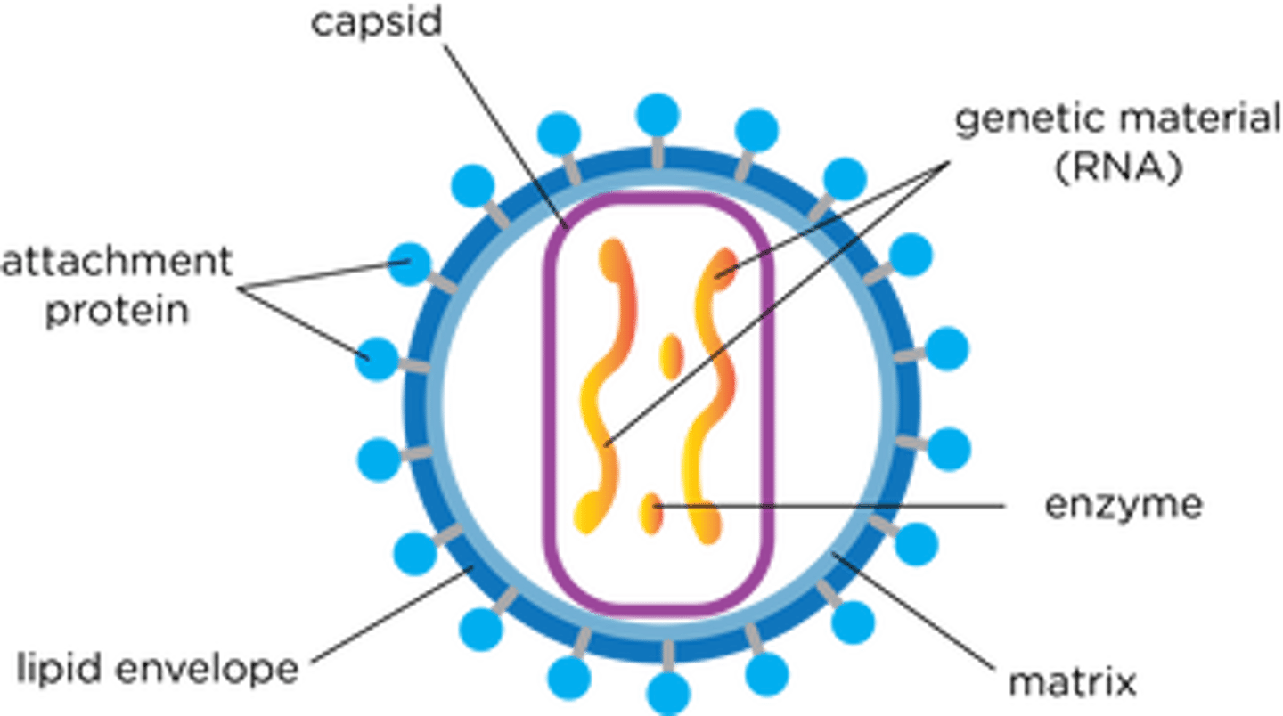
Draw the structure of a typical viral particle (3)
- Genetic material (DNA / RNA)
- Capsid (protein coat)
- Attachment proteins
What happens to eukaryotic cells in complex multicellular organisms? (2)
- Become specialised
- Through the process of differentiation
What is meant by the term "tissues"? (1)
Group of similar specialised cells that perform a specific function and have a common origin
What is meant by the term "organs"? (2)
- Structure that consists of different tissues which have a specific function
- E.g. stomach, which has a role in the digestion of food
What is meant by the term "organ system"? (1)
- Two or more organs which have a specific overall function (e.g. digestion)
- E.g. stomach, liver and pancreas all form the digestive system
What is meant by the term "resolution"? (1)
How well a microscope can distinguish between two points that are close together
What are the requirements for specimens in optical microscopes? (2)
- Be thin
- Be stained
Why must the specimen be thin in optical microscopes? (1)
So that light can pass through the specimen and a single layer of cells is visible
Why must the specimen be stained in optical microscopes? (1)
So that the structures are visible
What are the two main stains we use during microscopy? (2)
- Eosin is used to highlight the cytoplasm
- Iodine contained in potassium iodide solution highlights starch grains
What are the 5 structures that cannot be identified using an optical microscope? (5)
- Mitochondrion
- Ribosome
- Endoplasmic Reticulum
- Lysosome
- Cell-surface membrane
Describe how a transmission electron microscope produces a micrograph (4)
1. Specimen is stained with electron dense substances e.g. heavy metal salts
2. Beam of electrons are transmitted through the specimen
3. Staining substances deflect the electrons in the beam
4. The pattern that the remaining electrons produce as they pass through specimen is converted into an image
Describe how a scanning electron microscope produces a micrograph (3)
1. Specimen is coated with a thin film of heavy metal e.g. gold
2. Electron beam is scanned to and across the specimen
3. Electrons that are reflected from the surface are collected and produce an image on a viewing screen
Compare optical and electron microscopes (6)
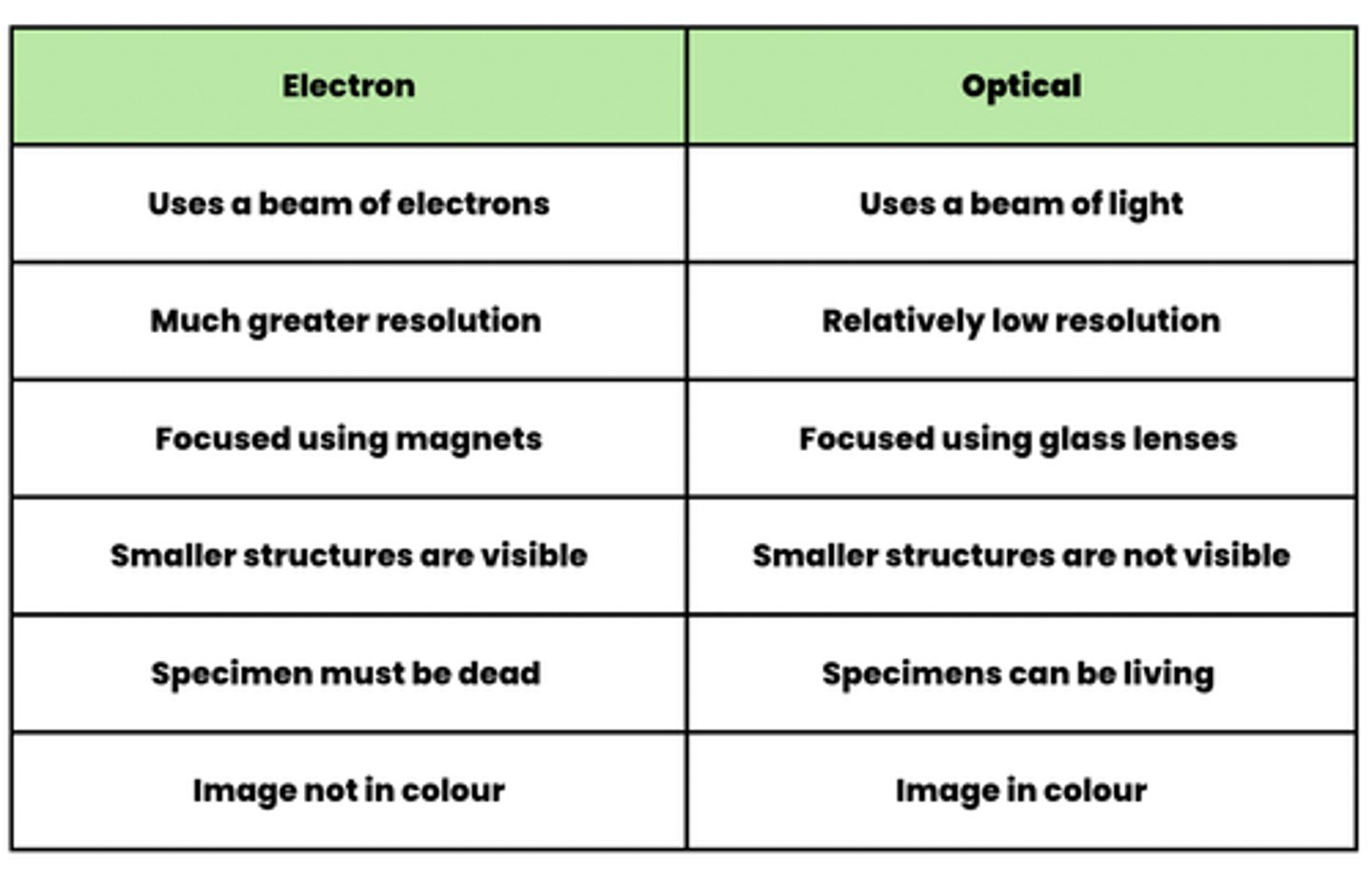
Why do electron microscopes have a higher resolution than optical microscopes? (1)
Electrons have a shorter wavelength
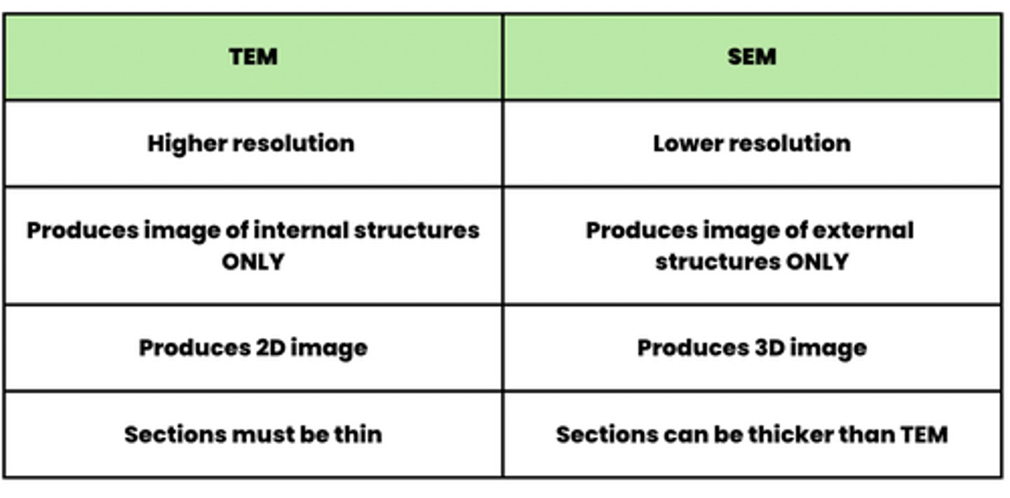
Compare transmission electron microscopes and scanning electron microscopes (4)
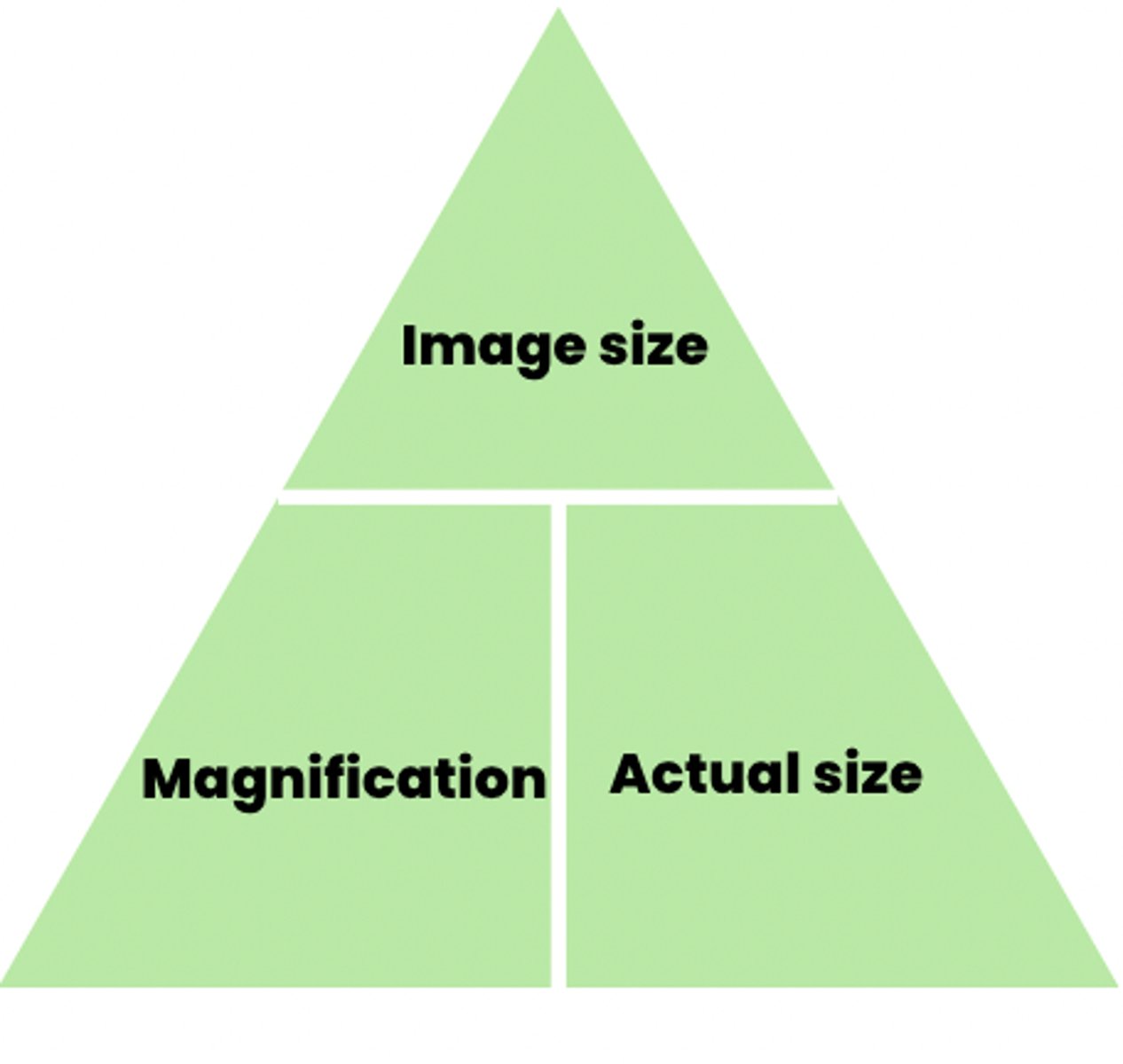
What is the formula triangle for magnification? (3)
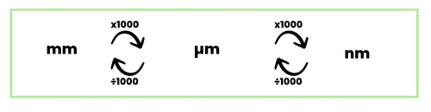
Draw a diagram to show the unit conversions needed in magnification (3)
What are the three stages of cell fractionation? (3)
1. Homogenisation - breaking the cells open
2. Filtration - Removing large debris
3. Ultracentrifugation - Seperating the organelles
What are the properties of the homogenising solution used in ultracentrifugation? (3)
- Isotonic
- Ice-cold
- Buffer solution
Why do we need to homogenise cells in an isotonic solution during ultra centrifugation? (2)
- Prevents the osmotic movement of water into and out of the organelles
- Which can cause them to burst or shrivel
Why do we need to homogenise cells in an ice-cold solution during ultra centrifugation? (2)
- Prevents the action of enzymes in the cell
- That may cause self-digestion (or autolysis) of organelles
Why do we need to homogenise cells in a buffer solution during ultra centrifugation? (1)
Maintains ph so that enzymes do not denature
Describe the method you would use to carry out cell fractionation and ultracentrifugation (4)
1. Cells are first broke open by homogenising the tissue in an ice-cold, isotonic buffer solution using a blender
2. This solution is then filtered to remove large debris
3. Homogenate is then centrifuged at a low speed and the densest organelle i.e. nuclei forms a pellet at the bottom of tube
4. The supernatant (the liquid above the pellet) can be spun faster for a longer period of time to isolate the other organelles
What is the order in which cell organelles are isolated during ultracentrifugation? (5)
1. Nuclei
2. Chloroplasts (if its a plant cell)
3. Mitochondria
4. Endoplasmic reticulum (SER and RER)
5. Ribosomes
Describe and explain how cell fractionation and ultracentrifugation can be used to isolate an organelle from a suspension of animal cells (7)
MODEL ANSWER
1. Homogenise the cells to break them open
2. Filter to remove large debris
3. Use an isotonic solution to prevent damage to the organelles
4. Keep the solution ice-cold to prevent damage by enzymes
5. Use a buffer solution to prevent protein denaturation
6. Centrifuge at a lower speed to separate the nuclei
7. Re-spin the supernatant at a higher speed to pellet the organelles at the bottom