Palmer- Physiology- Exam 3 worksheets 14-15
1/63
There's no tags or description
Looks like no tags are added yet.
Name | Mastery | Learn | Test | Matching | Spaced |
|---|
No study sessions yet.
64 Terms
Whare are the 4 major events that occur before and during cell division?
1. Reproductive signal
2. Replication of DNA
3. Segregation
4.Cytokinesis
What is the reproductive signal?
A signal to reproduce is received to initiate cell division
Growth Factor or Growth hormone!
What is cell segregation?
DNA is distributed to new cells (Mitosis/ Meiosis)
What is the major difference between meiosis and mitosis
Mitosis- division of somatic (body) cells. Create identical copies.
Meiosis- division of germ-line (sex) cells. create non-identical copies
which creates genetic diversity? Mitosis or meiosis?
Meiosis
What are the 2 stages of the cell cycle?
Mitosis (M- Phase)
Interphase
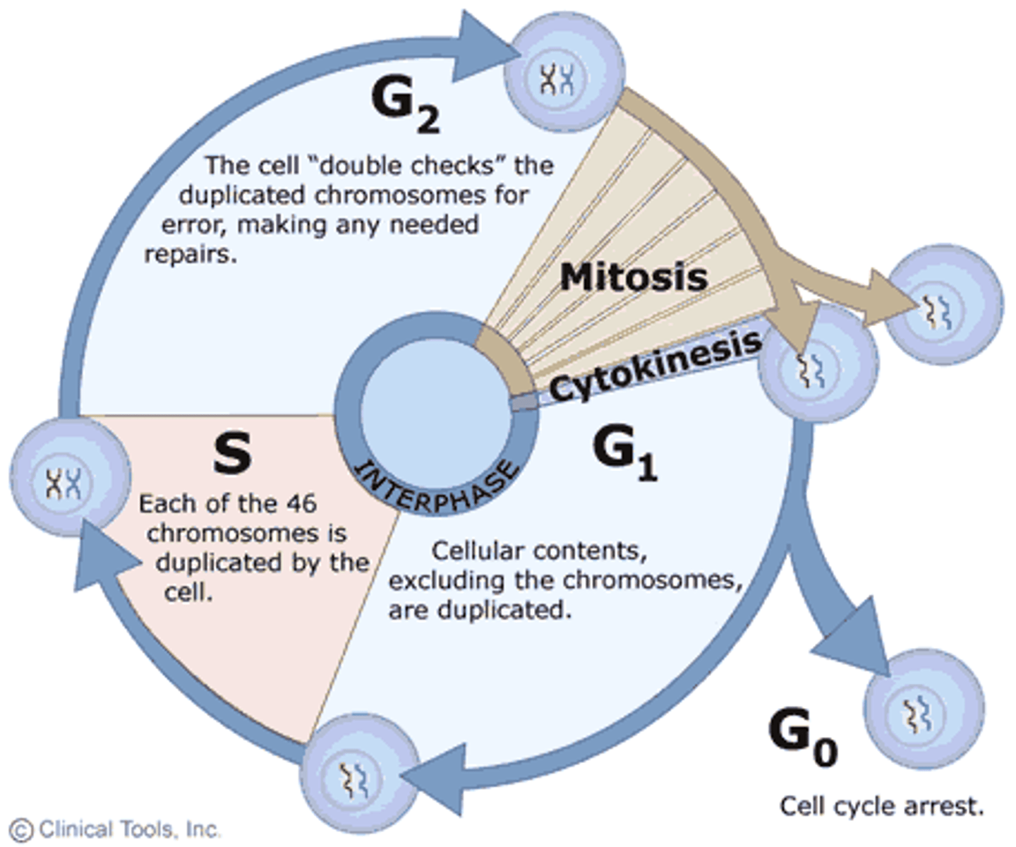
What is happening in M phase
the cell undergoes all stages of Mitosis and Cytokinesis, resulting in two new cells.
active dividing
Describe what is happening in each phase of interphase
G1 (Gap 1)- happens just after mitosis and before DNA synthesis. DNA is unreplicated. variable timeline
S Phase (Synthesis)- Cell' DNA is replicated, DNA synthesis
G2 (Gap 2)- DNA is duplicated at this point, cell makes preparations for mitosis like making microtubules for mitotic spindle
What 2 types of signaling molecules are used to stimulate cell division?
name examples
Growth Factors- ex: EPO, interleukins
Hormones- ex: Estrogen
Which type of pathway does EPO use to signal RBCs to mature and divide?
JAK/STAT pathway,
What types of pathway do interleukins use to stimulate transcription/translation of target genes?
JAK/STAT pathway, Ras activation and PI3K pathway
what type of pathway does estrogen use and what does it stimulate
steroid hormone superfamily of receptors and stimulates transcription
The events in DNA replication are ...?
a. Helicase - 'Unzips' the double helix allowing the parental strands to be exposed for copying
.b. DNA gyrase (topoisomerase) - removes supercoiling caused by helicase.
c. Single stranded binding proteins - prevent separated DNA from reannealing (rewinding), stabilizing the replication fork.
d. Primase - produces a short RNA primer, made of ribonucleotides, so that DNA polymerase can recognize where to start copying the strands.
e. DNA polymerase III - adds deoxyribonucleotides to the 3' end of the growing daughter strands
.f. DNA polymerase I - removes the RNA primers replacing them with the appropriate DNA nucleotides according to base pairing rules. Also plays a role in some DNA repair mechanisms.
g. Ligase - forms the phosphodiester bonds to seal the 'gaps' after the removal of the primers.
what phase of the cell cycle does DNA replication take place?
S phase
What are the structures of a duplicated chromosome
Duplicated Chromosome= 2 sister chromatids held together at the centromere.
Each sister chromatid is made of Chromatin

What does chromatin include?
proteins and DNA that make up sister chromatids
How many chromosomes do humans have?
46 (23 pairs)
When does DNA start to get packed into chromosomes
Begins in G2 of Interphase but not complete until prophase of mitosis
Explain the structure of a nucleosome and its parts
DNA wrapped around histones
Contains: 8 histone molecules, 146 base pairs of DNA and an H1 linker histone
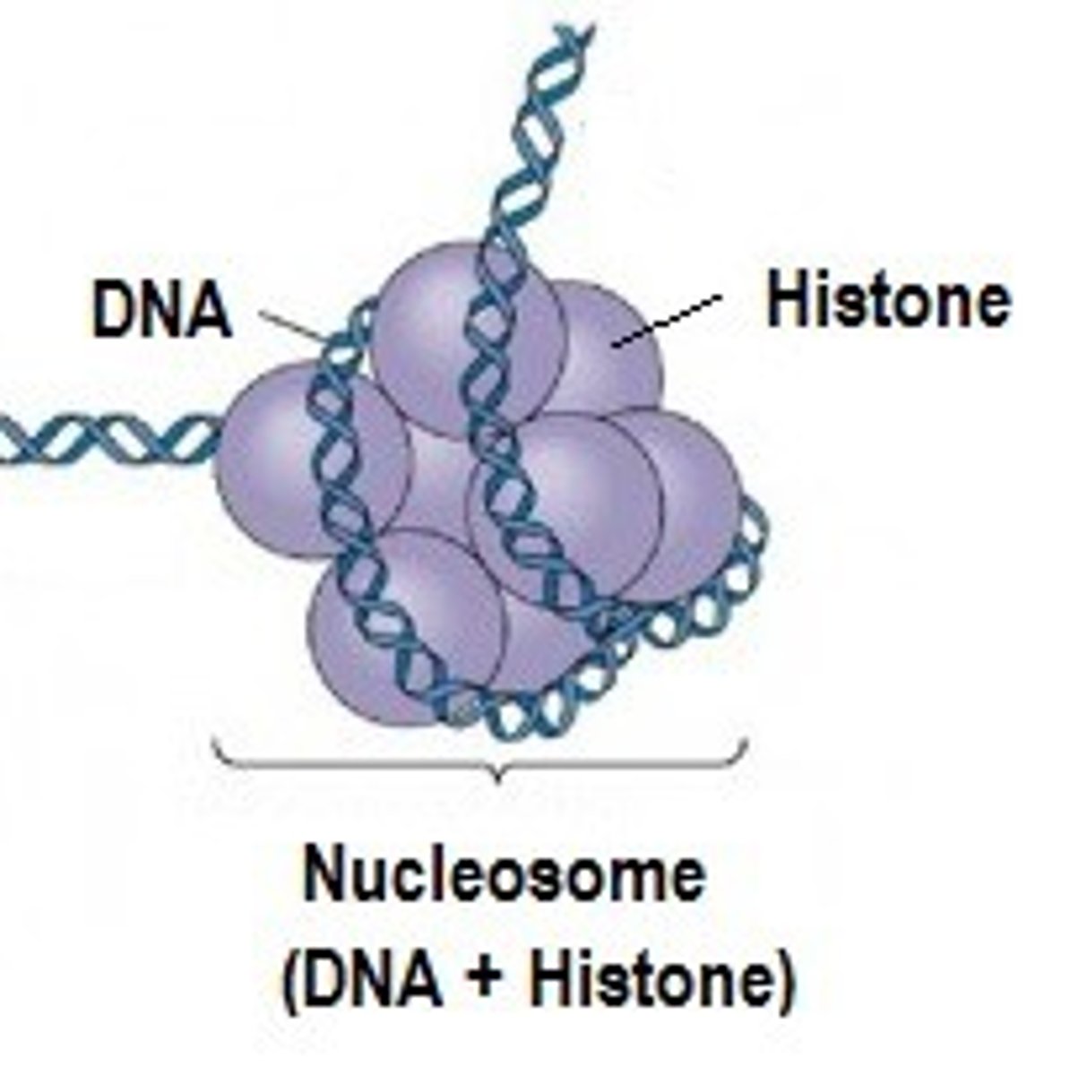
What are the 5 classes of histone proteins?
H1, H2A, H2B, H3, H4
*H1 is a linker histone!
What histone modification could I make if I wanted to increase the transcription of a gene? enzyme used?
Acetylation!
Histone acetyl transferases (HATs) add acetyl group to lysine residues and weakens DNA histone interactions, allowing more transcription
To decrease gene transcription, what histone modification would you make? enzyme used?
Remove an acetyl group
Histone deacetylases (HDACs) remove acetyl group making DNA and histone interaction tighter, resulting is decreased transcription
What is the difference between euchromatic and heterochromatin
euchromatin- "active DNA" genes are available to be transcribed. They are not wrapped tightly around a histone
heterochromatin- "DNA is inactive" and tightly condensed around a histone/
what phase of the cell cycle do the centrosomes replicate?
When do they move to opposite cell poles?
Replicated in S-phase
separate and move to opposite ends in G2-M transition
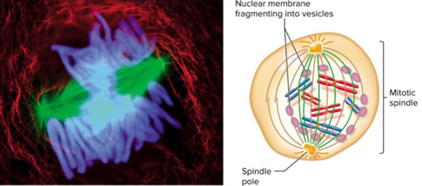
what is the most important structure in cell division?
mitotic spindle
What is the difference between the centromeres and the centrioles
Centromeres: region of the chromosome where spindle fibers attach via kinetochore proteins
Centrioles: two cylindrical cellular structures that are composed of nine triplet microtubules and form mitotic spindles.
what is the centrosome?
region where microtubules form, each produces 2 centrioles
What cytoskeletal protein makes the mitotic spindle?
What structure does it arise from
microtubules
the centrioles
What is the difference between the polar and kinetochore microtubules?
polar microtubules: form framework of the spindle. overlap and interact with a microtubule from other side
Kinetochore microtubules: attach to kinetochores on chromosomes
What is a kinetochore and where is it located
Where the kinetochore microtubules attach to the centromere of the duplicated chromosomes.
What happens in prophase
- Chromosomes condense
- DNA has been duplicated in S phase
-mitotic spindle develops
-Nucleolus disappears
What happens in Metaphse
- All centromeres at at the equatorial plate
- chromatids are now clearly connected to the poles by microtubules
What happens in anaphase
- separation of the chromatids toward the opposite ends of the spindle
-happens from kinetochore microtubules shortening
What happens in telophase
- disassembly of the mitotic spindle
- nuclear envelopes re-form and the chromosomes decondense
nucleolus will also reform in daughter cells
what happens in prometaphase?
sister chromatids become attached to the kinetochore (strandy part of the spindle), which are attached to the centrsome, part where nuclear envelope breaks
What are the 5 types of chromosomal mutations?
1. Nondisjunction
2. Deletions
3. Duplications
4. Inversions
5. Translocations
What happens in chromosomal nondisjunction
causes too many or too few chromosomes due to chromosomes failing to separate during anaphase
What happens in chromosomal deletions
segment of chromosome is removed
what happens in chromosomal duplication
chromosomal segment is doubled
what happens in chromosomal inversions
when a chromosomal segment is reattached upside down
what happens during chromosomal translocations?
when a segment is swapped with another chromosome segment
what phase of cell division does nondisjunction occur in?
anaphase
What phase of cell division does deletions, duplications, inversion and translocation all occur in?
during crossover in prophase 1 of meiosis 1
What are the phases of the cell cycle?
G1, S, G2, M
What is G0 and why would a cell enter this phase?
specialized resting phase
cells exit the cell cycle and pause division due to lack of nutrients
What is happening during mitosis
cells are actively dividing (PMAT-C)
Why are cell cycle checkpoints important?
ensures that the cell is healthy and has all components needed for proper division
What is being assessed at a cell cycle checkpoint?
- DNA and organelles are properly replicated
- no DNA mutations
- Chromosomes have attached to spindle fibers/properly separated during anaphase
What is the difference between phosphorylation and dephosphorylation?
What class of enzymes is used for each?
Phosphorylation is the addition of a phosphate group to a substrate.
Dephosphorylation is the removal of phosphate groups from a substrate
What are cyclins
proteins that CDKs are dependent on.
produced only in response to signals like growth factors and hormones
What are cyclin-dependent kinases?
specific protein kinases that trigger transitions from phase to phase in the cell cycle.
they are always present but not always active
how does the process of cyclins and CDKs work?
1. CDK is activated by binding to a cyclin, altering protein shape (exposing active site)
2. Cyclin-CDK complex acts as a protein kinase to phosphorylate regulatory proteins- triggering phase transition
3. Cyclin then breaks down to Cdk and becomes inactive
What cyclins/cdks are associated with G1-S transition?
Cyclin D-Cdk4 and Cyclin E-Cdk2•
Acts during the G1-S transition
Phosphorylates Rb- This moves the cell past the restriction point (R).
What cyclins/cdks are assocoiated with the S transition
Cyclin A-Cdk2
Acts during S•
Stimulates DNA replication
What cyclins/cdks are associated with G2-M transition
Cyclin B-Cdk1
Initiates mitosis
What is the process in which cell division is stimulated (inhibition of Rb)
Cyclins D and E activate Cdk 4 and 2
in turn inactivates Rb by phosphorylating it.
When Rb becomes inactivated, the cell can progress past G1 into S phase."Inhibiting the inhibitor
What is the difference between tumor suppressor genes and oncogenes?
Tumor suppressor gene- brake pedals of the cell cycle
(Proto) Oncogenes- gas pedal of cell cycle
What are examples of tumor suppressors and (proto) oncogenes
Tumor suppressors - Rb, p21, p53(Proto)
Oncogenes - Cyclins and CDK
When UV radiation is encountered by a skin cell, a cascade of events occurs to keep the cell from dividing.
Describe this cascade
1. UV rays cause DNA Damage
2. DNA damage activates protein kinase that phosphorylates and activates p53
3. active p53 binds to regulatory region of p21 gene
4. p21 gene is transcribed/translated making tumor suppressor p21.
5. p21 inhibits cyclin and CDK resulting in cell cycle arrest (cell cannot divide)
What are the two main ways cells can die?
Necrosis and apoptosis
what is necrosis and when/how does it occur
- cell death due to injury or hypoxia (pathological)
- cell swells (inflammation) and bursts destroying neighboring cells
-process takes several days
what is the process of apoptosis? How is it stimulated
-genetically programmed cell death
-Membranes bleb, DNA cut up by caspases
- no inflammation caused, no damage to neighbor cells
- occurs in a few hours
Describe the internal apoptotic pathway
Internal signaling pathway - p53 activation -> Bax -> cytochrome c release -> activation of caspases -> breakdown of chromatin, proteins and membranes.
Describe the external apoptotic pathway
External signaling pathway - FasL binds to Fas receptor on target cell surface -> activation of caspases -> breakdown of chromatin, proteins and membranes.