Anatomy Exam 3
1/91
There's no tags or description
Looks like no tags are added yet.
Name | Mastery | Learn | Test | Matching | Spaced | Call with Kai |
|---|
No analytics yet
Send a link to your students to track their progress
92 Terms
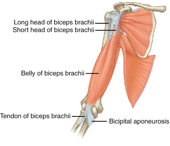
Origin
The attachment of a tendon to the more stationary bone
-More stationary/less movable
Insertion
The attachment of the muscle’s other tendon to the more movable bone
-Movable
Belly of origin
“Head” of origin

Action
The main movements that occur during contraction
Ex: Flexion or Extension
Tendon
Connects muscle to bone
Aponeurosis
A thin, flattened sheet of tendon
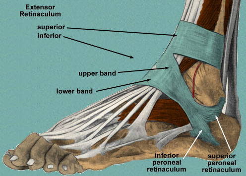
Retinaculum
a band of thickened deep fascia around tendons that holds them in place. It is not part of muscle. Its function is mostly to stabilize a tendon.
Prime Mover (agonist)
Contracts to cause an action
Synergists
Work with prime mover, prevent unwanted movement or otherwise aid with movement
-Ex: Biceps brachaii & Brachalis (In elbow flexion)
-Ex; Triceps brachaii (In elbow extension)
Fixator
Stabilize the origin of the prime mover
Antagonist
Opposes the action of the prime mover
-Ex: Triceps brachaii (In elbow flexion)
-Ex: : Biceps brachaii & Brachalis (In elbow extention)
Anatagonistic pairs
Most muscles are arranged in opposing pair at joints
-Depending upon the movement, many muscles may act as prime movers, antagonists, synergists, or fixators
Fascicle Arrangement (Power & range of motion)
-The longer the fibers in a muscle, the greater the range of motion it can produce
-The power of a muscle depends not on the length but on its total cros- sectional area
Fusiform
Fascicles nearly parallel to longitudinal axis of muscle; terminate in flat tendons; muscles tapers toward tendons, where diameter is less than at belly
-Ex: Digastric muscle
Parallel
Fascicles parallel to loghitudinal axis of muscle; terminate at either end in flat tendons
-Ex; Stylohyoid muscle

Convergent
Fascicles spread over broad area converge at thick central tendon; gives muscle a triangular appearance
-Ex: Pectoralis major muscle

Unipennate
Fascicles are arranged on only one side of tendon
-Ex: Extensor digitorum longus muscle
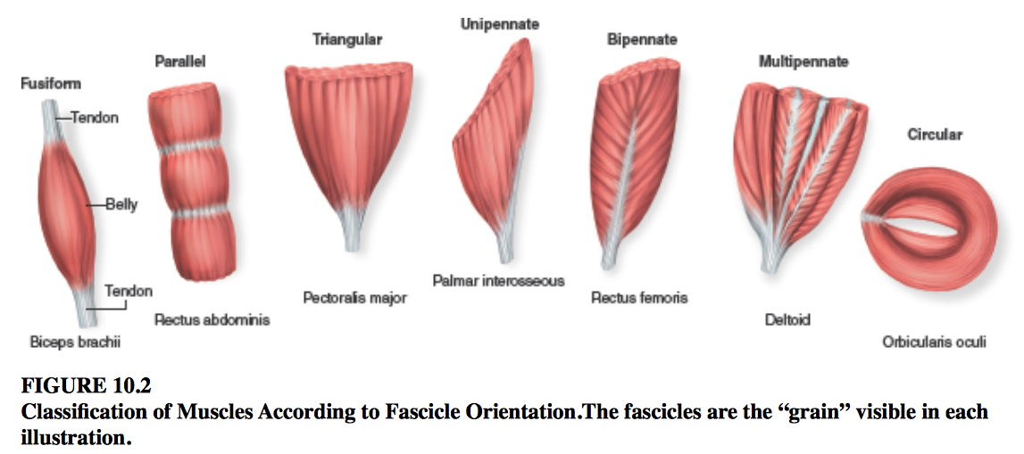
Bipennate
Fascicles are arranged on both sides of centrally positioned tendons
-Ex: Rectus femoris muscle

Multipennate
Fasclicles attach obliquely from many directions to several tendons
-Ex: Deltoid muscle

Circular
Fascicles in concentric circular arrangements form sphincter muscles that enclose an orifice(Opening)
-Ex: Orbicularis oculi muscle

Intrinsic muscle
Extrinsic muscle
Originate on a different body part than where they insert and act
Anterior compartment
Muscles dorsiflex the foot at the ankle joint
Lateral compartment
Muscles plantar flex and evert the foot at ankle
Posterior compartment
Muscles are split between superficial and deep groups. Superficial muscles share a common tendon of insertion
-Ex: calcaneal tendon
Hypertrophy
Increase in muscle fiber size
Atrophy
Reduction in muscle size
Fibrosis
The overgrowth, hardening, and/or scarring of various tissues
Myoblast
Are cells that fuse to form single muscle fibers
Satellite Cells
Are myoblast cells that do not fuse with muscle fibers and remain adult skeletal muscle
Moter neuron axon
Is the branch of a moter unit and branches to innervate a number of muscle fibers
Sarcolemma
The plasma membrane of a skeletal muscle fiber
Transverse Tubules
Are deep invaginations of the sarcolemma that extend into the sarcoplasm of muscle fibers as a network of narrow membranous tubules
Sarcoplasmic Reticulum
Stores calcium ions needed to initiate muscle contraction
Terminal Cisternae
Blind sacs of the Sarcoplasmic Reticulum, they are reservoirs and specific sites for calcium ion release
Triad
Two terminal cisternae and the centrally placed t-tubule form a structure
Myofibrils
Are long, cylindrical structures that extend the length of the muscle fiber
-Are 80% of the volume of a muscle fiber
Myofilaments
Are bundles of protein filamentsin a myofibril
Thin Filament
Are 5-6 nanometers in diameter and are primarily composed of two strands of the protein actin twisted around to form a helical shape
Actin
A protein
G-Actin
Is a singular spherical molecule in a strand of actin
F- Actin
Is a filament composed of a strand of G-actin molecules
Tropomyosin
A short, thin, twisted filament that covers small sections of the actin strands
Troponin
Attaches to actin and tropomyosin, provides a binding site for calcium ions
Thick Filaments
Are assembled from bundles of 200-500 myosin protein molecules
Myosin
Consists of two strands, each strand has a free, globular head and an attached, elongated tail. The heads are Actin and ATP binding sites
Titin
A “cablelike” protein that extends from the z discs to the M line through the core of each thick filament
-Stabilizes the position of the thick filament and maintains thick filament alignment within a sarcomere
Dystrophin
Part of a protein complex that anchors myofibrils that are adjacent to the sarcolemma to proteins within the sarcolemma
-Links internal myofilament proteins of a muscle fiber to external proteins
Muscular Dystrophy
Collective term for hereditary diseases in which the skeletal muscles degenerate, lose strength, and are gradually replaced by adipose and fibrous tissue
Myoglobin
Is a protein found in the muscle cells of animals. It binds iron and oxygen, functioning as an oxygen-storage unit to provide oxygen to working muscles
Glycogen
A multibranched polysaccharide of glucose that serves as a form of energy storage in animals, fungi, and bacteria.
A-band
Dark band in the middle of the sarcomere: composed of entire thick filaments and in it’s lateral end regions of overlapping thin filaments
I-band
Extends from both directions of a Z-disc and are bisected by the Z-discs. Contain only thin filaments and titin proteins
Z-disc
Dark proteins called titins in the center of the I-band where thin filaments attach
Sarcomere
Is the functional contractile unit of a skeletal muscle fiber
-Myofilaments within myofibrils are arranged in repeating microscopic cylindrical units
H-zone
Is the most central portion of the A-band in a resting sarcomere
-Only thick filaments are present
M-line
A thin transverse protein meshwork structure in the center of the H-zone
-Serves as an attachment site for the thick filaments and keeps thick filaments aligned during contraction and relaxation
Somatic Motor Neurons
Extend from the brain and spinal cord to innervate skeletal muscle fibers
-Control voluntary actions
Motor Unit
Is composed of a single motor neuron and all of the muscles fibers it controls
-Involved in muscle contraction
Neuromuscular Junction
The point where a motor neuron meets a muscle fiber
Synaptic Knob
Is an expanded tip of an axon
-A nerve impulse transfers here from the axon
-Houses numerous synaptic vesicles
Moter-end plate
Is a specialized region of the sarcolemma. It has folds and indentations to increase the membrane surface area
-Has ACh receptors
Synaptic Cleft
IS a narrow space separating the synaptic knob and the motor end plate
Acetylcholine (ACh)
An organic compound that functions in the brain and body of many types of animals (including humans) as a neurotransmitter.
Synaptic Vesicles
Are small membrane sacs filled with molecules of the neurotransmitter acetylcholine (ACh)
Acetylcholinesterase
Resides in the synaptic cleft, rapidly breaks down molecules of ACh that are released into the synaptic cleft
-It is needed so ACh is not continuously stimulating a muscle
ACh Receptors
In the motor neuron plate and act like doors that are normally closed and only ACh is allowed in
Sliding filament mechanism
When a muscle contracts, thick and thin filaments slide past each other and the sarcomere shortens
Excitation-Contraction Coupling
The stimulation of a muscle fiber by a nerve impulse results In a series of events that culminates in muscle fiber contraction
-CA++ release channel in SR
Contraction
When contractile proteins within muscle cells slide past one another and the muscle cell shortens
-Allows muscle movement
Relaxation
Ca++ pumps pn SR
Contraction Cycle
A signal is sent from the brain or the spinal cord to the muscle via neurons
An action potential is generated in the neuron, releasing Ca++ in the neuromuscular junction
The influx of caalcium ions causes acetylcholine (AcH) to be released in the synaptic cleft
AcH binds to the AcH receptors present in the sarcolemma, increasing its permeability
Na++enter the sarcolemma, changing its polarity, and creating an action potential
Ca++ are released by the sarcoplasmic reticulum, as the action potential travels down the T-tubules in the muscle fiber
Ca++ bind with troponin C, causing the tropomyosin to shift, and expose the myosin binding sites on actin
ATP is hydrolyzed into ADP and phosphorus, releasing energy for myosin power stroke
Myosin binds to actin
Myosin head bends and actin slides over the myosin surface
Myosin releases the ADP molecule
As the myosin head swivels, another ATP molecule binds to myosin, breaking the actin-myosin bridge.
When the nervous impulse stops, the calcium gates close, and the sarcoplasmic reticulum is no longer permeable. The Ca++ return to the sarcoplasmic reticulum, and troponin and tropomyosin are reverted to their original positions. With the binding sites blocked, myosin cannot form cross-bridges with actin, and the muscle relaxes.
Muscle tone
Is the resting tension in a skeletal muscle
Isometric Contraction
The length of the muscle does not change because the tension produced by this contacting muscle never exceeds the resistance.
Isotonic contraction
The tension produced equals or is greater than the resistance , and then the muscle fibers shorten resulting movement
Concentric Contractions
Actively shorten a muscle
-Ex: lifting something up
Eccentric Contraction
Actively lengthen a muscle
Ex: Putting something down
Slow Twitch
slow oxidative (SO), red, or “Dark Meat”
Abundant mitochondria, myoglobin and capillaries - deep red color
Adapted for aerobic respiration and fatigue resistance
Ex: soleus of calf and postural muscles of the back
Fast Twitch
Fast glycolytic (FG), “White Meat”
QUICK & STRONG response, but fatigue very rapidly
Rich in enzymes that generate lactic acid causing fatigue
Poor in mitochondria, myoglobin, and blood capillaries which gives pale
appearanceEx: extrinsic eye muscles, gastrocnemius and biceps brachii
Intermediate Fibers
Fast myosin ATPase
Produce a fast and powerful contraction
Used for medium duration like walking or biking
Has a high resistance
Endurance Exercise
Long distance running or maintaining posture
Resistance exercise
Short duration, intense movement
Ex: sprinting, lifting weights
Functions of muscle tissue
1. Body movements → moving the skeleton
2. Stabilizing body position → keeping the skeleton stable
3. Storing, and movement, of materials within the body
4. Generating heat
Excitability
The ability for a cell to respond to a stimulus
Conductivity
Involves an electrical change that travels along the plasma membrane during a muscle or nerve impulse
Elasticity
the tendency to return to their initial shape
Extensibility
the ability to be stretched without
damage
Skeletal Muscle
-Voluntary
-Striations
-Attached to bones
Cardiac Muscle
Voluntary control
Short, branching cells
Striated appearance
Cells are attached at their ends by intercalated discs:
Gap Junctions – “pores”; electrically link cells together, thus action potentials
spread from cell to cell
Desmosomes = strongest cellular junctions!
Smooth muscle
Smooth muscle fibers have thin and thick filaments that are not
arranged in sarcomeres
Contractions are also activated by Ca2+ ions, but are slower and last
longer
Two Types/Arrangements of Cells & How They’re Innervated:
Single Unit or Visceral (sheets of cells, all cells act together)
Multiunit smooth muscle (cells individually innervated so
individual cells act independently of one another)
Two types differ in the pattern of innervation
Smooth muscle fibers shorten more than striated muscle fibers
Smooth muscle is under involuntary control by autonomic neural
input, as well as multiple other factors (hormones, paracrines, stretch
response)