ECTODERM, MESODERM, ENDODERM (copy)
1/88
There's no tags or description
Looks like no tags are added yet.
Name | Mastery | Learn | Test | Matching | Spaced | Call with Kai |
|---|
No analytics yet
Send a link to your students to track their progress
89 Terms
Ectoderm
the outermost germ layer in animals.
gives rise to the skin, nervous system, and sense organs. In the early embryo, it is the first layer to form from a fertilized egg.

Epiblast
What is 1?
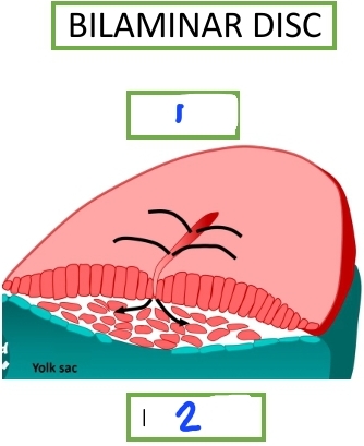
Hypoblast
What is 2?

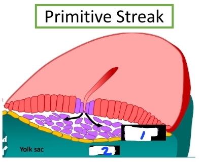
Intraembryonic mesoderm
What is 1?

Endoderm
What is 2?
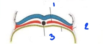
Ectoderm
What is 1?

Mesoderm
What is 2?

Endoderm
What is 3?

Mesoderm
The notochord must arise from what germ layer?
Chordata
an embryonic midline structure common to all members of the phylum ________, providing both mechanical and signaling cues to the developing embryo
Notochord
an embryonic midline structure common to all members of the phylum Chordata, providing both mechanical and signaling cues to the developing embryo
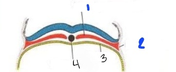
Ectoderm
What is 1?

Mesoderm
What is 2?
Endoderm
What is 3?

Notochord
What is 4?

Neural tube
What is 5?

Amnios
What is 6?

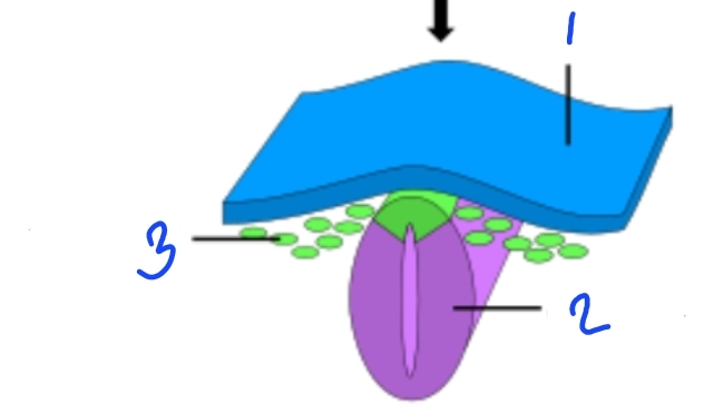
oropharengeal membrane
what is 1?

notochordal process
what is 2?

cloacal membrane
what is 3?
skin
nervous system
What does the ectoderm form?
muscles
bones
notochord
what does the mesoderm form?
gut lining
internal organs
what does the endoderm form?
prechordal plate
chordamesoderm
Some mesodermal cells migrate forward toward the embryo’s midline. These cells become:
Prechordal plate
This mesodermal cell becomes the anterior organizer
Chordamesoderm
This mesodermal cell becomes the precursor of the notochord
Chordamesoderm
This extends forward from the primitive node (Hensen’s node in birds/mammals)
Intraembryonic mesoderm
What is 1?
Extraembryonic mesoderm
What is 2?

Endoderm
What is 3?

Notochord
What is 4?
Sonic hedgehog (Shh)
Notochord secretes factors like ____ to induce the neural plate and pattern the somites (future vertebrae, ribs, muscles)
Ectoderm
Mesoderm
Endoderm
Gastrulation forms these germ layers
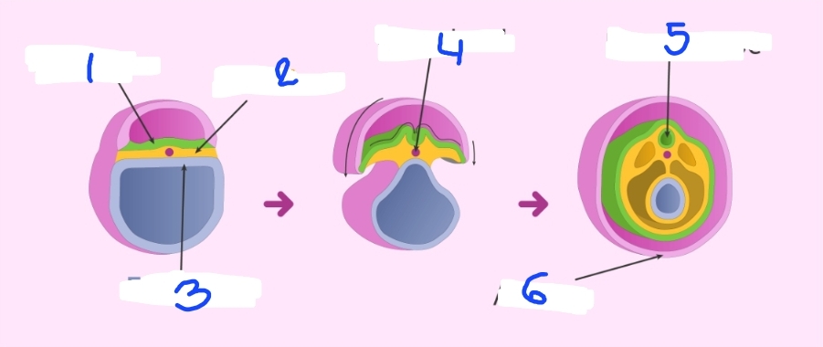
Neural plate border
What is 1?
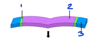
Neural plate
What is 2?

Epidermis
What is 3?

Convergence
What is 1?
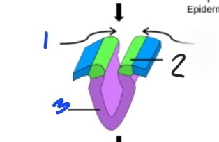
Neural fold
What is 2?

Neural groove
What is 3?

Epidermis
What is 1?

Neural tube
What is 2?

Neural crest
What is 3?
Neural tube
the developing brain and spine
neural crest
is a transient, multipotent population of cells that arises during early vertebrate development
fourth germ layer
what is the neural crest often called?
Multipotent
Cell’s ability to develop into multiple—but limited—cell types within a specific lineage or tissue family. Neural crest cells are ________.
Totipotent
can form all cell types, including embryonic + extraembryonic
pluripotent
can form any body cell
multipotent
can form several related cell types
unipotent
can orm one type
Brain (anterior neural tube)
spinal cord (posterior neural tube)
CNS
motor neuron axons
interneurons
neuroglia of CNS (except microglia)
astrocytes
oligodendrocytes
ependymal cells (lining ventricles)
other structures derived from neural tube
craniofacial cartilage and bone
odontoblasts (tooth dentin)
cranial nerves ganglia (V, VII, IX, X)
connective tissues of the face
middle ear bones
cranial neural crest
melanocytes
dorsal root ganglia (sensory neurons)
sympathetic chain ganglia
adrenal medulla (chromaffin cells)
schwann cells and peripheral glia
trunk neural crest
aorticopulmonary septum
smooth muscle of great arteries
ouflow tract of the heart
cardiac neural crest
vagal and sacral neural crest
enteric nervous system (Auerbach’s - smooth muscle layers of the GI wall and Meissner’s plexuses - in the submucosa, just beneath the mucosal layer) - defects →Hirschsprung disease (congenital aganglionic megacolon) occurs when neural crest cells fail to migrate to the distal colon
olfactory epithelium
receptor cells
supporting cells
olfactory placode
lens
far vision, near vision
lens placode
inner ear
cochlea
vestibule
semicircular canals
otic placode
nodose ganglion - CN 10 - Vagus
trimeginal ganglion - CN 5 - trigeminal
geniculate ganglion - CN 7 - facial
cranial nerves
epithelial cells - epidermis, nails, hair, sweat glands, nasal cavity, oral cavity, ear canal, inferior anus
surface ectoderm
Mesoderm
germ layer present in animal embryos that gives rise to specialized tissue types
paraxial mesoderm
intermediate mesoderm
lateral mesoderm
somatic layer
splanchnic layer
4 types of mesoderm
Paraxial mesoderm
immediately beside the notochord and neural tube
somites
somitomeres
Derivative structures of paraxial mesoderm
sclerotome
dermatome
myotome
somites differentiate into:
sclerotome
somite that become vertebrae, ribs, skull base
dermatome
somite that turns into dermis of the back
myotome
somite that turns into skeletal muscles of the body wall and limbs
Somitomeres
Paraxial mesoderm derivative that forms the axial skeleton, true skeletal muscles, and dorsal dermis
somites
Are blocks of mesoderm that are located on either side of the neural tube in the developing vertebrate embryo.
Sclerotome
is derived from a ventromedial part of the somite and is formed by epithelial—mesenchymal transition
Dermomyotome
is derived from the epithelial dorsolateral part of the somite
spinal meninges
dermis, subcutaneous tissue
Dermatome
epiaxial muscles
hypaxial
myotome
vertebrae
intervertebral disc
ribs
sclerotome
Intermediate mesoderm
Between paraxial mesoderm and lateral plate mesoderm
kidneys
ureters
Renal system
testis/ovaries
epididymis/vas deferens
fallopian tube/uterus
gonads
Lateral mesoderm
Type of mesoderm that has 2 layers
somatic layer
splanchnic layer
two layers of the lateral mesoderm
Endoderm
foregut
midgut
hindgut
primitive pharynx
pharyngeal pouches
foregut pouches
cloacal membrane
pectinate line
pectinate line
endoderm-derived
2/3 anal canal
ectoderm-derived
1/3 anal canal
cloacal membrane
anterior
urogenital canal sinus
bladder, prostate, urethra (males)
posterior
anal canal
foregut pouches
first
respiratory tract
second
hepatic (liver, gall bladder)
third
pancreatic (pancreas)
pharyngeal pouches
first
middle ear
auditory tube/eustachian tube
second
tonsils (tubal, pharyngeal, lingual, palatine)
third/fourth
superior parathyroid gland
inferior parathyroid gland
c-cells
thyroid
thymus
hindgut
distal 1/3 of transverse colon
descending colon
sigmoid colon
rectum and anal canal
midgut
last 2 parts of duodenum
jujenum
ileum
cecum
ascending colon
proximal 2/3 of transverse colon
foregut
pharynx
esophagus
stomach
first 2 parts of duodenum