Veterinary Parasitology CH6 - Tapeworms of Domestic Animals and Humans
1/28
Earn XP
Description and Tags
Study material for Chapter 6 of Diagnostic Parasitology for Veterinary Technicians. For class BIO225 at MWCC.
Name | Mastery | Learn | Test | Matching | Spaced | Call with Kai |
|---|
No analytics yet
Send a link to your students to track their progress
29 Terms
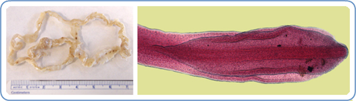
Hymenolepis nana, Hymenolepis diminuta
“Dwarf tapeworm” (nana), “Rodent tapeworm” (diminuta)
Hymenolepis nana: 1 mm by 25-40mm
Hymenolepis diminuta: 3 mm by 20-60 mm
Affects: Mice, rats, gerbils, hamsters (definitive) dogs and humans (H. nana more common - incidental)
Adults: small intestine
Found: Worldwide
Cause of infection: Ingestion of infective fleas, beetle, cockroach (H. diminuta) which is intermediate host; Ingestion of infective egg or autoinfection (H. nana) no intermediate host
Hymenolepis nana life cycle
Eggs pass in feces — direct life cycle – only tapeworm that does not need intermediate host
Swallowed by host, hexacanth enters villus of small intestine
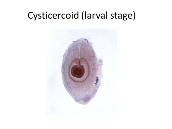
Matures into nontailed cysticercoid
Moves back to lumen, attaches to lining and matures to adult stage
Hymenolepis diminuta life cycle
Eggs pass in feces
Ingested by arthropod intermediate host
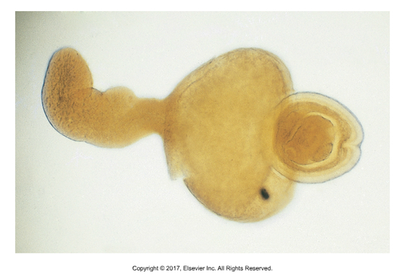
Hexacanth embryo matures into tailed cysticercoid
Attaches to lining of small intestine and matures into adults
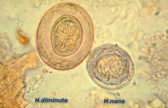
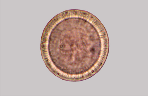
Hymenolepis sp. eggs
Eggs are detectable in fecal floatation, but shed intermittently
Proglottids do not float
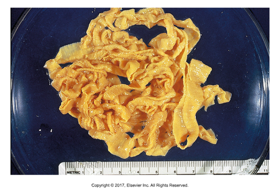
Moniezia expansa
“Ruminant tapeworm”
Affects: Cattle, sheep and goats (Moniezia benedini in cattle only)
Found: Worldwide
Adult in small intestine
Cause of infection: Ingestion of infected grain
Much larger than Hymenolepis (up 1.6 cm by 6m)
Unarmed scolex
Short, wide proglottids
Unique eggs with square (M. benedini) or triangular (M. expansa) shells
Three-layered egg shell, inner-most is pyriform apparatus — pear shaped
Prepatent period: 40 days
Eggs seen on fecal flotation
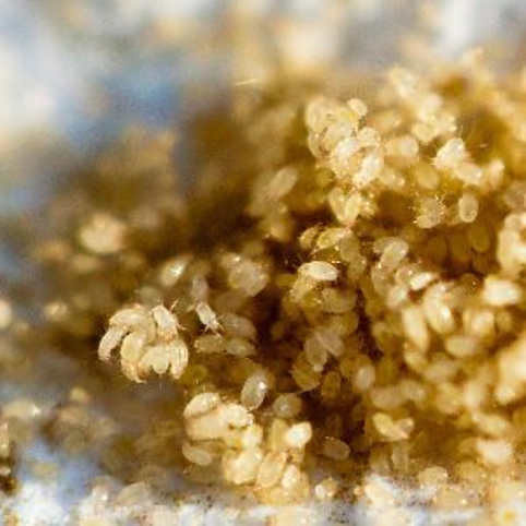
Moniezia metacestode stage
Proglottids and eggs in feces of infected ruminants
Cysticercoid found inside oribatid grain mites that have ingested hexacanth embryo
Ruminants infected by eating infected grain
Each cysticercoid forms one adult
Large numbers of adults can block intestines, especially in young animals
Pasture rotation as well as management of grain mites can help with reinfection rates
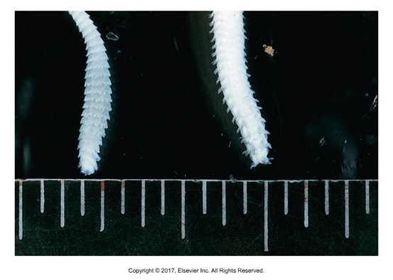
Thysanosoma actinoides
Affects: Sheep, goats, cattle
Adults in lumen of bile duct, pancreatic duct, small intestine – can cause obstructions
Intermediate host: unknown, possibly psocids (“booklice” that feed off mold, lichen, grains and wallpaper and book- binding glue)
Derivation: “fringed body”– unique fringe on the posterior half of each proglottid
Unarmed scolex, form egg packets (6-12), no pyriform apparatus
Taenia saginata (adult), Cysticercus bovis (larval stage)
“Beef tapeworm”
Adult: small intestine
Found: Worldwide
Definitive host: Human
Cause of infection: Ingestion of raw or undercooked intermediate host, beef cattle
Only member of Taenia with unarmed scolex, 14-32 branches of uterus in gravid
Taenia solium (adult), Cysticercus cellulosae (larval stage)
“Pork tapeworm”
Adult: small intestine
Found in: Mostly underdeveloped countries - Latin America, India, Africa, Far East
Definitive host: human
Cause of infection: Ingestion of undercooked intermediate host, pigs
Armed scolex, 7-16 branches of uterus in gravid
Can develop cysticercus in human muscle, brain, eye, spinal cord
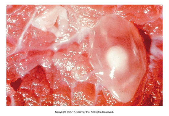
Taenia hydatigena (adult), Cysticercus tenuicollis (larval form)
“Canine/dog bladder worm”
Affects: Dogs, adults in small intestine
Intermediate host: cattle, sheep, goats
Found in: Worldwide
Cause of infection: Ingestion of omentum of ruminants with golf-ball size cysticercus (not pathogenic to ruminants)
Armed scolex, proglottids have single lateral genital pore
Eggs can be seen in fecal flotation
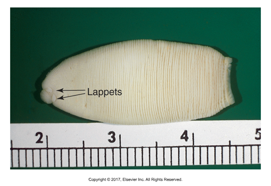
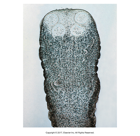
Anoplocephala sp.
A. perfoliata, A. magna, Paranoplocepha mamillana
Found in: Worldwide
Intermediate host: Grain mites
Unarmed scolex
Cause of infection: Ingestion of mites
Unique morphology: unarmed oblong scolex, two round lappets behind each of the four suckers, only one pair of reproductive organs per glottid
Pyriform apparatus in egg with one or more flattened sides
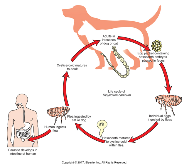
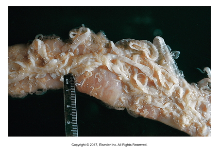
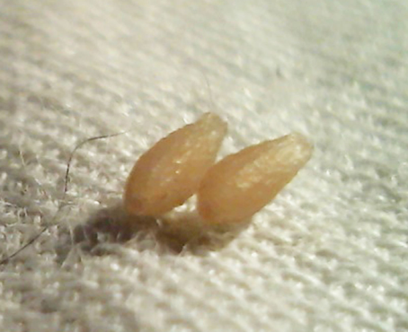
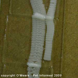
Dipylidium caninum
“Double pore” or “cucumber seed tapeworm”
Most common tapeworm of dogs and cats
Affects: Dogs and cats
Intermediate host: fleas
Cause of infection: Ingestion of fleas
50 cm long
Long proboscis on armed scolex
Gravid proglottids can be motile when shed in feces, coat hair, bedding
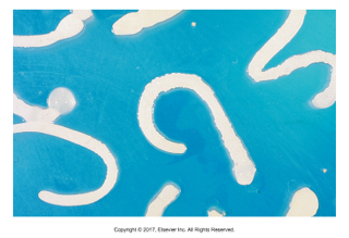
Each gravid proglottid contains thousands of egg packets
20-30 eggs per packet, each with hexacanth embryo inside
Prepatent period: 14-21 days
Dipylidium caninum life cycle and prevention
Metacestode stage is cysticercoid in intermediate host: flea
Larval fleas ingest hexacanth embryos
Dogs and cats ingest the infected fleas
Children are most likely humans to ingest fleas and get zoonotic infection
Must include good flea control in order to prevent reinfestation
Egg packets found in fecal flotation
Proglottids found in gross feces
Taenia sp.
Affects: Dogs
Adults in small intestine
Intermediate hosts: Rabbits and hares (T. pisiformis), ruminants (T. hydatigena), sheep (T. ovis)
Cause of infection: Ingestion of intermediate host
1-2 cm (T. ovis) to 200 cm (T. pisiformis) to 500 cm (T. hydatigena)
Armed scolex, motile gravid proglottids– if found in feces, hydrate in saline on slide to see eggs inside
Single lateral pore
T. pisiformis has pea sized cysticercus, attached to greater omentum of rabbit
T. hydatigena has ping pong ball sized cysticercus, attached to greater omentum of ruminant
T. ovis has cysticercus found in muscle of sheep
Preventing dogs and cats from eating viscera or musculature of intermediate hosts is best way to prevent Taenia infections
Taenia taeniaeformis (adult), Cysticercus fasciolaris (larval form)
Affects: Cats
Intermediate host: Rats and mice
Found in: Worldwide
Cause of infection: Ingestion of rats and mice
60 cm long
Armed scolex, bell shaped proglottids, taeniid egg
Taenia taeniaeformis life cycle
Unique in that larval stage is strobilocercus
Rodents ingest hexacanth eggs from cat feces, develop into cysticercus inside liver for 42 days
On day 42, cysticercus changes shape into strobilocercus with scolex and long neck attached to bladder (Cysticercus fasciolaris)
Cat infected by eating rat or mouse liver
Scolex attaches to cat intestine
Multiceps sp.
“Many headed worm”
Affects: Dogs, not zoonotic
Adult in small intestine
Intermediate host: Sheep (M. multiceps) or rabbit (M. serialis)
40 to 100cm long
Double row armed scolex, taeniid eggs
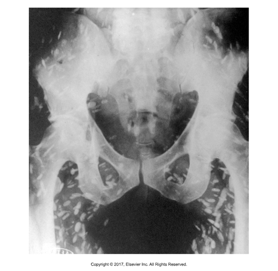
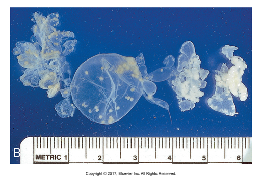
Multiceps sp. metacestode stage
Forms coenurus that is found in sheep or rabbit intermediate host
Coenurus cerebralis (M. multiceps) or Coenurus serialis (M. serialis)
Single large bladder with several scolices attached to the wall
Unique in that many tapeworms mature from each coenurus, one for each scolex
In sheep, hexacanth embryos are ingested and larvae (5cm) migrate to nervous system (brain or spinal cord) and can produce neurological symptoms in infected sheep
Dogs infected by ingesting sheep parts
In rabbits, hexacanth embryos are ingested in dog feces, hatch and migrate into subcutaneous tissues which can interfere with movement, slow rabbits more likely to be caught and eaten by dogs
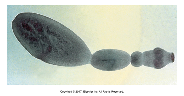
Echinococcus granulosus (dogs) and Echinococcus multilocularis (cats)
“Spiny berry”
Intermediate host: Sheep, cattle, herbivores (E. granulosus), rats, mice and voles (E. multilocularis),
Both are highly zoonotic
Cause of infection: Ingestion of intermediate host
Form unilocular (E. granulosus) or multilocular (E. multilocularis) hydatid cysts
Adults are tiny: 1 to 7mm in length with only three proglottids: 1 immature, 1 mature, 1 gravid
Very difficult to diagnose as gravids are seen in feces, adults never get to numbers that cause obstruction
Diagnosis of Echinococcus
Can find adults in intestinal tract at necropsy
Suspected infections can be diagnosed by purging animal of all feces using arecoline hydrobromide
All feces should be handled with caution using gloves and incinerated
Current ELISA tests for Echinococcus available
Cases in dogs, cats or intermediate hosts must be reported to state and federal public health officials
Taeniid eggs similar to Taenia and Multiceps sp.
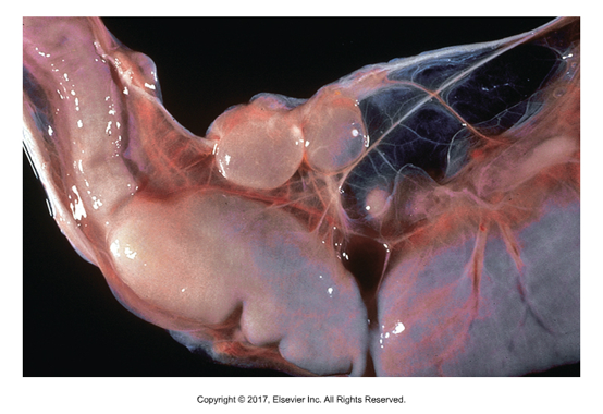
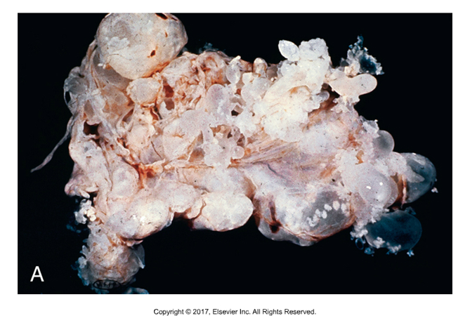
Echinococcus metacestode
Larval stage is hydatid cyst
E. granulosus forms unilocular cysts within liver, lung, other organs of ruminant
Thick cyst wall, thin germinal membrane; “Brood capsules” contain protoscolices that bud from germinal membrane; Each protoscolex forms one adult worm)
E. multilocularis forms multilocular or alveolar cysts within liver, lungs of rodents
No thick wall, grapelike cysts that are very invasive, replacing normal tissue almost like cancer; Thin germinal membrane; “Brood capsules” with protoscolices bud from membrane; Praziquantel can treat adult Echinococcus but will not kill cyst stage
Mesocestoides sp.
Affects: Dogs, cats, other carnivores
12cm to 2m in length
Oblong scolex, four acetabula, unarmed
Only true tapeworm with two intermediate hosts
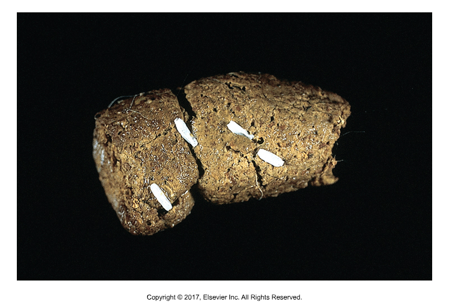
Mesocestoides sp. metacestode stage
If ingested by oribatid mite, develops into cysticercoid inside mite
If cysticercoid ingested by mouse or reptile, develops into tetrathyridium which can multiply asexually to make large numbers that can infect dogs, cats
Tetrathyridia multiply heavily in intermediate host, mostly in serous cavities like peritoneum or liver causing large increase in girth
When ingested by definitive host, tetrathyridium develop into adults in small intestine

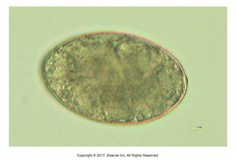
Pseudotapeworms Cotyloda
Look similar to true tapeworms but have centrally located reproductive organs
Unembryonated operculated eggs (almost identical to trematodes)
Most release eggs directly from uterus passed in feces of definitive host
Sometimes short chains of proglottids are released
No acetabula or rostellum but two slitlike organs called bothria
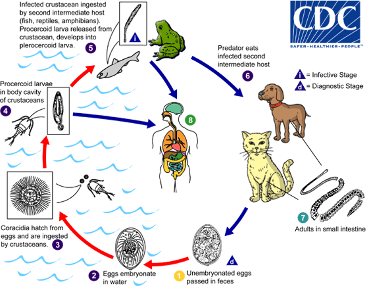
Pseudotapeworms Cotyloda life cycles
All use two intermediate hosts
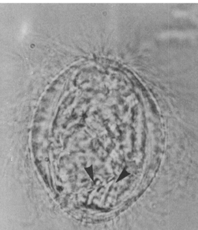
Eggs that come into contact with water release ciliated hexacanth embryo called coracidium
Swims in water, ingested by microscopic crustacean and develops into procercoid
Then ingested by fish or amphibian, matures in muscle into metacestode larva called plerocercoid or sparganum
Definitive host eats intermediate host to become infected
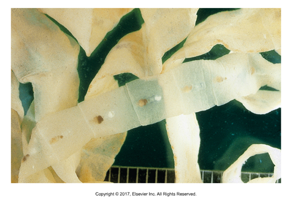
Spirometra sp.
“Zipper tapeworm” (due to mature proglottids pulling apart or unzipping)
Affects: Dogs and cats
Found in: North and South America, Far East, Australia, most prevalent in Florida and Gulf Coast
Adults in small intestine
Intermediate host: crustacean, then fish or frog
Rare to release gravid proglottids
Sparganosis
Spirometra sp. metacestode stage
Sparganum larval stage, solid-body with slit-like mouthparts
Found in muscle of intermediate host
Diphyllobothrium latum
“Double leaf” or “Broad fish tapeworm”
Affects: Dogs, cats, humans
Adults in small intestine
Intermediate host: crustacean, then fish
Found in: Scandinavia, Ukraine, N. America
2m to 12m long
Four acetabula and two bothria
Can cause pernicious anemia in host due to B12 absorption, gravids detach in small chains
Largest human tapeworm