E3
1/72
There's no tags or description
Looks like no tags are added yet.
Name | Mastery | Learn | Test | Matching | Spaced | Call with Kai |
|---|
No analytics yet
Send a link to your students to track their progress
73 Terms
anatomy of eye
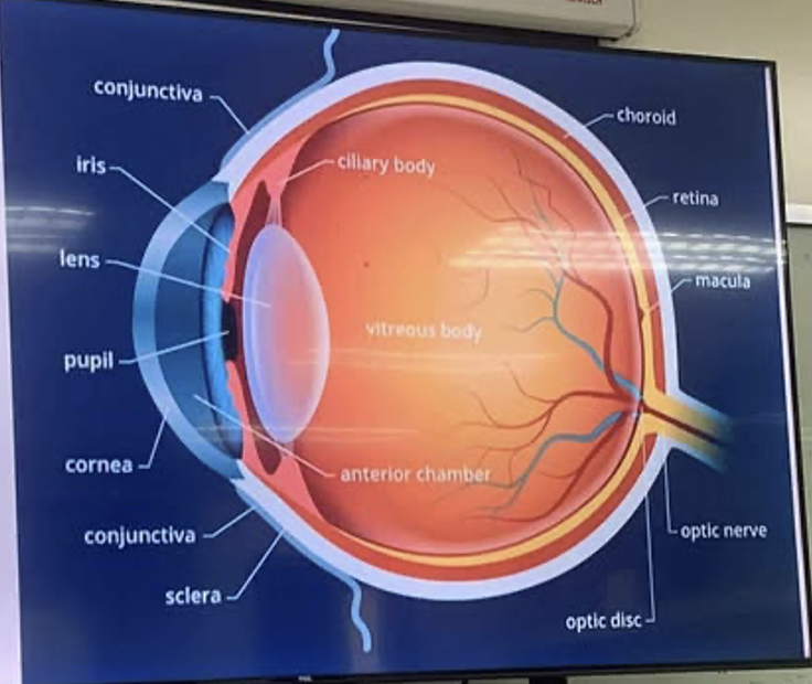
tunica fibrosa
cornea
sclera
tunica vasculosa (uvea)
iris
ciliary body
choroid
3 chambers of eye
anterior chamber
posterior chamber
vitreous chamber
Anterior chamber
– In front of the cornea, the first chamber
Contains aqueous solution/ fluids
Together with the aqueous solution joints with the posterior chamber
Posterior chamber
– also produces aqueous solution
Vitreous chamber/ body
– biggest chamber, contains vitreous fluid
Used in necropsy procedure
other parts of the surrounding eyeball
palpebra
conjunctiva
teargland
nictating membrane
extraocular mm
superior and inferior obliques
recti
muscle
dorsal
ventralatteral
medial
retractobulbi
etiologies of eye disorder
bacterial
moraxella, Neisseria catarrhalis, mycoplasma conjunctivae, B. canis
chalmydia
(conjunctivitis in cat) and reckettsiae (ticks and mites vector)
parasitic
(eyeworms: thelazia, oxyspirura, philopthalmus and toxoplasma)
viral
herpesvirus
mechanical and chemical irritations
allergic
mycotic infection
bacterial
Moraxella bovis
– Infections bovine keratoconjunctivitis
Better known as pinkeye
Transmitted by face flies
While feeding on lacrimal secretions, they transmit Moraxella from one bovine to another
bacterial
Neisseria catarrhalis
– normal flora of the oral and upper respiratory tract of companion animals (dogs & cats)
Lx caused – coming from wound bites
bacterial
Mycoplasma conjunctivae
– common causative agents of respiratory diseases
Common Lx – specific to this, respiratory problems à eye Lx
bacterial
Brucella canis
– brucellosis common in bovines, goats, livestock causing mastitis;
In canines, causes Lx in the reproductive system
Can also be seen in the eyes
chlamydiae
Causes conjunctivitis in cats
Rickettsiae – transmitted by ticks and mites (vector)
parasitic
Caused by eyeworms such as:
Thelazia
oxyspirura
Philophthalmus
toxoplasma
Nematodes ((Roundworms, whipworms, hookworms)
Thelazia (roundworm)
Moves like a worm/serpent in the eyes of the dog
Zoonotic to humans; eyeworm in humans as well
Regular deworming renders this not a threat to the owner
In companion animals specifically:
In the US: Theilazia californiensis
In Asia: Theilazia callipaeda
Manually take off can be a solution – sedate first the dog for risks
Pyrantel – drug
T. guloSA, t. skrjabini, and T, rhodesii – livestock
Nematodes ((Roundworms, whipworms, hookworms)
Oxyspirura petrowi
– found in quail
trematodes (flukes)
Philopthalmus
found in wild birds
Genus Philopthalmidae
protoza
Sheds oocyst in the feces
In the body will undergo reproduction (in the cat)
Dispersed in the different parts of the body
Cat continuously sheds the oocyst in their feces
This can transmit this disease to other animals – dogs, humans
Can stay in the dog/ humans; the oocyst can burrow in the tissues such as skeletal, alimentary in the form of cysts and remain dormant as such
When immunosuppressed, it can become lethal
Can burrow in the tissue of the brain as a cyst as well
Toxoplasma gondii – Uveitis, retinitis, choroiditis,
Specific host: only the CAT family can be hosts
Asymptomatic
virus
Herpesvirus – is usually at the genital organs but can cause eye Lx
In dog – Herpesvirus 2 (FHV-1)
In cat – herpesvirus 1
Can cause secondary infections such as papillomavirus (papillomas)
Mycoses
Superficial Lx of the skin, usually external.
If left untreated, commonly in stray dogs, the eyes and ears are also affected
Crusty, flaky skin; likely with Lx in the ear
Due to this, other causative agents can enter the skin of the eyes and ears causing disease
E.g. Dermatophycosis – can make the skin very dry
If the eye is infected, the ear will always be paired (mycoses specific)
tear deficiency can happen because of the following causes:
Most common and #1 cause: age
Tear production lowers as one ages
2nd cause is medication that can cause reduced tear production
Antihistamines
Antidepressants
Decongestant
3rd cause is medical condition causing reduction of tears
Distemper (practically closed eyes because of the lack of tear production)
Arthritis
lupus
mechanical & chemical
irritations such as when one scratches their eyes
tear film together with corneal epithelium become damaged from excessive scratching
lining of epithelium of the eye
is the eyelid
made of non-keratinized squamous epithelium
associated with columnar cells with goblet cells
secretes mucin/ mucous (muta/luha)
exception include (in the dog)
THIRD EYELID (with upper and lower eyelid)
humans used to have a third eyelid and lost it from evolution; it is simply a remnant
TEAR FILM –
In front of the eye, supposed to be clear,
hydrates and keeps on moisturizing the eyes – prevents infection
dryness of eyes = more prone to infection
3 layers of tear film
lipid layer
aqueous layer
mucin layer
lipid layer (1st layer)
Produced by Meibomian gland
aqueous layer (2nd layer)
Contains: protein, water, electrolytes – those being produced by the lacrimal glands
mucin layer (3rd layer)
Produced by the goblet cells of the columnar epithelium
mucin
is the compensatory mechanism of the eye to compensate for the loss of tears
Is not enough to lubricate the eyes and thus becomes sticky
entropion
inversion of palpebral margin
Infection of this if left untreated will worsen
First harming the tear film, then the corneal epithelium, and eventually the corneal stroma
Can eventually cause ulcerations of the cornea
Tx: TACKING – surgical procedure
Typically a triangle-shaped incision due to the coverage of the ligament covering their nose
Common in short-nosed breed of dogs
Prone to cephalic breed of dogs
ectropion
eversion of palpebral margin (esp. lower eyelid)
Must be corrected surgically
Causes chronic irritation if left untreated – can eventually become a neoplasm - squamous cell carcinoma
Less common than entropion
Ectropion – maybe due to the overcorrection of entropion
Common in large breed of dogs that has many eye folds
e.g. Bernard, hounds, chowchow (prone to entropion and ectropion)
Trichiasis
condition where normally positioned hairs, often around the eyes, grow in a way that they rub against the cornea or conjunctiva, causing irritation
Common in brachycephalic breeds of dogs
Distichiae
are hairs that grow in an abnormal location on the eyelid margin (specifically emanating from gland ducts)
Grows outside the margin
e.g. in the hair glands – typically plucked but must be sterile to avoid infections in the oil glands
Ectopic cilia
growth of hair in the conjunctiva that grow specifically through the conjunctival surface
common again in brachycephalic dogs such as the Pekingese
Coloboma
absence of eyelids (may be partial or complete)/ portion of the eye tissue or iris is missing or underdeveloped
Exopthalmos (propoptosis)
protrusion of the eyeball
has many causes such as:
due to injury, bacterial, fungal, medical conditions such as hypo & hyperthyroidism
enophthalmia/ enophthalmos
bilateral retraction of eyeball
sunken eyes, displaced backward within the orbit
pairs with exophthalmos
happens because there is a loss of retrobulbar fat
Hyopyon
presence of pus cells in the anterior chamber of the eye
characterized by the accumulation of inflammatory cells in the anterior chamber of the eyes
if there is no other solution, it must be drained
the pus can come from Pseudomonas aeruginosa
Some anti-inflammatory cells will secrete myeloperoxidase – giving the pus green color
Keratoglobus
increased convexity of the cornea
Tx is via surgery
Nystagmus
abnormal oscillatory movement of the eyeball
Indicative of nervous problems – vestibular
Distemper – neuronal type
The head is still & with no stimulus, the eyes move either:
Horizontally (side-to-side)
Vertical (up-down-up-down)
Rotary (round in circles)
Uveitis
inflammation of uvea (uvea/ tunica vasculosa): iris, ciliary body, and choroid
Must be differentiated from nuclear sclerosis
There is no cure for this ds but simple acceptance
Keratitis
inflammation of the cornea
E.g. IBK of bovine
Blepharitis
inflammation of the eyelids
In tagalog is kuliti
Nyctalopia/ xerophthalmia
night blindness; likely caused by deficiencies in Vit A/ retinol
Anisocoria
Refers to the mismatched constriction of pupils
Wherein one is dilated, and the other is constricted
Mydriasis
Both pupils are dilated
Difficult to treat
In surgery, when dilated indicates lack of optic nerve reaction – meaning likely non-responsive brain activity
Miosis
Constriction of pupils
Indicative in surgery because it can tell the stage of anesthesia; indicates that the animal is just asleep
cataract
clouding (opacity) of the lens, due to the clumping of protein in the lens
Not always white, can be blue
Sclerosis
has green margins
From age
Hordeolum
infection of the eyelids (oil producing glands of the eyelashes)
Reason for carefulness: plucking hair from distichiae
Dacryocystitis
inflammation of the lacrimal sac
Seen in distemper, parvo virus, could simply be blocked from canaliculi, from constricted duct
If the lacrimal ducts are destroyed, they have to be removed; maintenance with artificial tears
pannus
increased vascularization of the cornea
Pinkeye, overuse of cellphone, etc.
Strabismus
inability of one eye to attain binocular vision due to the imbalance of eyeball muscles
synecchia
adhesion (esp. of iris) to other parts of the eye
normal ear anatomy
Like humans but have muscle tissues – allows for ear mobility
Continuously works for 24 hours – even in sleep, hearing does not stop
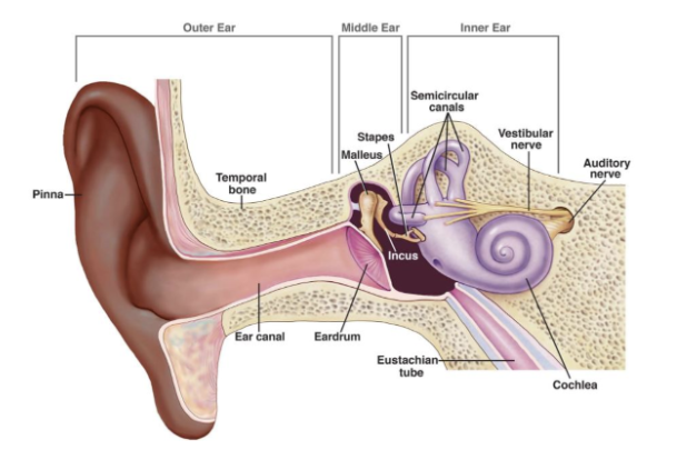
How sounds are processed
Enters from the external ear, into the ear canal, moving inwards
Once sound touches the tympanic membrane, sounds are amplified
Amplified sounds are transferred into the ossicles
Smallest bones in the body which are components of the middle ear (there are 3: malleus, incus, stapes)
Malus à incus à stapes
Stapes will prepare the sounds to be transferred into the semicircular canal – organs of the vestibule; connects to the cochlea
Vestibule is the organ of balance
Transfers to the cochlea
Inside the cochlea, sounds will traverse with fluid (endolymph)
Once enters the Organ of Corti (which is inside the cochlea), it will travel with the hair cells of the organ of corti
Once the hair cells perceive the sound, they will interpret the sounds as electrical impulses to the vestibulocochlear nerve (acoustic nerve)
Which are then sent to the brain, which interprets the sounds
Semicircular canals
are responsible for the sense of balance due to the displacement of perilymph (NOT endolymph)
Commonly dogs with distemper cannot balance properly because of this.
Cochlea
is the organ with 3 turns, looking like a snail; containing endolymph, the Organ of Corti, and hair cells
parasitic
Otodectes mites
– with Lx looking like sand
Burrows under the skin of the ear
parasitic
Sarcoptic mange (also known as scabies)
Zoonotic to people, and very common in stray animals
Highly contagious skin condition caused by the Sarcoptes scabiei mite
Causes rubbery skin that may cover the animal all over
Can also go to the ear
bacterial
Pseudomonas otitis
– in animals appear to always have pus
Wet, moist, and is filled with pus
mites of livestock
Raillieta auris
– would burrow in papilloma and warts of the ears
mites of livestock
Raillieta auris
– would burrow in papilloma and warts of the ears
viral infections in ears
Distemper and parvo
also emit Lx in the ears
Tx is to focus on the systematic infection and not the ear
others include:
Viral & mycoplasmal pathogens
Mycotic such as Malassezia pachydermatis
Antibiotics such as:
Aminoglycosides (with ototoxic, hepatotoxic & nephrotoxic effects)
deafness
lacking/ deficient in the sense of hearing due to damage of cochlear parts;
cochlea has most of the components where vibrations are converted into impulses
otitis
general term for inflammation of ear structures
Otitis externa – affects the pinna and ear canal
Otitis media – affects the ear drum, stapes, malleus
Otitis interna – affects the cochlea
Otosclerosis (hereditary)
bone surrounding the middle and inner ear (stapes bone) grows excessively & therefore sounds are not transmitted properly à leads to deafness
Perichondritis
infection of cartilage of the outer ear
Myringitis
inflammation of eardrum
tinnitus (symptom)
noise originates in the eardrum rather than from the environment
Dogs hear tinnitus
Seen are:
Tendency to tilt head (compensatory mechanism)
Sudden stoppage of barking and looking at barking at an empty space