female reproductive system lab models
1/66
There's no tags or description
Looks like no tags are added yet.
Name | Mastery | Learn | Test | Matching | Spaced |
|---|
No study sessions yet.
67 Terms
mons pubis
a

clitoris
h

labia majora
i

labia minora
j

urinary bladder
b

uterine tube
c
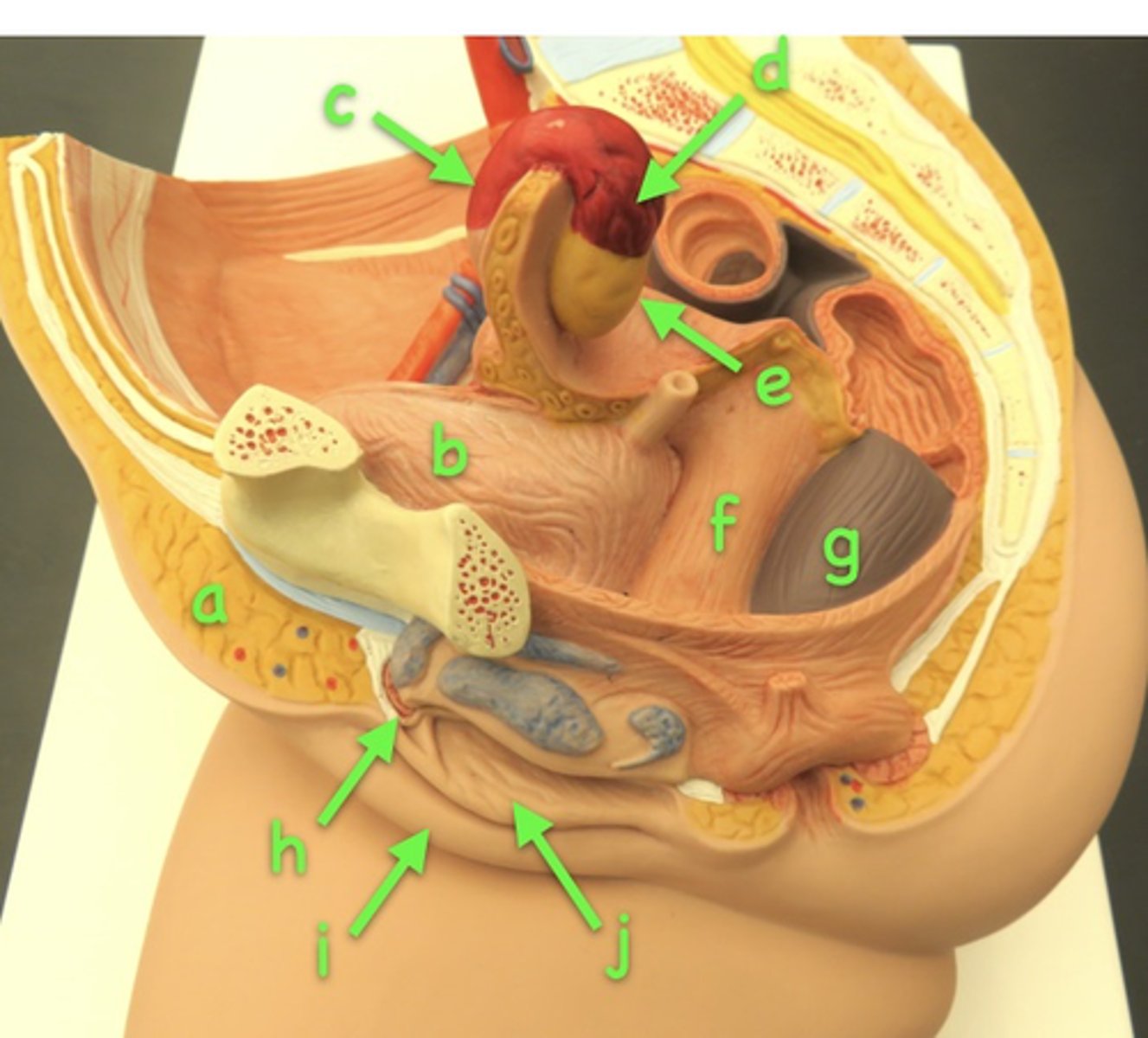
fibriae
d

left ovary
e

external walls of the vagina
f

rectum
g

Midsagittal mons pubis
a

Midsagittal clitoris
h

Midsagittal labia majora
i

Midsagittal labia minora
j

Midsagittal urinary bladder
b

Midsagittal uterus
c

Midsagittal fundus
d

Midsagittal body
e
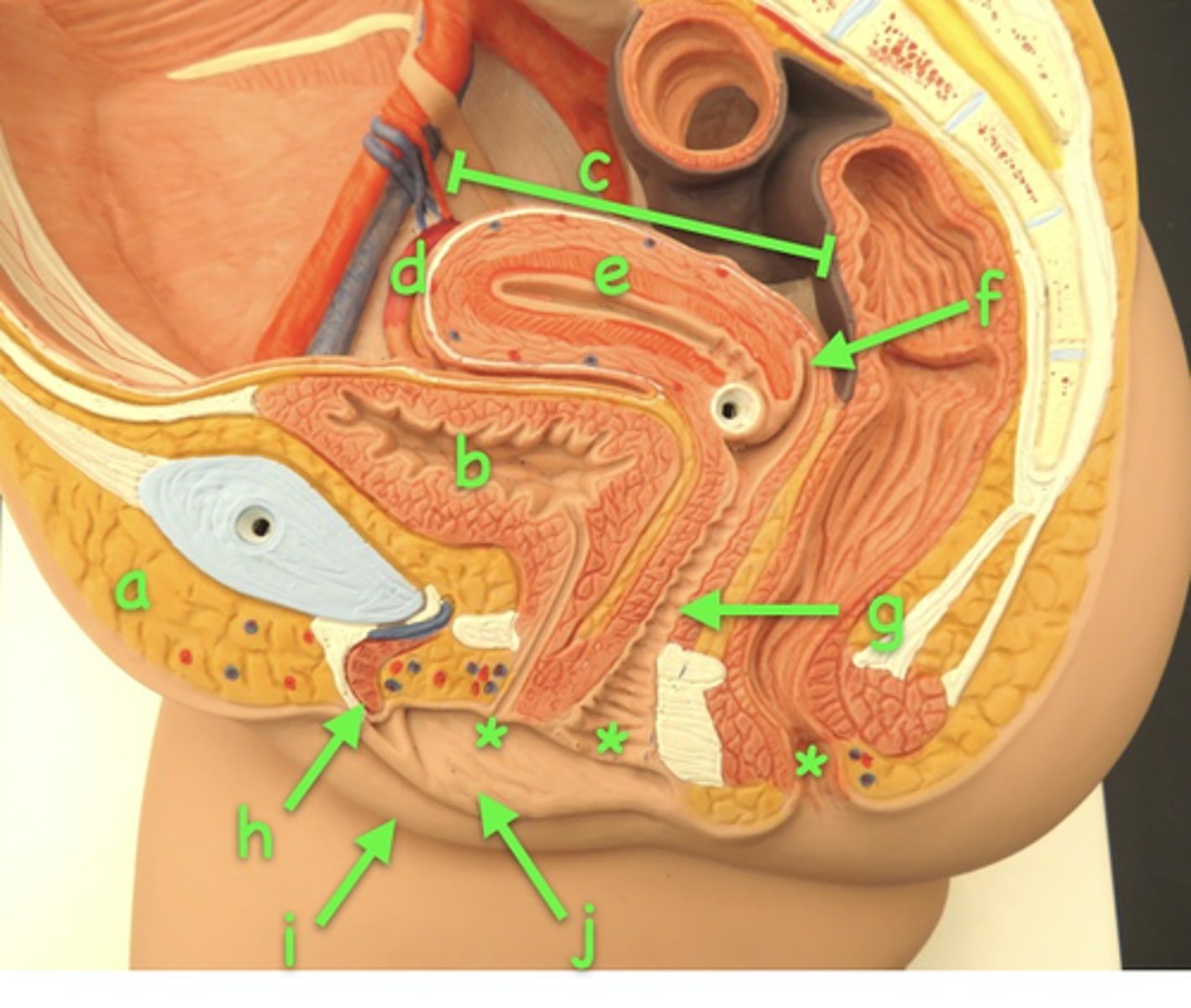
Midsagittal vagina
g

Midsagittal fornix
f

Midsagittal three openings in the female pelvic floor.
from left to right, the urethra, vagina, and anal canal.
*

cortex
to the right, and below the lines

ovarian follicles
arrows
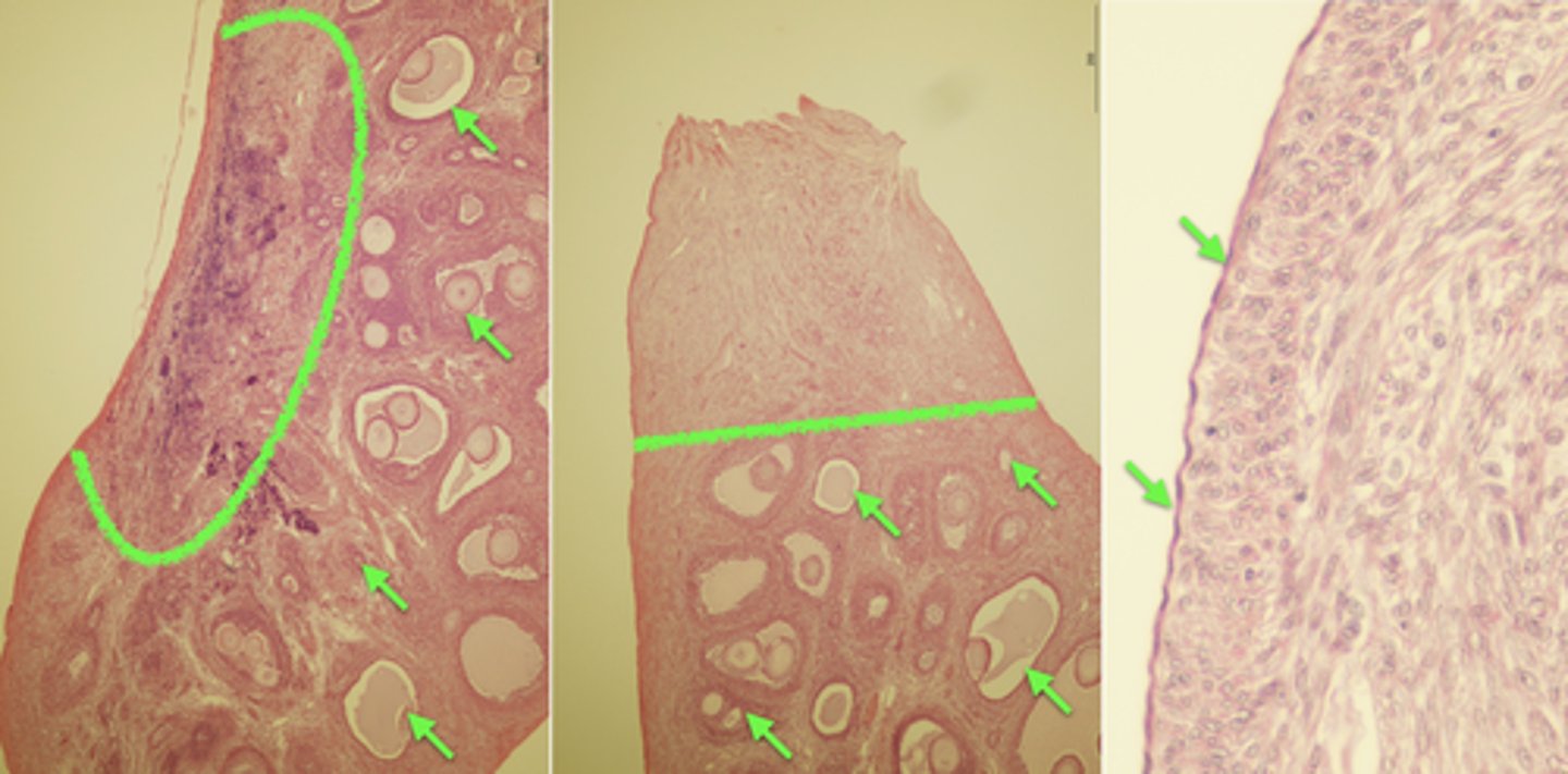
purple-staining germinal epithelium
right arrows

the egg of a primordial follicle
*

follicular cells of a primordial follicle
a

the darker nucleus within the egg of a primary follicle
*
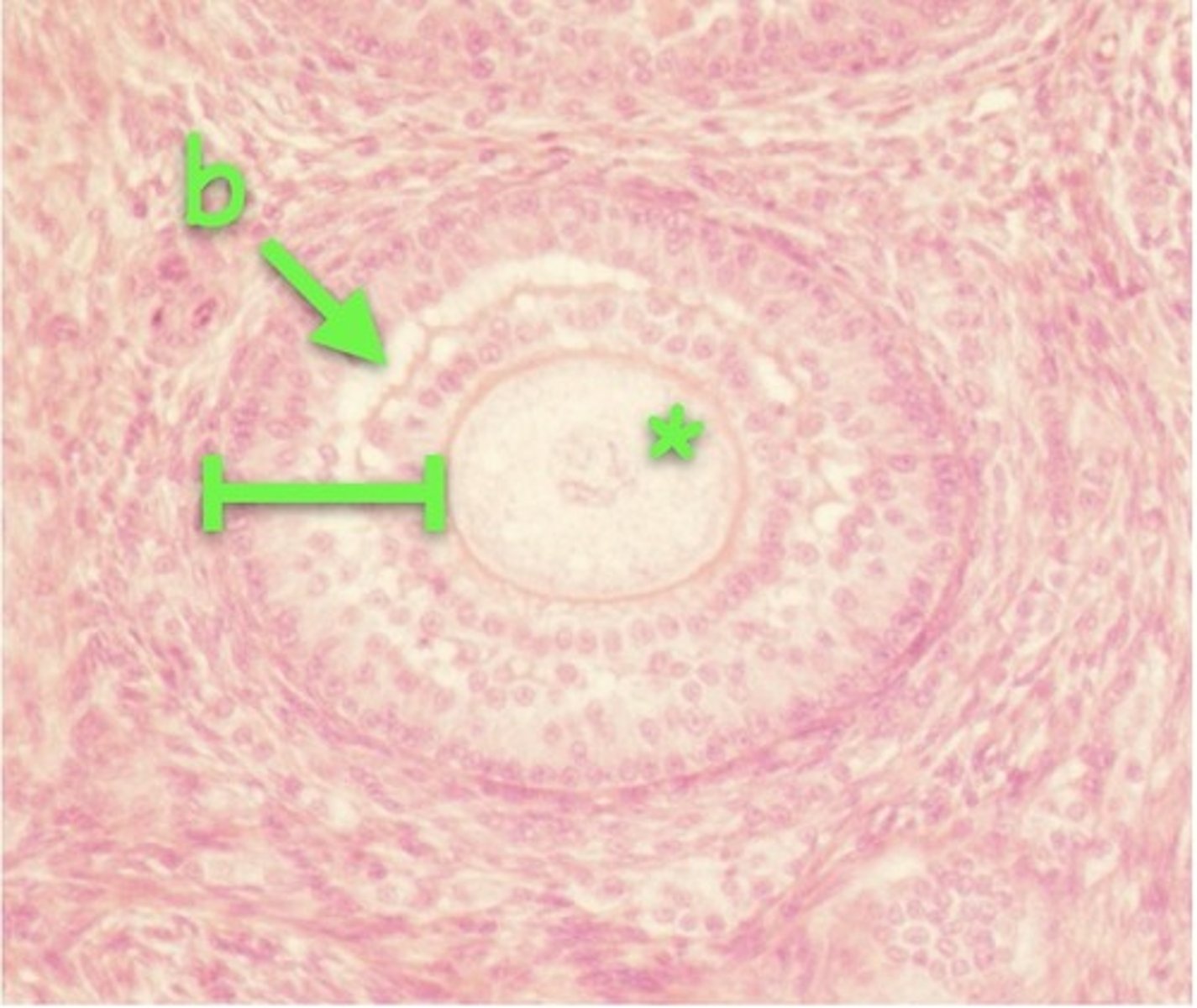
granular cells
bracket

cavitation
b

secondary follicle with its egg cell
* (upper right)

secondary follicle containing a darker nucleus, granular cells
brackets (upper right)
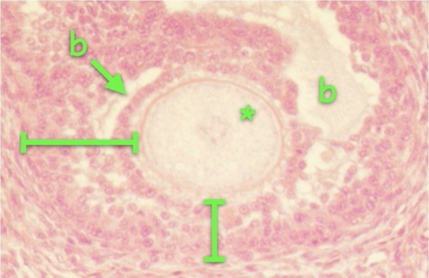
antra of secondary follicle
b

early tertiary (graffian) follicles
what is this

oocyte with its nucleus and very dark nucleolus
*
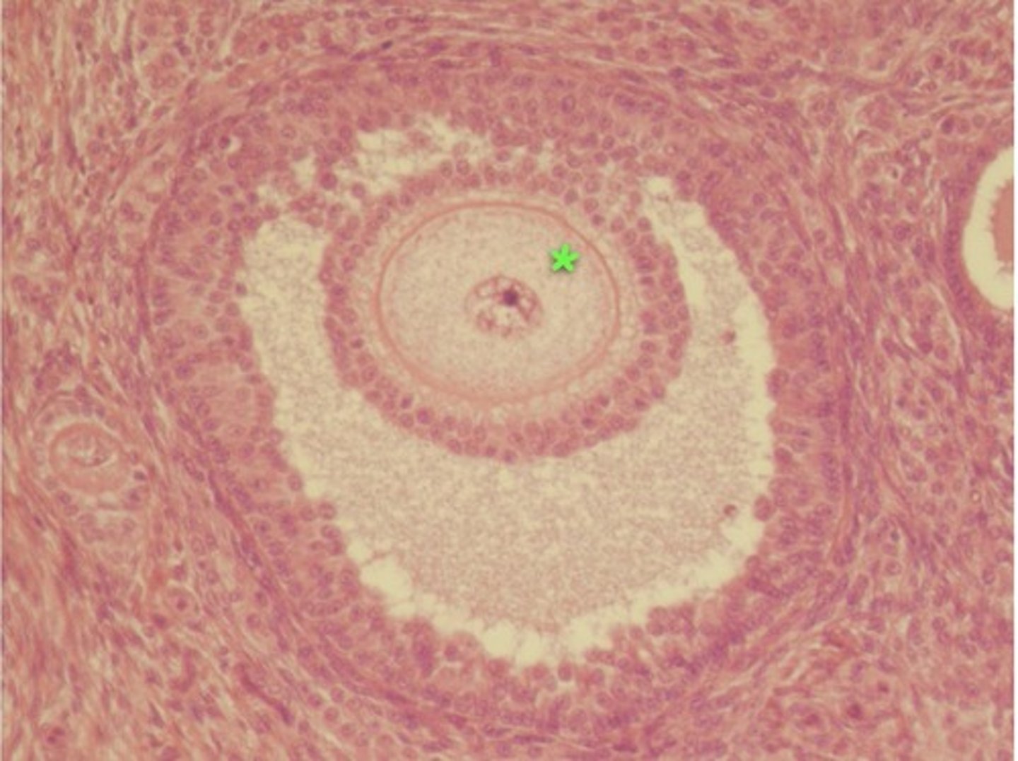
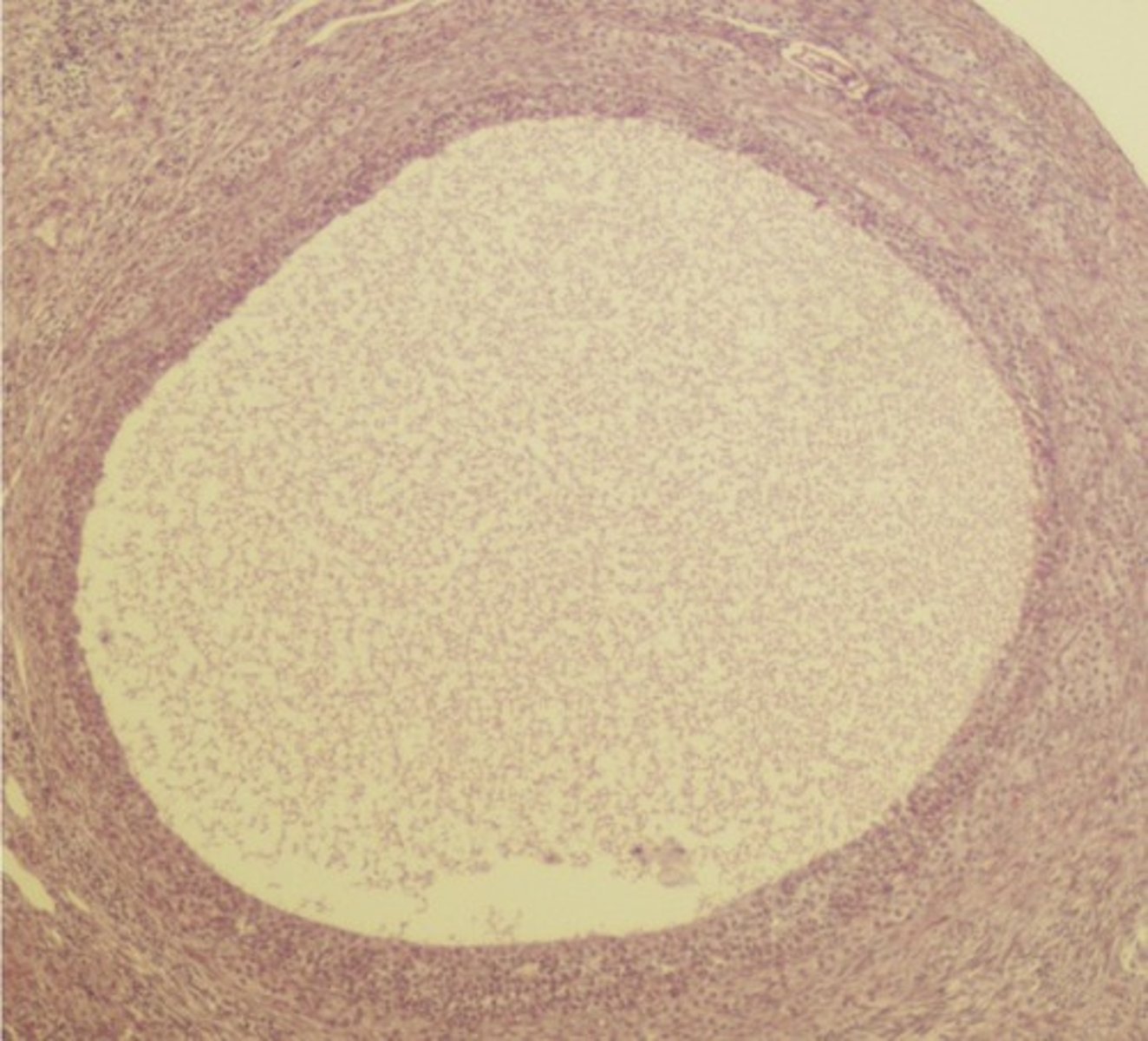
later tertiary (graffian) follicles
what is this

fluid-filled antrum
what is this?
outer perimetrium made of visceral peritoneum
a

thick middle myometrium made of smooth muscle
b

inner endometrium
c

anterior/superior dome-like fundus
d
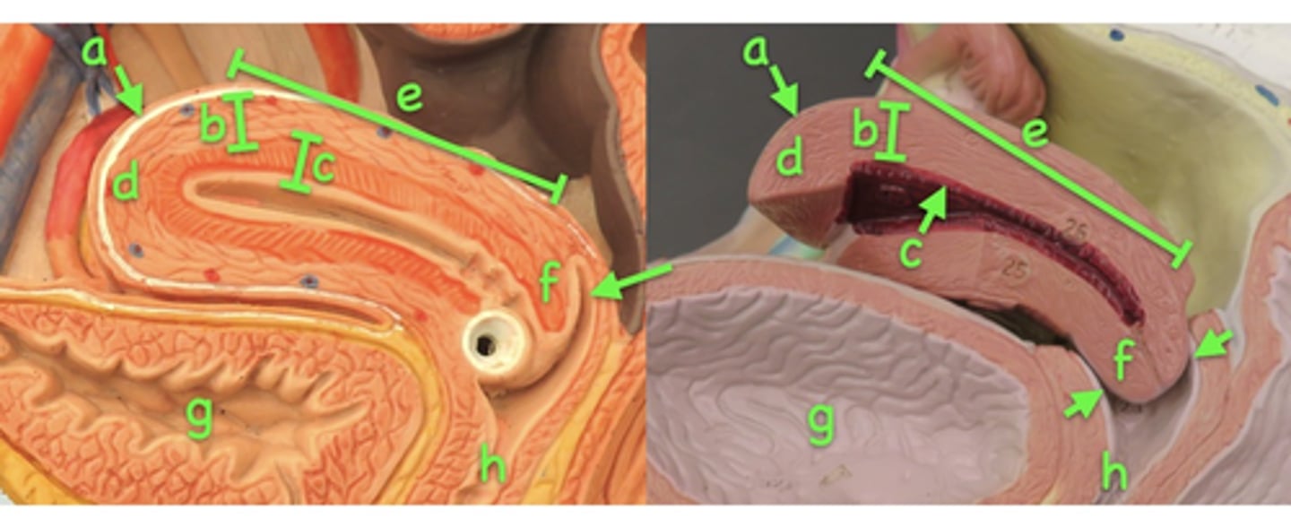
body
e

narrow cervix
f

vagina
h

the fornix
arrows

the fundus
a

body
b

cervix
c

vagina
g

fornix
h
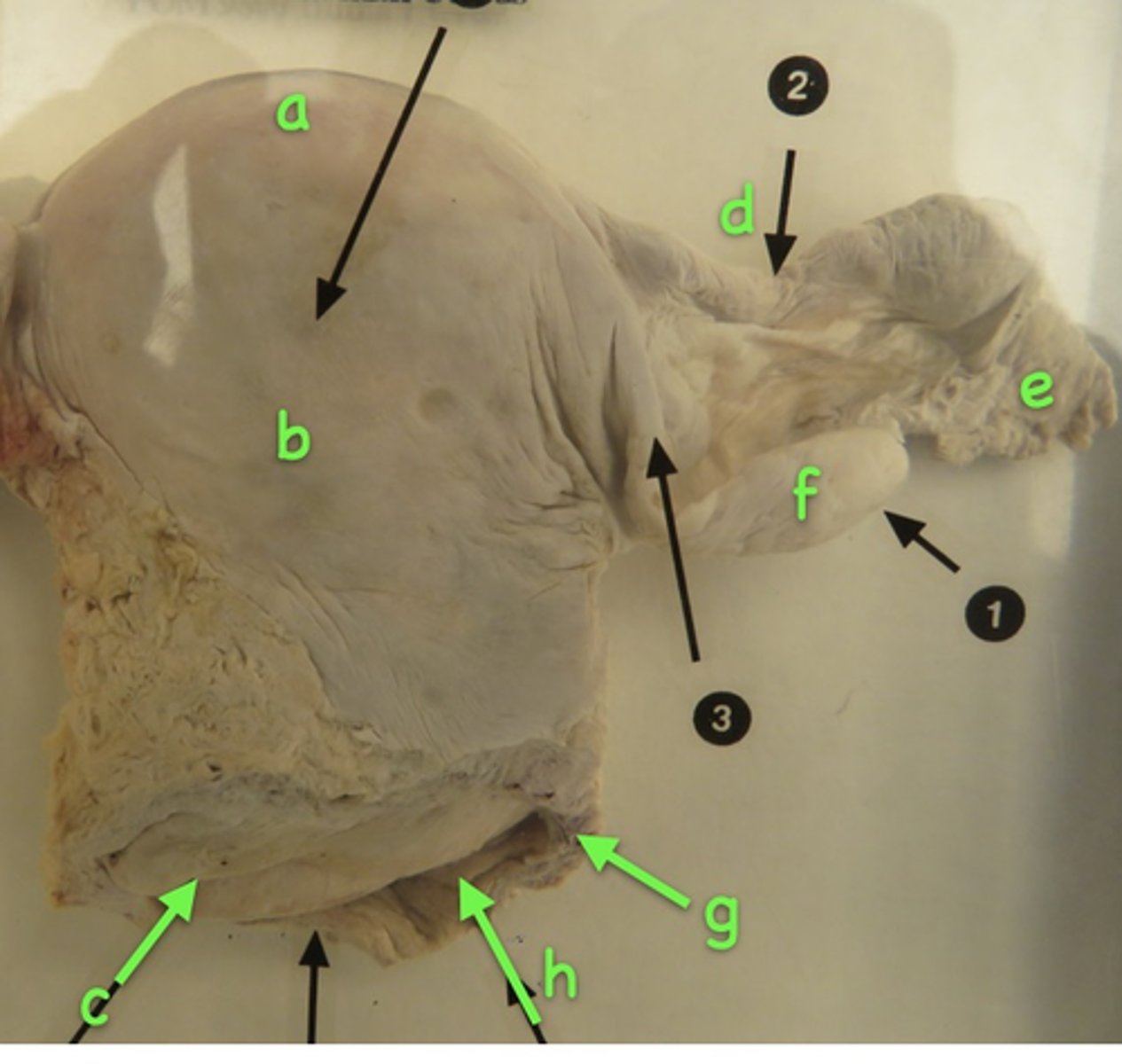
uterine tube
d

fringe-like fimbriae
e

ovary
f

myometrium
*
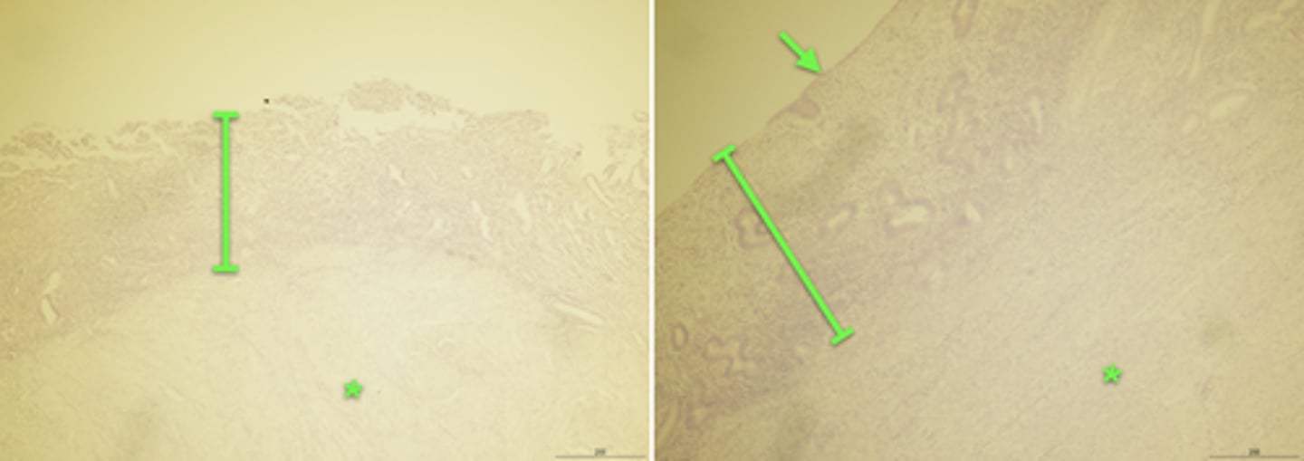
endometrium
brackets

The endometrium
(bracket on left image, and entire right image)

enlarged glands
arrows
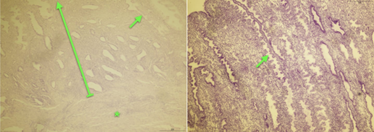
myometrium
* (left image)

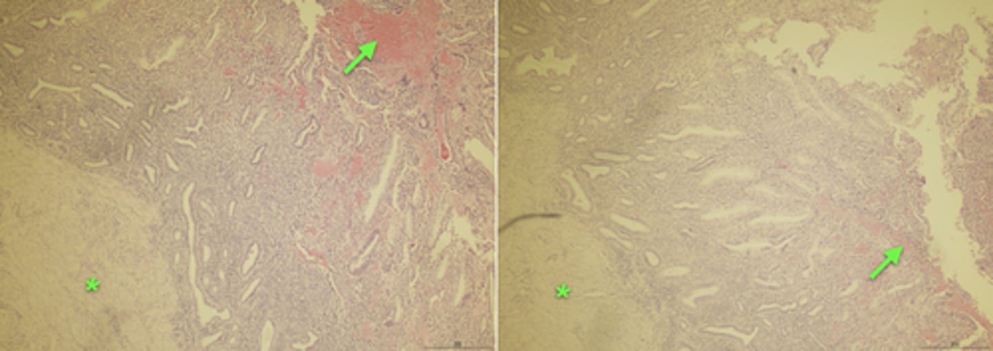
myometrium
(*)

The darker-staining endometrium is beginning to deteriorate
(especially right image)

blood
arrows
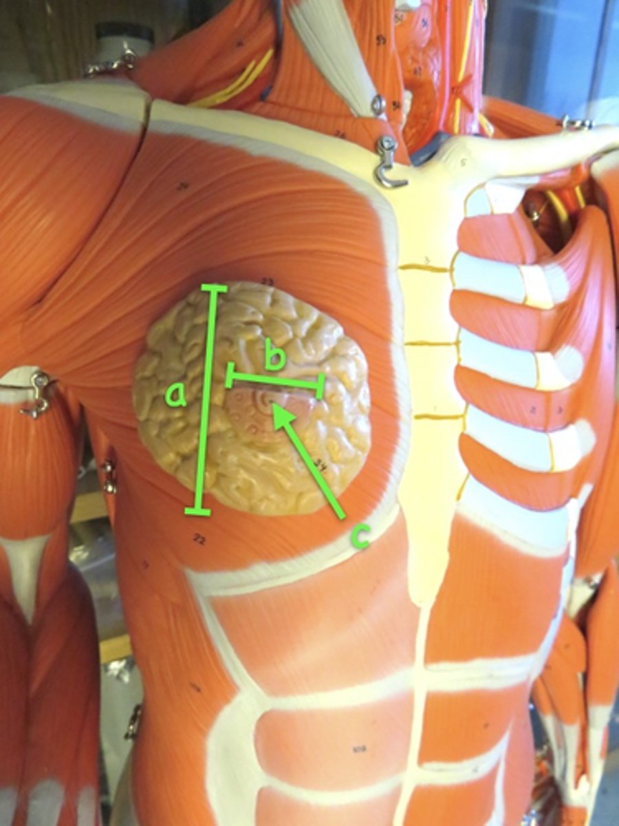
breast
a

areola
b

nipple
c
resting breast
left

active breast
right
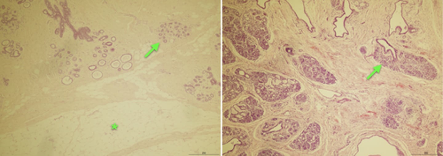
the mammary glands, much enlarged in the lactating breast (right)
arrows

adispose tissue (more visible in the left resting breast)
(*)
