Behavioural Neuroscience PSY 3301 Visual System
1/64
There's no tags or description
Looks like no tags are added yet.
Name | Mastery | Learn | Test | Matching | Spaced | Call with Kai |
|---|
No analytics yet
Send a link to your students to track their progress
65 Terms
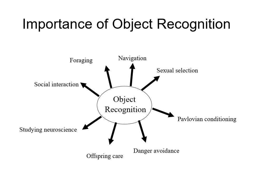
importance of object recognition
essential for perceiving, navigating, and interacting with the environment by identifying and categorizing objects from sensory input. It is crucial for survival, allowing humans and animals to distinguish between, for example, threats and useful tools, while also serving as a foundation for cognitive processes like memory, reasoning, and language.
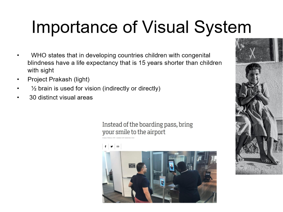
Importance of visual system
essential for survival and daily functioning, acting as the primary sensory system to construct a mental representation of the world.
half the brain is used for sight directly and indirectly
30 distinct visual areas
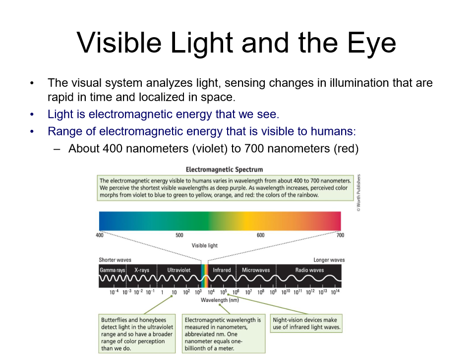
Visible light to the eye
we see from about 400 nanometers (violet) to 700 nm (red)
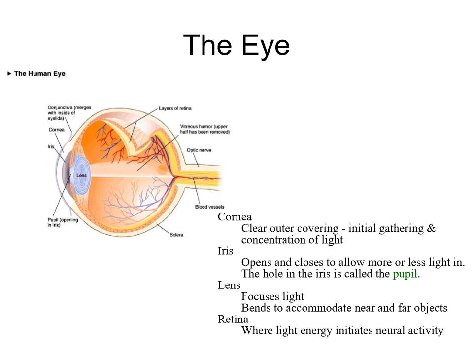
the eye 4 main parts
Cornea - the eye's clear, dome-shaped front surface, acting like a windshield to protect the eye and focus light, providing about two-thirds of the eye's focusing power, essential for sharp vision by directing light onto the retina
Iris - the colored, muscular diaphragm in the front of the eye that controls the size of the pupil, regulating the amount of light entering the eye for clear vision, functioning like a camera's aperture.
Lens- a clear, flexible structure behind the iris and pupil that focuses light onto the retina,
Retina - the light-sensitive tissue lining the back of the eye that converts light into neural signals,
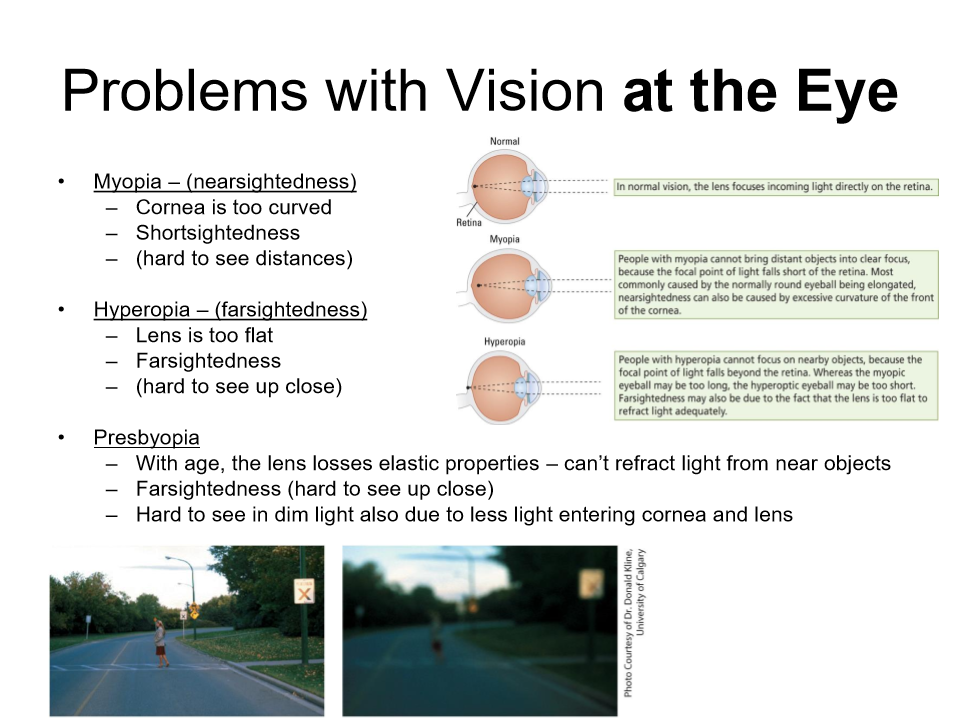
problems with vision at the eye
Myopia - (nearsightedness)
cornea is too curved
short sightedness, hard to see distances
Hyperopia - (farsightedness)
lens is too flat
farsightedness, hard to see up close
Presbyopia
with age the lens losses elastic properties, cant refract light from near objects
far sightedness
harder to see in dim light due to less light entering cornea and lens
Review
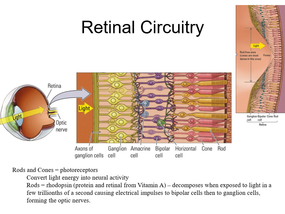
bipolar molecules go to ganglion cells create the optic nerve
ganglion cells transparent
Bipolar cells
A retina neuron - gets input from photoreceptors and creates action potentials in the 2nd layer of cells or ganglion cells
Horizontal cells
A type of retinal neuron which links photoreceptors and bipolar cells
Amacrine cells
A type of retinal neuron that links bipolar cells and ganglion cells
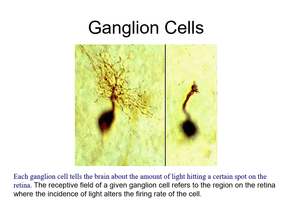
ganglion cells
final output neurons of the vertebrate retina, collecting visual information from bipolar and amacrine cells to transmit it to the brain via the optic nerve.
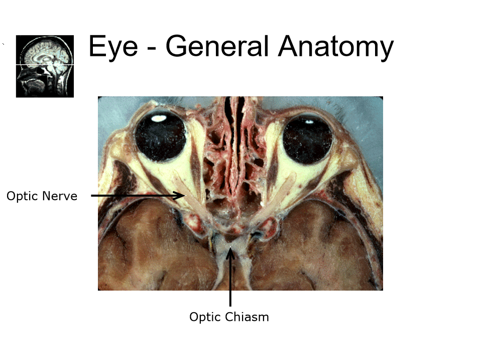
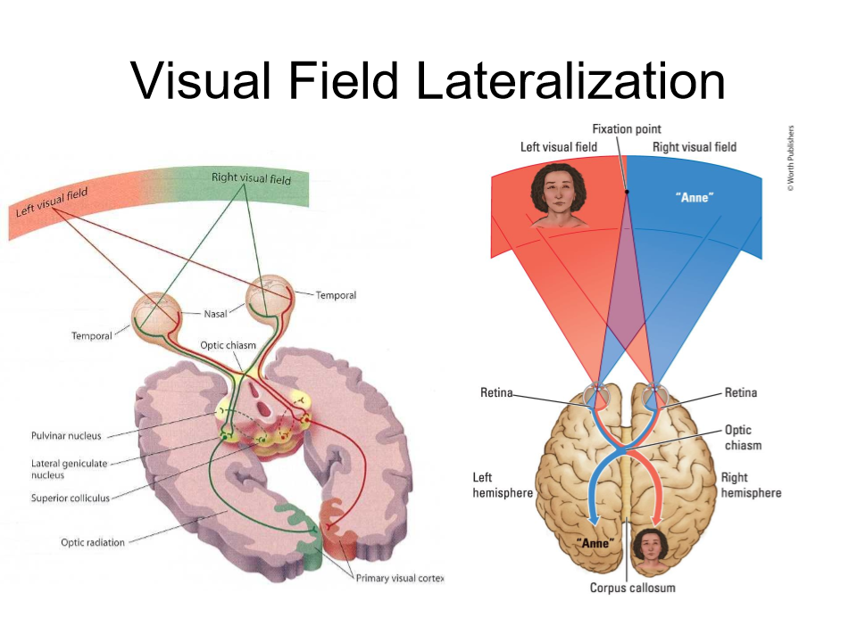
optic nerve and optic chiasm
cross over at the optic chiasm
The optic nerve (cranial nerve II) transmits visual information from the retina to the brain, while the optic chiasm, located at the base of the brain near the pituitary gland, is the X-shaped junction where medial (nasal) nerve fibers decussate (cross) to the opposite side.
REVIEW
REVIEW
2 types of ganglion cells
magnocellular cells (M-cell) , Parvocellular cells (P-cell)
M cell properties
large
receives input primarily from rods (no colour)
sensitive to light and moving stimuli
100,000 in periphery mostly for movement processing
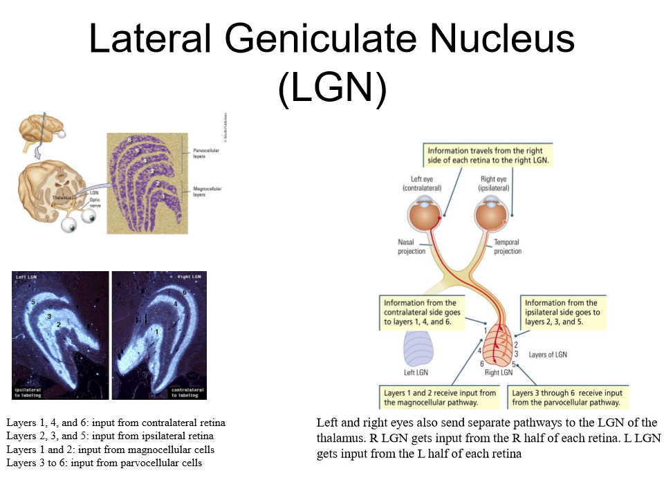
send projections to the magnocellular cells in the thalamus (layers 1 and 2 of the lateral geniculate nucleus LGN)
P cell properties
small
recieves unput from cones so sensitive to colour
1 million
mostly in fovea - fine details
sends projections to the parvocellular cells in the thalamus (layers 3-6 of the lateral geniculate nucleus.
Lateral Geniculate Nucleus
A paired, six-layered, bean-shaped relay station in the thalamus that acts as the primary gateway for visual information traveling from the retina to the primary visual cortex (V1).
6 layers m-cells and p-cells go to different layers
right eyes info goes to certain layers same with the left
very separated and organized well
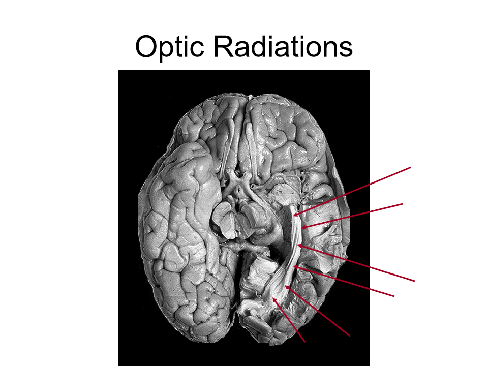
Optic radiations
group of axons that are organized and travel back to primary visual cortex
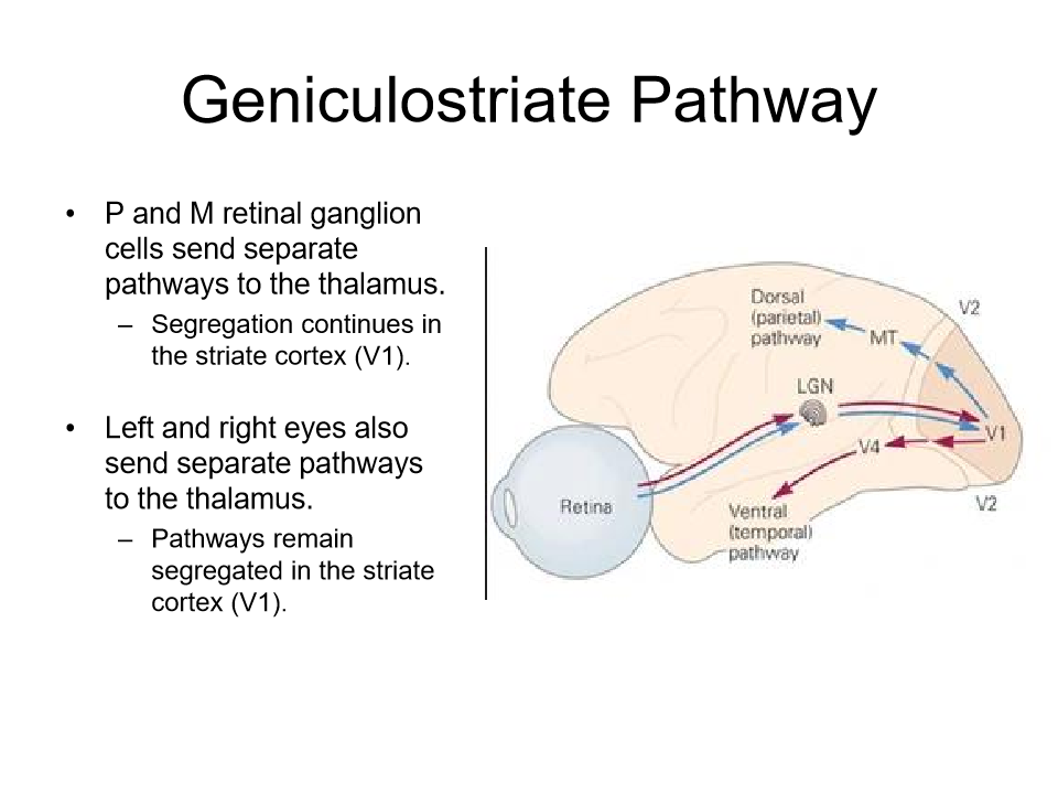
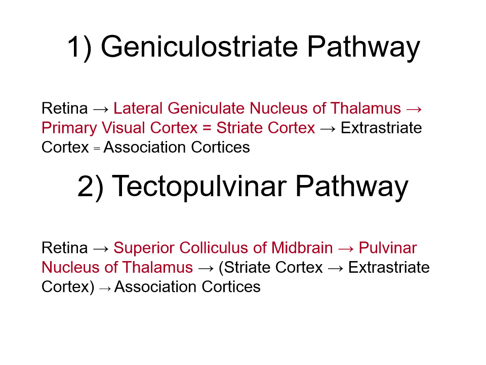
Geniculostriate pathway
starts in lateral geniculate nucleus thalamus to the striate cortex which is the visual cortex (V1)
mostly p cells (conscious visual processing pathway) more evolved
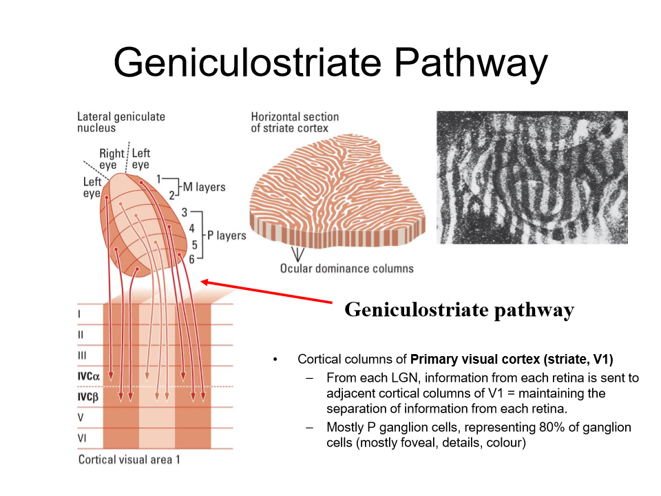
Review layer four is where visual info comes in
REVIEW
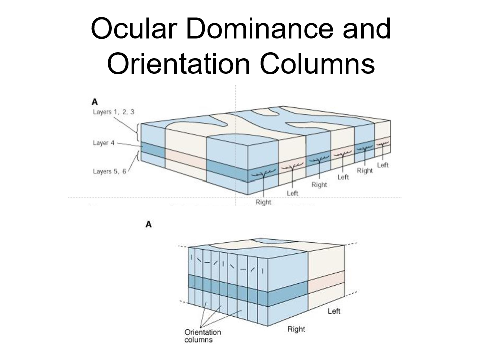
occular dominance columns
right info goes to orange part and left info goes to the light parts
review
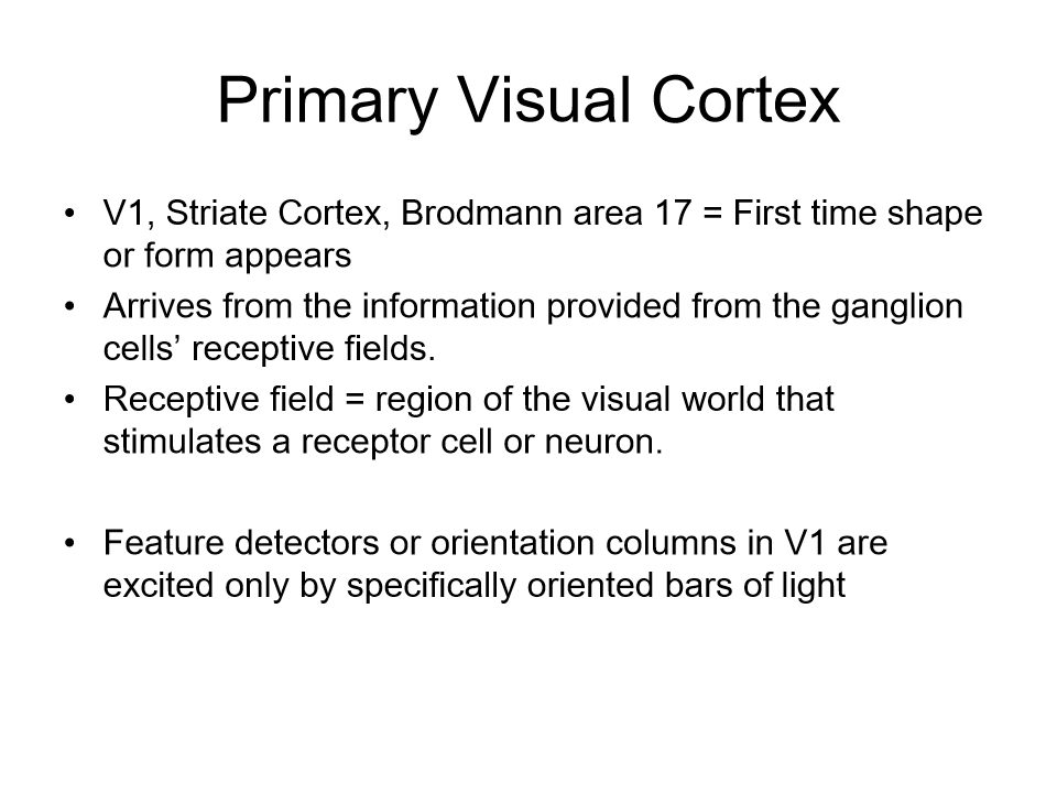
the V1 visual cortex striate cortex
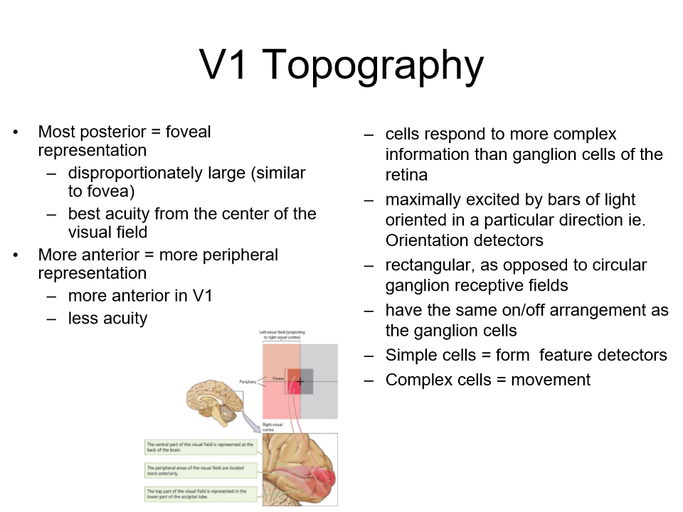
V1 Topography
most visual aquity will be in the foveal in the middle. Most posterior only edges and shadows demonstrated
More anterior = peripheral; representation
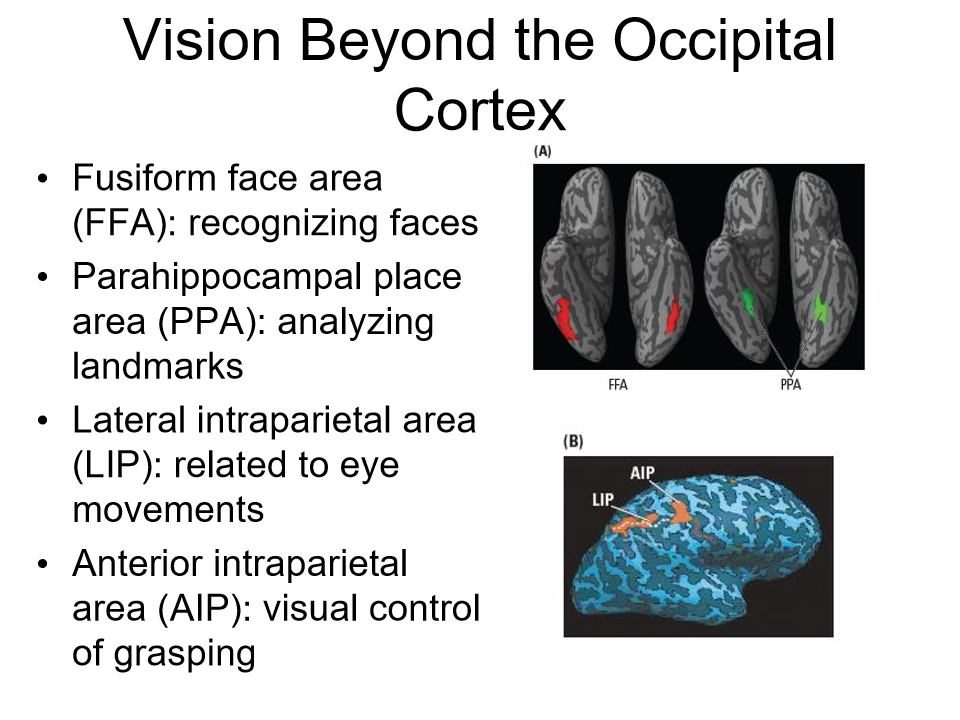
occipital cortex
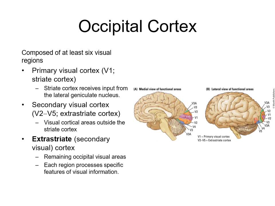
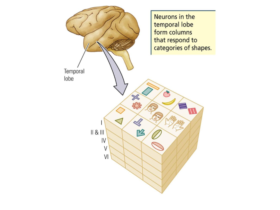
extrastriate cortex
more details in visual areas. V2, V3, V4 and V5
Review
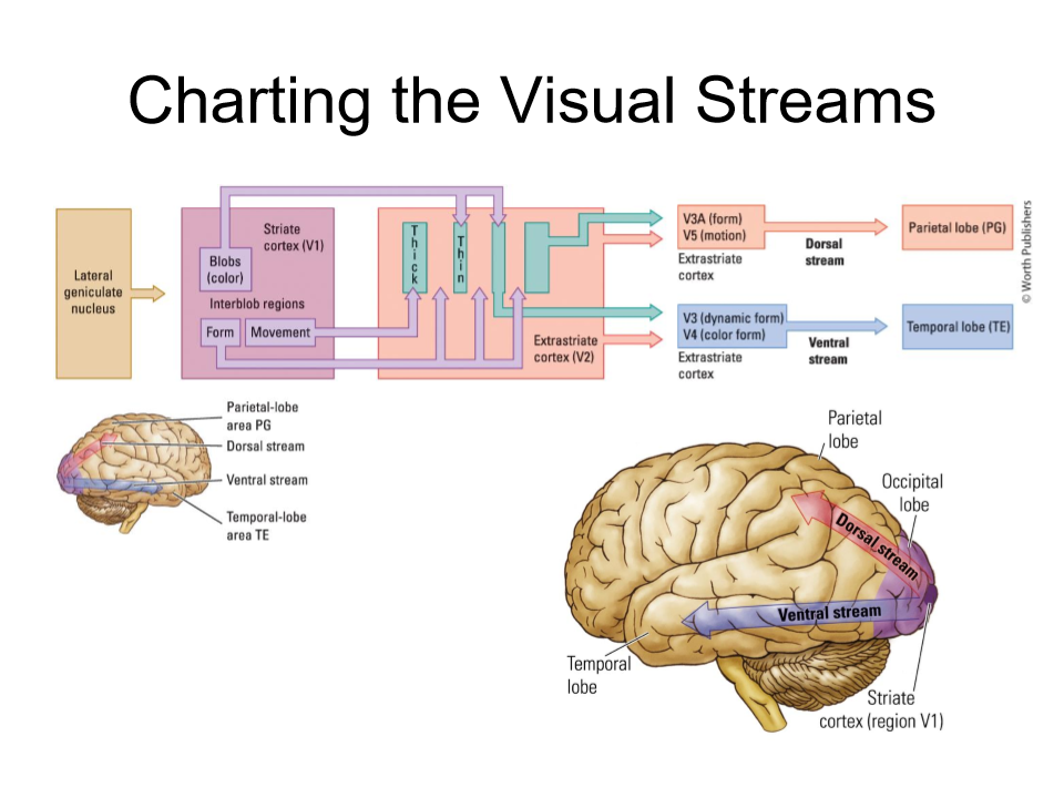
Ventral stream - object recognition (knowing the item)
dorsal stream - visual stream that allows you to pick up items and conduct movement
Theories of colour vision 1
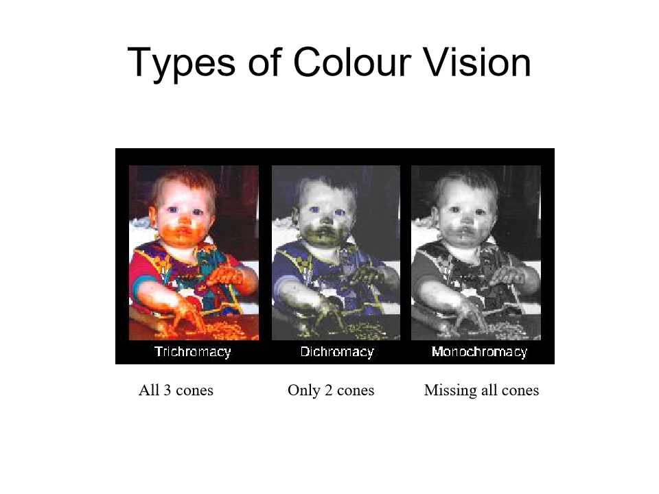
Trichromatic
genetics for colour blindness are on the x chromosome more common in men to have colourblindness and red and green
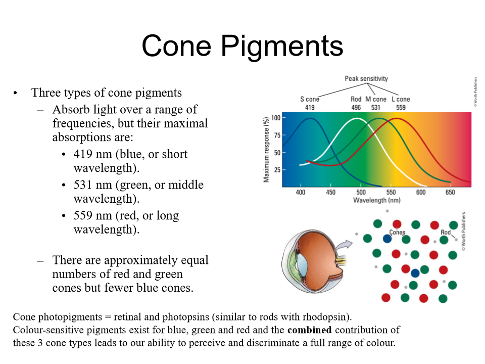
Three types of cones
red, green, blue cones more red and green cones than blue
cone pigments
Types of colour vision
complementary after images
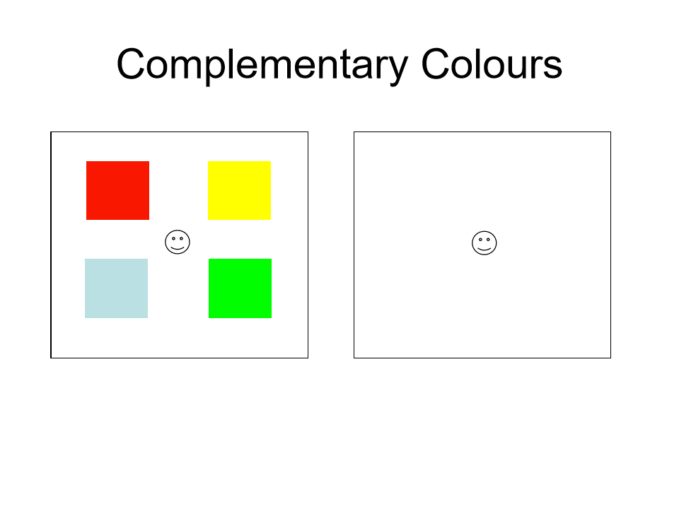
2 colour vision theory opponemt process theory
1) theory one that 3 types of cones allow us to see the world in all combinations if these wavelengths
2) opponent process theory in the retina on-off cells are specific to excititation for a colour and an inhibitory for another colour. explains opposit colours like red vs green
colour blobs in V1
Blob- region in the visual cortex that contains colour sensitive neurons
interblob- region between blobs that participates in the perception of form and motion
tectopulvinar pathways
very innate, M cells goes from retina to superior colliculus which sends info to the pulvinar region of the thalmus
medial pulvinar - sends connections to parietal lobe
Lateral Pulvinar- sends connections to temporal lobe
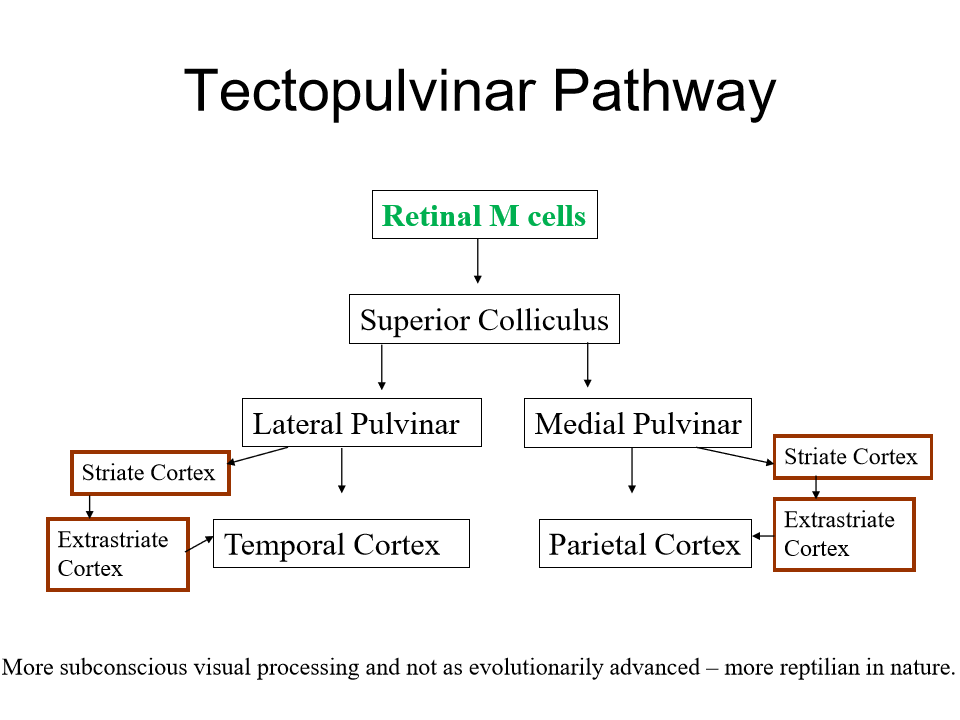
superior colliculus
where visual input enters
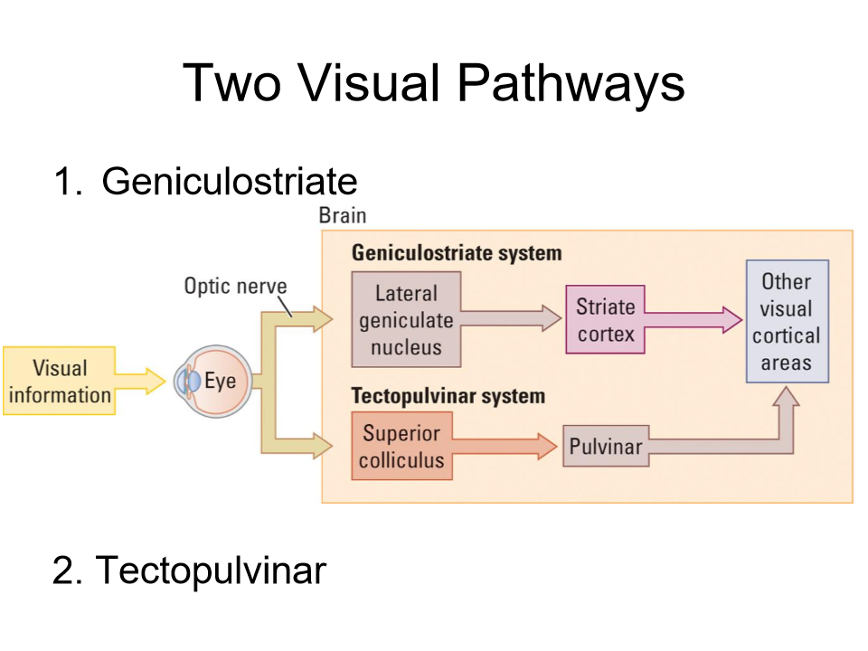
KNOW these pathways
Visual info- to eye and optic nerve- THEN there are 2 paths
1) GENICULOSTRIATE SYSTEM lateral geniculate nucleus - striate cortex, olther visual cortical areas
2) TECTOPULVINAR SYSTEM - superiour colliculus- pulvinar-other visual cortical areas
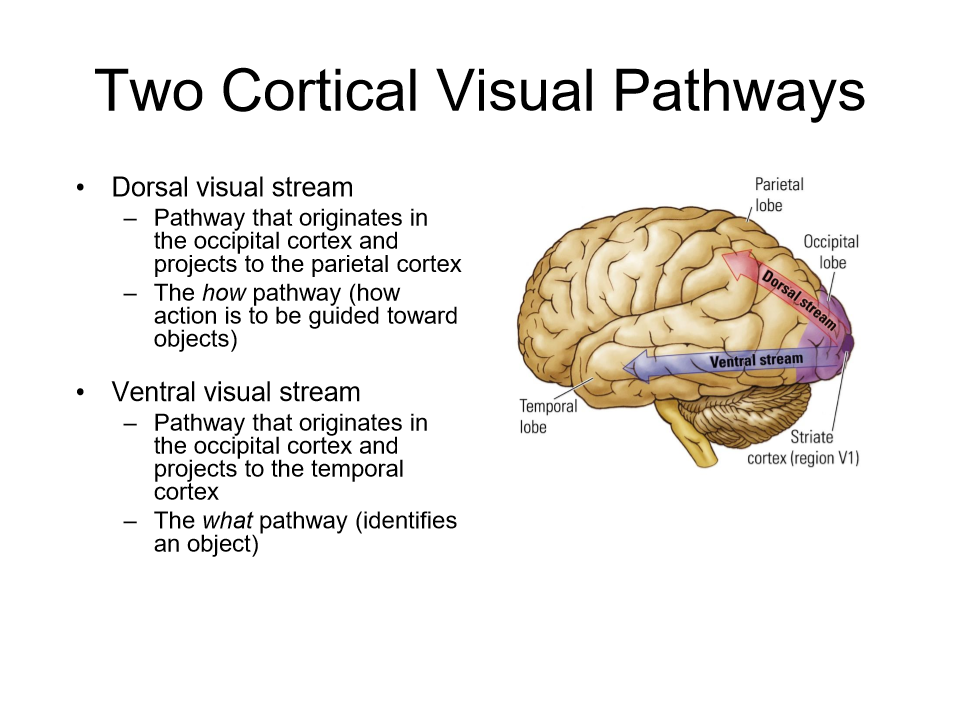
the two streams
dorsal the HOW pathways - starts in occipital cortex, projects to parietal cortex (how action is guided toward objects)
ventral the WHAT pathway - starts in occipital cortex projects to temporal cortex (identifies an object) jjjijujiujjujijijjijiuyjhuyjujujhyjilljklkjlklllkljlkllollkljjujyjhuyhujjhjhjuhuyjhjujuyhkjkjjkjkjuuyjhjkjjjjkjjijjkjjkjjujkjjkjjuyjhjuhhhuyjjuhyjhuyjuhyhjuyjhuyhjyujhuyhjyujhuyjhuyhjyujhuyjhuyujhuyhjuyjuhyjhuyhyjjuhyhjyuhyjujhuyhjuyjhuyhjyuhjuhyhyjuhyjujhuyhjyuhyujjhuyhjyuhyjuhjuyhjyuhyjhuyhyjuyjujhuyhyjujhuyhyjuhyjhuyuhyjujhuyhyjuhyjhjyyuhyjujhuyhjuyjhuyyhjyuyjujhuyhjuyjuhyhjyhyjuhyjyjhyhghyhyhghghyjyhujhyjuyhyhhyjhhyjhyhyjuhyhyhyjhyhhyhyjhyjhyjthyhyhyjhyhyjuhyhyhjuyhyjhyjjhyuhyjhyjjhuyhjyhyjjhuyjyhyjhjuyhyhyhyjuhyhyhjyuhyhjyuhyhyhyhyjhyhyhyjhhyjhhyjhyhyjjuhjjuhyjlkjkhjjhjlklkjjlklklkkllklllk
if you have damage to any part of the pathways
you will get different types of visual problems
Photoreceptors rods and cones
Light information travels
pupil- lens and cornea
retinal photoreceptors = rods and cones
retinal neurons = horizontal cells- bipolar cells - amacrine cells - ganglion cells m and p
optic chiasm - geniculostriate m and p or tectopulvinar (m) pathways
1) geniculostriate pathway
2) tectopulvinar pathway

Primary visual cortex
Cortical pathways
illusions
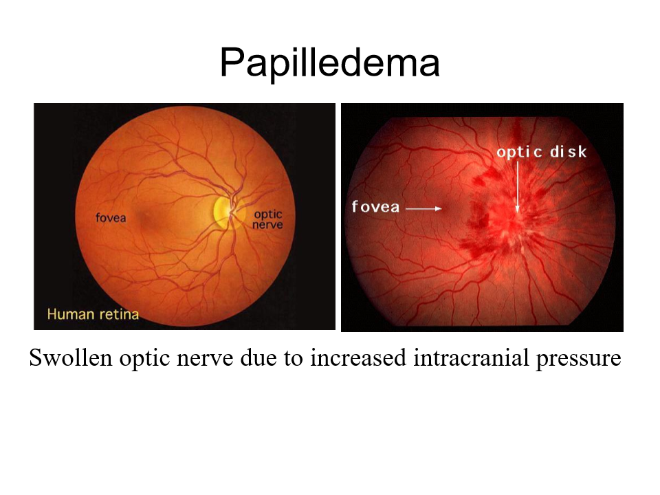
papilldema
swollen optic nerve, due to increased intracranial pressure. A symptom of something having gone wrong. One of most common symptoms signaling signs of MS, arterial malformation

Retinal Detachment
Very painful, retina comes off the back of the eye
Retinitus Pigmentosis
retina looks like a different colour

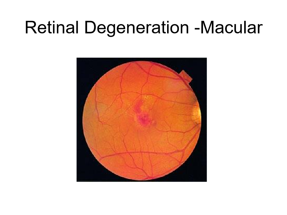
Retinal Degeneration - Macular
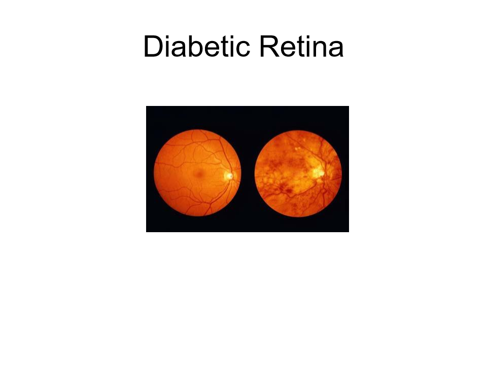
Diabetic Retina
Problems with Visual system
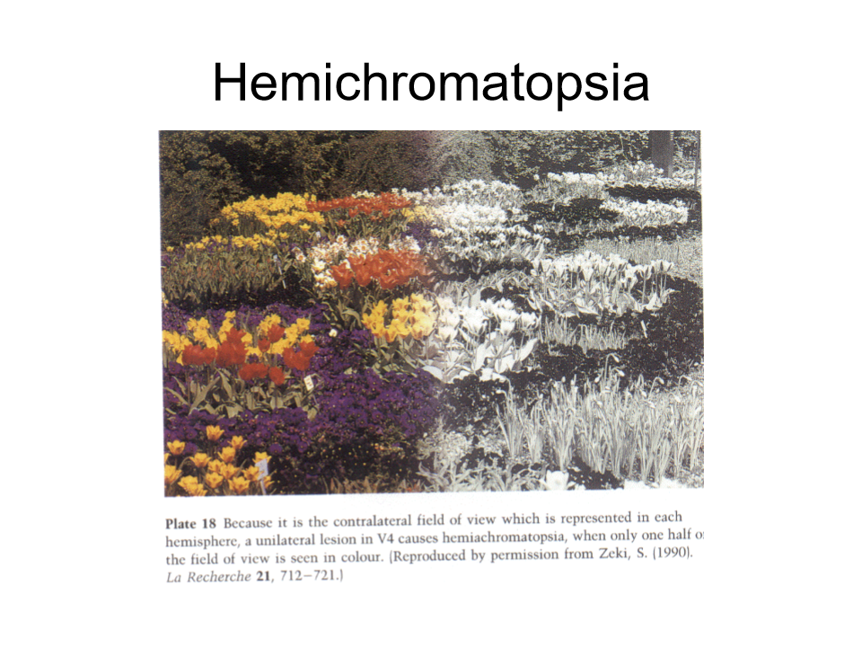
Hemichromatopsia
half the visual field is not observed in colour, extrastriate lesion V4.
Problems with visual system
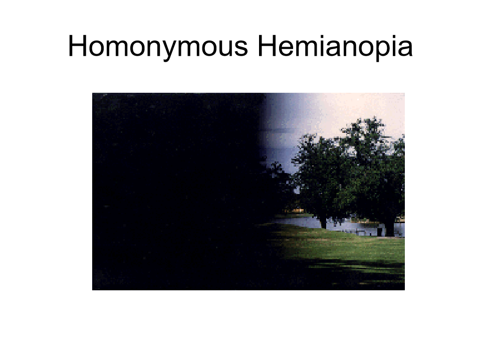
Homonymous Hemianopia
Nothing wrong with the persons eyes but the brain doesn’t display half of the visual field.
Where the injuries are to the brain
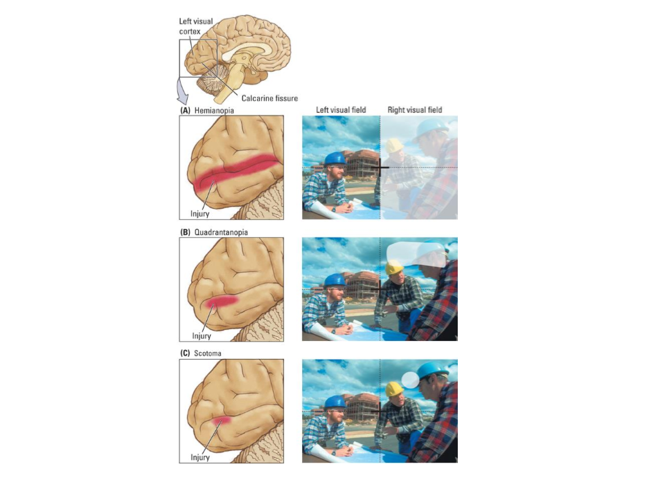
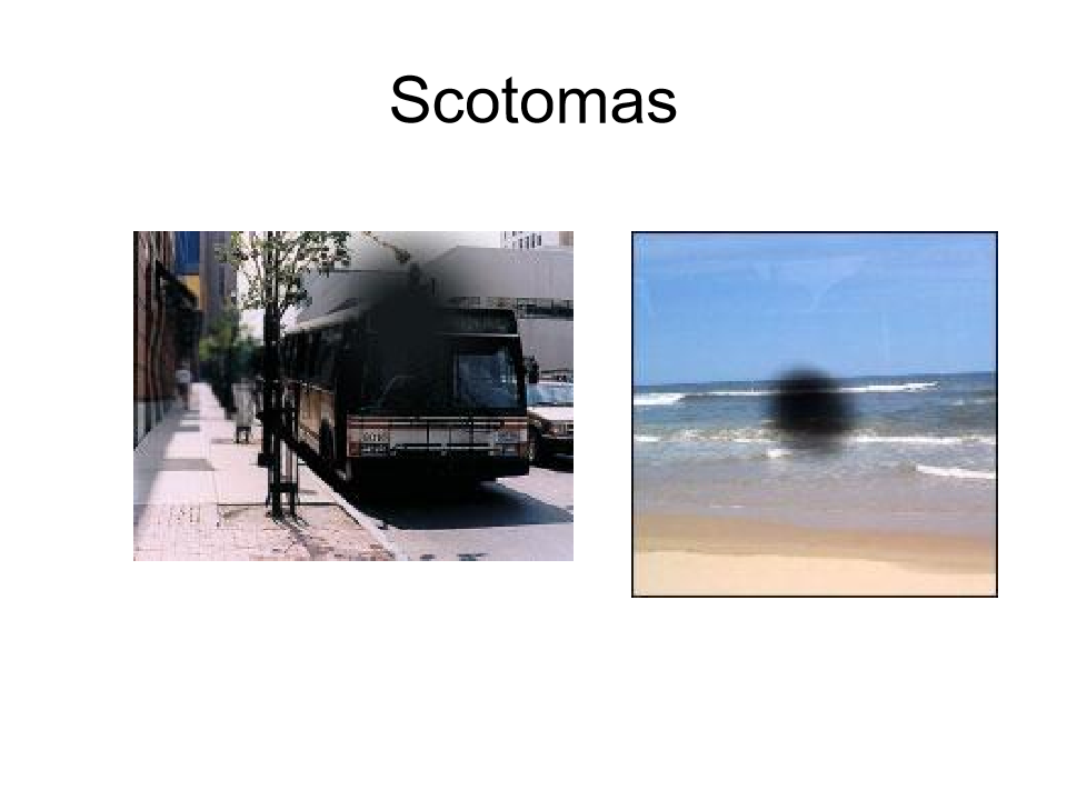
Scotomas
Achromatosia
seeing in black and white, no action potential happening,
optic ataxia
dorsal stream nit good, inability to move the hand to an object by using vision.
Balints Syndrome
Cant really reach out to objects or see more than one object at a time
photoreceptors
specialized cells in the eye's retina, primarily rods and cones
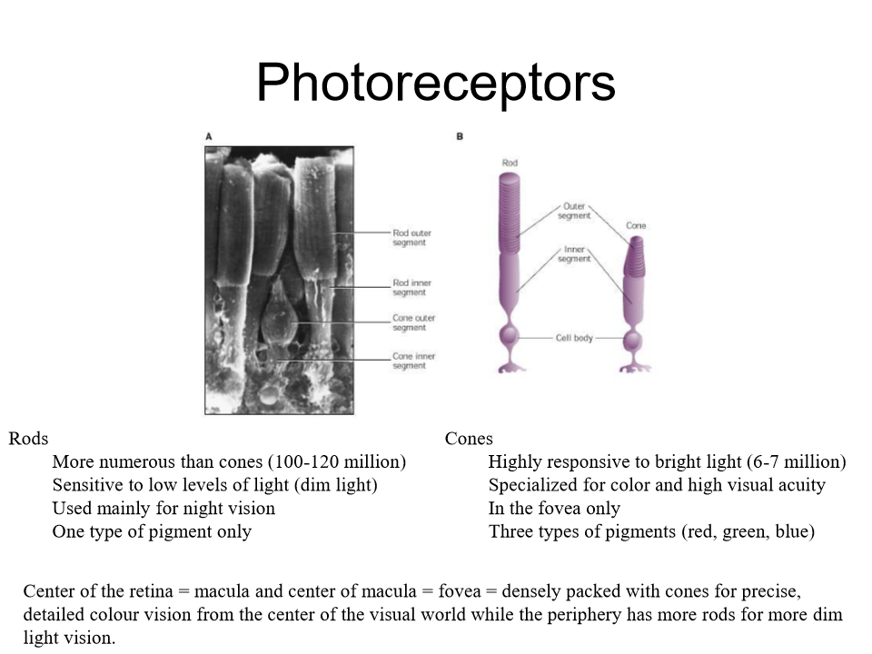
Rods
Rods - more numerous than cones (100-120 million) sensitive to low levels of light (dim) used mainly for night vision, one type of pigment only
Cones
Cones - highly responsive to bright lights 6-7million, specialized for colour and high visual aquity. In the fovea only. 3 types of pigment red blue green
Center of retina is called
Macula
Center of macula is
the fovea - densely packed with cones for precise detailed colour vision from center of the visual world while the periphery has more rods for more dim light vision