4b: Nervous System - Brain
1/74
There's no tags or description
Looks like no tags are added yet.
Name | Mastery | Learn | Test | Matching | Spaced | Call with Kai |
|---|
No analytics yet
Send a link to your students to track their progress
75 Terms
how many dura layers are in the brain? and spinal cord?
2 layers in the brain, 1 layer in spinal cord
which cranial meninge is the toughest layer? what are the 2 layers of this meninge called?
dura mater
periosteal (outer), meningeal (inner)
how is the dural venous sinuses formed in the brain?
from the separation of dura layers
what do the dural venous sinuses contain? where does it drain to?
collects venous blood and CSF
internal jugular veins via the arachnoid granulations
what separates the L/R hemisphere of the cerebrum? what about L/R hemispheres of cerebellum? what about separation cerebrum from cerebellum?
(meningeal layer of dura mater)
falx cerebri
falx cerebelli
tentorium cerebelli
what space holds CSF?
subarachnoid space and the 4 ventricles
what is the function of CSF?
bathes and protects the brain and spinal cord by absorbing shock
which meninge layer directly adheres to the brain and is the thinnest layer?
pia mater
what is the function of pia mater?
directly covers the brain, anchors blood vessels that supply the brain and spinal cord which prevent excessive mvmt and damage to brain
how many ventricles are there in the brain? name them
4
2 lateral ventricles (side horns), the 3rd ventricle (middle one with a hole), and 4th ventricle (triangular, superior end)
what connects the lateral ventricles to the 3rd ventricle
interventricular foramen
what connects the 3rd ventricle to the 4th ventricle
cerebral aqueduct
where does the CSF flow through after the 4th ventricle
central canal of spinal cord
what structure in the ventricles produce CSF? where can you find these structures?
the choroid plexus
there is a choroid plexus in each of the ventricles
describe the flow of CSF starting from lateral plexus until the heart/lungs (include the ducts)
lateral ventricles > (interventricular foramen) > 3rd ventricle > (cerebral aqueduct > 4th ventricle > central canal OR sub arachnoid space (via lateral aperture) > subarchnoid granulations > drain into dural venous sinus > drains into internal jugular vein > back to heart/lungs > gets oxygenated > back to ventricles
how does CSF get to subarachnoid space and into internal jugular vein?
from 4th ventricle > subarachnoid space/arachnoid granulations > dural venous sinuses > internal jugular vein
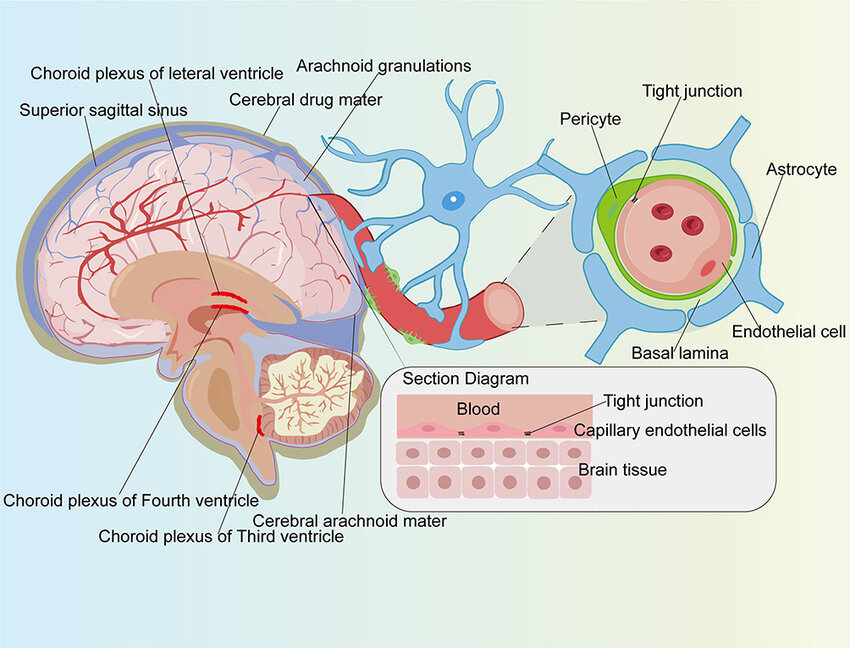
what creates the blood-brain-barrier? what exactly is it
the tight junctions of the capillary’s endothelial cells (between blood vessels and the brain)
what makes the tight junctions so tight in the BBB?
astrocytes (glial cell - supporting cell of neurons)
- they push up against the capillaries
what substances are allowed to pass through the BBB?
there are quite a lot actually
lipid soluble substances: O2, H2O, Co2, alcohol, barbiturates (depressants/sedative), nicotine, caffeine
glucose via facilitated diffusion
some ions can diffuse very slowly
what kind of substances are not allowed to pass thru BBB?
proteins, most anti-biotics, pathogens
which part of the brain is the major relay station between sensory and motor signals? and plays a role in maintaining consciousness?
thalamus
which part of the brain regulates homeostasis?
hypothalamus
how does the hypothalamus regulate homeostasis? (6)
communicates with autonomic NS: (heart rate, smooth muscle for stomach, urinary bladder contractions, glands)
produces hormones
regulates emotional and behaviour patterns with limbic system (emotional expression and sexual arousal)
regulates eating and drinking (hunger, satiated, thirsty)
regulates body temperature
regulates circadian rhythm
which hormones does the hypothalamus have an effect on via the pituitary gland
releasing hormones, inhibiting hormones, oxytocin
which part of the brain is the “emotional brain”? what is included in this system ?
limbic system: amygdala, hippocampus, thalamus, hypothalamus, cingulate gyrus
what part of the brain plays a role in emotions, memory, motivation, and smell?
for example: when you smell a nice cinnamon candle it provokes a certain feeling/memory
limbic system
what part of the brain is in control of the start/stop of movement + start/stop of cognitive processes (ie: attention, memory, planning) + autonomic subconscious mvmts (ie: arm swings when walking)
basal nuclei aka corpus striatum
what is makes up the basal nuclei/corpus striatum?
caudata nucleus, putamen, globus pallidus
which part of the brain controls for coordination during movement, balance, and posture? how does it do this?
cerebellum
-evaluates voluntary mvmts and controls for subconscious muscle
how does the cerebellum attach to the brainstem? what is contained in this?
cerebellar peduncles: axons that connect cerebellum to brainstem and cortex (used for coordination and balance)
what is contained in the midbrain of brainstem?
cerebral peduncles, tectum, substantia nigra, red nuclei
what is the function of the midbrain’s cerebral peduncles?
axons of motor pathway
what is the function of the midbrain’s tectum? give an example
reflex center for audio & visuals, and startle reflex
ex: sudden mvmt when you’re surprised by unexpected presence
what is the function of the midbrain’s substantia nigra?
release dopamine and help control subconscious muscle
what happens if there is a loss of neurons in the substantia nigra (midbrain)?
parkinson’s disease
what is the function of the midbrain’s red nuclei?
this is the synapse spot for the cerebral cortex and cerebellum that controls for mvmt
what part of the brainstem coordinates voluntary motor output and breathing?
pons
which part of the brainstem contains the axons that connect L/R of the cerebellum AND connects the cortex to cerebellum (contralaterally)?
pons
what part of the brain stem regulates heart beat and normal breathing? and autonomic reflexes of: swallowing, vomiting, sneezing, coughing and hiccuping
medulla oblongata
what 2 things are part of the medulla oblongata?
pyramids and olive
what is the “pyramid” in the medulla oblongata?
decussation of cortico-spinal tracts (crossing over)
→ tracts (axons) that control for voluntary mvmt of the limbs and trunk
what is the “olive” in the medulla oblongata?
communicates with cerebellum to provide instructions when learning new motor skills → adjusts movements
the cerebellum communicates with what part of the brain stem when leanring new motor skills?
olive part of the medulla oblongata
which brain lobe is in charge of executive functioning, motor control, personality
frontal lobe
which brain lobe is in charge of sensory perception and integration?
parietal lobe
which brain lobe is in charge of auditory processing, memory, and emotion?
temporal lobe
which brain lobe is in charge of visual processing?
occipital lobe
identify these structures in the picture:
central sulcus
longitudinal fissure
pre-central gyrus
post-central gyrus
where do you find the primary MOTOR cortex?
pre-central gyrus
where do you find the primary somato-sensory cortex?
post central gyrus
what is an association area?
area that combines motor and sensory info to make sense of things
somatosensory association area recognizes what
recognizes objects just by touching it
(can have eyes closed and hold something and know what that object is)
which lobe stores facial recognition?
inferior, temporal lobe
what does the orbito-frontal lobe cortex recognize? where is this area?
discriminate among different odours,
lateral frontal lobe
which association area allows for the formation of thoughts based on all incoming sensory inputs? where is this area
common integrative around the parietal lobe
(sensory, visual, auditory info)
what is the pre-frontal cortex in charge of?
higher cognitive functioning: personality, intellect, complex learning, recall info, initiative, judgement, reasoning, planning, conscience, mood
where is wernicke’s area and what is it for?
language comprehension (ability to understand what is being said)
temporal/parietal lobe
dominant in left hemisphere
where is broca’s area and what is it for?
speech production (ability to speak)
frontal lobe, anteriorly
dominant in left hemisphere
think: broca’s = speech = closer to tongue = anterior frontal lobe
name the 3 spaces in the spinal cord
epidural space, subdural space, subarachnoid space
what attaches the spinal cord to the arachnoid and dura mater in order to protect the spinal cord from shock/displacement? hint: it’s part of the meninges mater
denticulate ligaments (extensions of pia mater)
why is there a cervical and lumbo-sacral enlargement in the spinal cord?
these have the nerves that innervate the upper and lower limbs
what is the difference between conus medularis, cauda equina, and filum terminale in the spinal cord?
conus medularis: end of the cord (around L1)
cauda equina: horse’s tail, nerves of lumbar, sacral, and coccygeal regions
filum terminale: the final end of the cord, attaches to the coccyx
the dorsal root ganglion contains what kind of cell bodies ?
sensory neuron cell bodies
motor neurons are in the ___ nerve root
sensory neurons are in the ___ nerve root
motor: anterior/ventral
sensory: posterior/dorsal
gray mater and white mater contains what part of the nueron
gray mater: cell bodies
white mater: myelinated axons
where would you find the cell bodies (gray mater) of autonomic motor neurons in the spinal cord? < this would be the pre-ganglionic neuron
lateral gray horn
what is meant by “tract” in the spinal cord
a tract is a bundle of myelinated axons
“track” = road = highway of neurons
what information does the anterior and lateral spinothalamic tracts carry to the cns?
pain, temperature, itch, tickle (the “uncomfortable” feelings)
describe the anterior and lateral spinothalamic tracts
tracts = white mater = axons
spine → thalamus = ascending pathway = sensory neurons
anterior and lateral spinal column
what information does the posterior column-medial lemniscus tracts carry? is this an ascending or descending pathway?
info about touch, pressure, vibration, proprioception
ascending pathway
describe the anterior and lateral corticospinal tracts
tracts = white mater = myelinated axons
cortico-spinal = descending pathway = motor neurons
anterior and lateral white column on spinal cord
what do the anterior and lateral corticospinal tracts innervate?
somatic/voluntary skeletal muscle
what makes a reflex a spinal reflex
when synapse happens only within the spinal cord
define these components of a spinal reflex:
sensory receptor
sensory neuron
integrating center
motor neuron
effector
sensory receptor: detects stimuli
sensory neuron: sends signal from receptor to spinal cord
integrating center: synapse between sensory neuron to motor nueron (may contain an interneuron)
motor neuron: sends impulse from spinal cord down to effector muscle
effector: muscle that responds
give an example for each component of a spinal reflex for touching a hot object:
sensory receptor
sensory neuron
integrating center: this is a polysynaptic reflex
motor neuron
effector
sensory receptor: pain receptors in skin
sensory neuron
integrating center: interneuron
motor neuron
effector: finger/arm muscles = move hand away