exam 1 medchem class 4
1/21
There's no tags or description
Looks like no tags are added yet.
Name | Mastery | Learn | Test | Matching | Spaced | Call with Kai |
|---|
No analytics yet
Send a link to your students to track their progress
22 Terms
what happens after GPCR is activated
G-protein trimer (has alpha, gamma, beta subunits) relays the signal: then secondary messengers are activated (eg cAMP, Ca2+, PI) which lead to diverse biological responses
downstream response options after GPCR
short term responses from protein kinases, immediate early gene mRNA after transcription, immediate early gene protein after translation, dimerization and post-translational modification, long-term gene responses
how does the g-protein trimer relay the signal
alpha subunit can activate target protein, activated beta-gamma complex can open a K+ channel, alpha subunit can stop the signal also (with intrinsic GTPase activity- by hydrolysing its bound GTP to GDP, inactivating and reassociating with the beta-gamma dimer)
g protein cycle
basal state - agonist binding leads to association and GTP-GDP exchange which - leads to dissociation where the alpha and beta-gamma subunits are effectors - then GTPase activity of alpha subunit returns it to basal state
Galpha-s mediated signaling
adenylate cyclase system - activates adenylyl cyclase - cAMP - PKA - multiple physiological effects - inc. smooth muscle relaxation
bonds that histamine can have
Ionic w/ NH3+ they become protonated
Can form an amino compound w/ negative charge aa (aspartame and glutamate)
N and HN can do H bonds
Aromaticity can do van der waals bonding
what are actions of cAMP
cell-type dependent, depend on different PKA protein substrates
eg of physiological actions of cAMP following GCPR activation
adrenaline increases heart rate
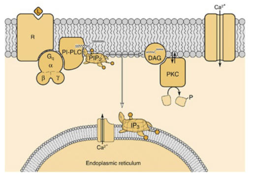
Galpha-q mediated signalling expl
Once Gq protein gets activated
Alpha subunit is activating enzyme - PI-PLC - Is breaking down a phospholipid
PIP2 - polar head of phospholipid, part of membrane
Very polar head that has been cleaved off
Now have IP3
Rest of phospholipid - DAG -
Activates protein kinase (PKC)
Phosphorylation of proteins
Goes on till Gq protein stops being activated
How can you measure PLC activation?
via Ca2+ levels inside the cell
3 notes of intracellular Ca2+
very important 2nd messenger in cells, many proteins sensitive to its concentration in the cytoplasm, its concentration is tightly regulated
example of an important Ca2+ sensitive protein
Calmodulin - can bind Ca2+ and then change conformation and eg bind target proteins
CaM-kinase dependent NO synthesis (4 steps)
Ach binds muscarinine receptor (GPCR) - signalling via PLC gives Ca2+, Ca2+ binds calmoduline, calmoduline activates CaM-kinase, CaM-kinase activates NO synthase, etc.
molecular basis of sight
chromophore changes upon illumination - GPCR is the mediator from proton to “molecular movement”
from photon to electric signal
membrane outer segment has cation selective ion channel - open in the dark, Na+ influx, depends on cGMP levels; proton inhibits Na+ influx (activation of cGMP-specific PDE via G protein, membrane hyperpolarization); hyperpolarization transferred to synaps
how do we see different colors
3 kinds of cones - diff proteins
how to do signal termination with a: transmitter
breakdown/re-uptake
how to do signal termination with a: g protein
intrinsic GTPase activity
how to do signal termination with a: second messenger
breakdown
how to do signal termination with a: protein
dephosphorylation
how to do signal termination with a: receptor
regulation of function and localization
GC and AT pairing in DNA - how many bonds, which
GC - 3 H bonds
AT - 2 H bonds