Para: Exam 4 Labs & Kahoot
1/100
There's no tags or description
Looks like no tags are added yet.
Name | Mastery | Learn | Test | Matching | Spaced | Call with Kai |
|---|
No analytics yet
Send a link to your students to track their progress
101 Terms
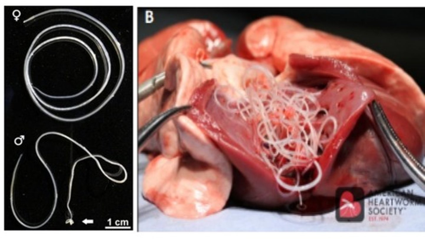
Males, females
Dirofilaria immitis Adults
-The _____ are shorter & their tail is coiled
-The ______ are longer & their tail is straight.
*Infects canids (DH) with mosquitoes as IH
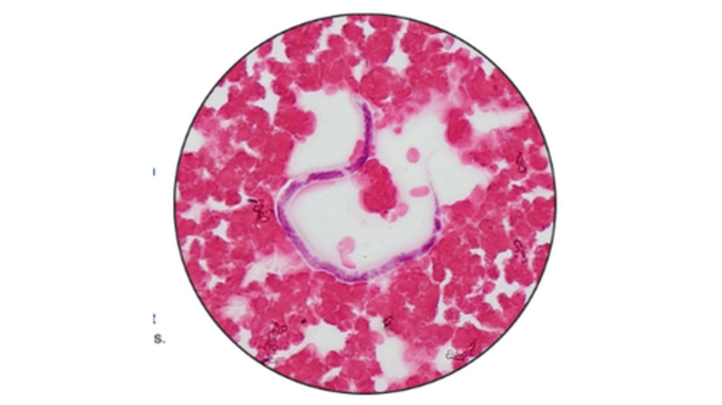
Microfiliariae
Stage of Dirofiliria immitis that are released from adult females & circulate in the blood
*Can be detected in blood smears from an infected canine host
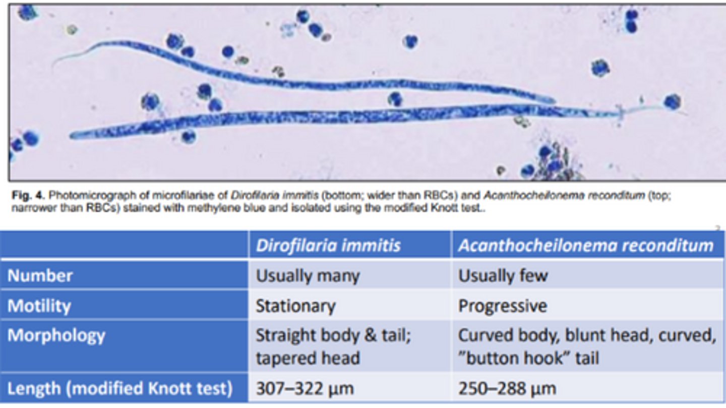
Acanthocheilonema reconditum
Does the following describe Dirofilaria immitis & acanthocheilonema reconditum?
-Curved body, blunt head, curved, "button hook" tail
-Progressive motility
-Usually few in #
-Narrower than RBC

Dirofilaria immitis
Does the following describe Dirofilaria immitis & acanthocheilonema reconditum?
-Straight body & tail, tapered head
-Stationary motility
-Usually many in # in infections
-Wider than RBCS

Modified Knott test
Microfilariae concentration test that's recommended by for
evaluating morphology to differentiate D. immitis from Acanthocheilonema reconditum
SNAP test
ELISA that detects Heartworm antigen (antigen-capture)
-Can provide somewhat quantitative results
Onchocerca cervicalis
Filariid that infects equids (DH) with biting midges as the IH
-Adults (slender & white) are located in the nuchal ligament
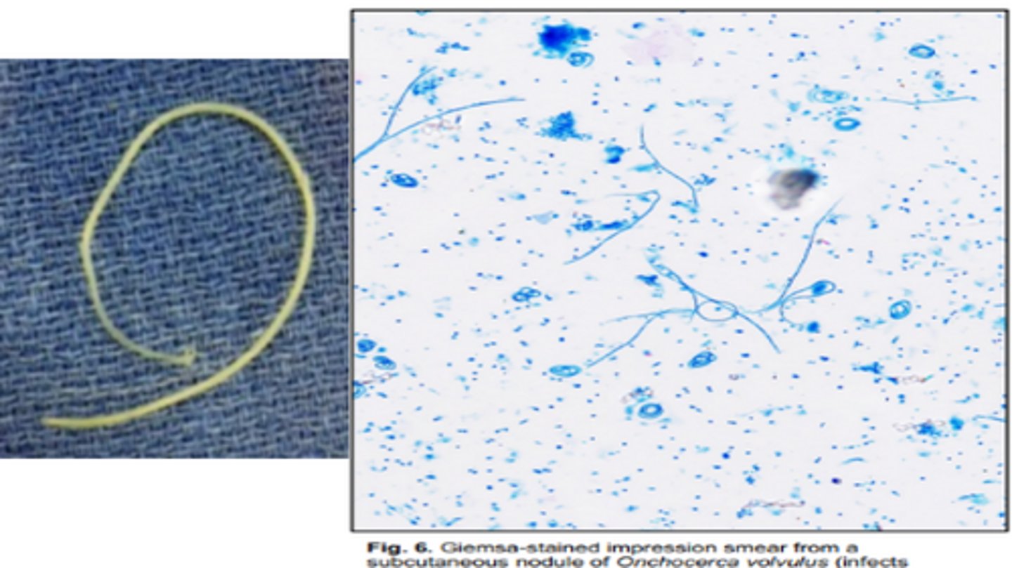
Onchocerca cervicalis
Filiariid in equine whose microfiliariae can be found in the ocular conjunctiva, dermis, & connective tissues

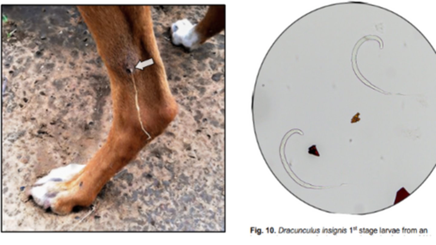
Dracunculus insignis
Guinea worm that infects canids (mainly) & cats with copepods being the IH
-Long, slender & white adult females often protrude from nodular lesions of subcutaneous tissue of limbs and L1s are released into water
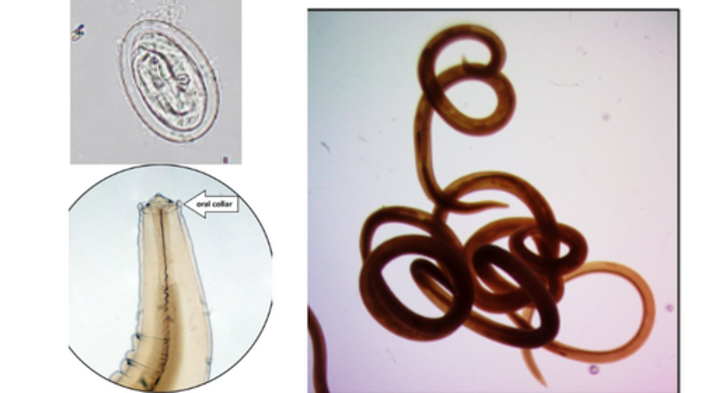
Physaloptera
Nematode that infects canids (DH), cats, racoons, etc.
-Have a prominent cuticular collar (cephalic alae) around the mouth
-Adults may be expelled in vomitus or embedded in stomach mucosa at necropsy

Physaloptera
Nematode in dogs & cats with thick shelled eggs that contain L1 larvae
-Length of eggs is 2x their width
*Best recovered using a fecal sedimentation technique due to their high density; adults found on endoscopy or in vomit
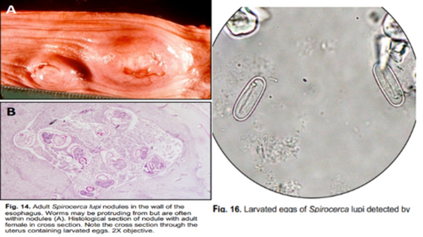
Spirocerca lupi
Nematode that infects the esophagus of canids (mainly) & felines
-IH = coprophagous beetles
-Migrating juveniles may be found within nodules in the wall of the aorta
Draschia, Habronema
Stomach nodule that infects equids (DH) with musca domestica (House fly) & Stomoxys calcitrans (stable fly)
-Adults form nodules in the stomach
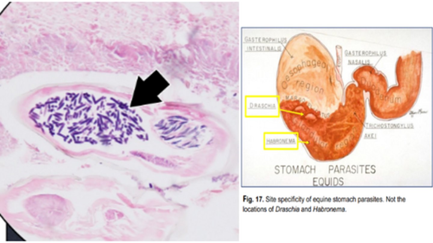
L3
_____ (stage larvae) of Draschia & Habronema spp. are deposited by flies on various parts of the body & can be associated with summer sores (cutaneous habronemiasis).

Margo plicatus, glandular stomach
Nematode Nodules in Equine Stomach Vary by Location:
-Draschia forms in the _____ _____.
-Habronema forms in the ____ _____.

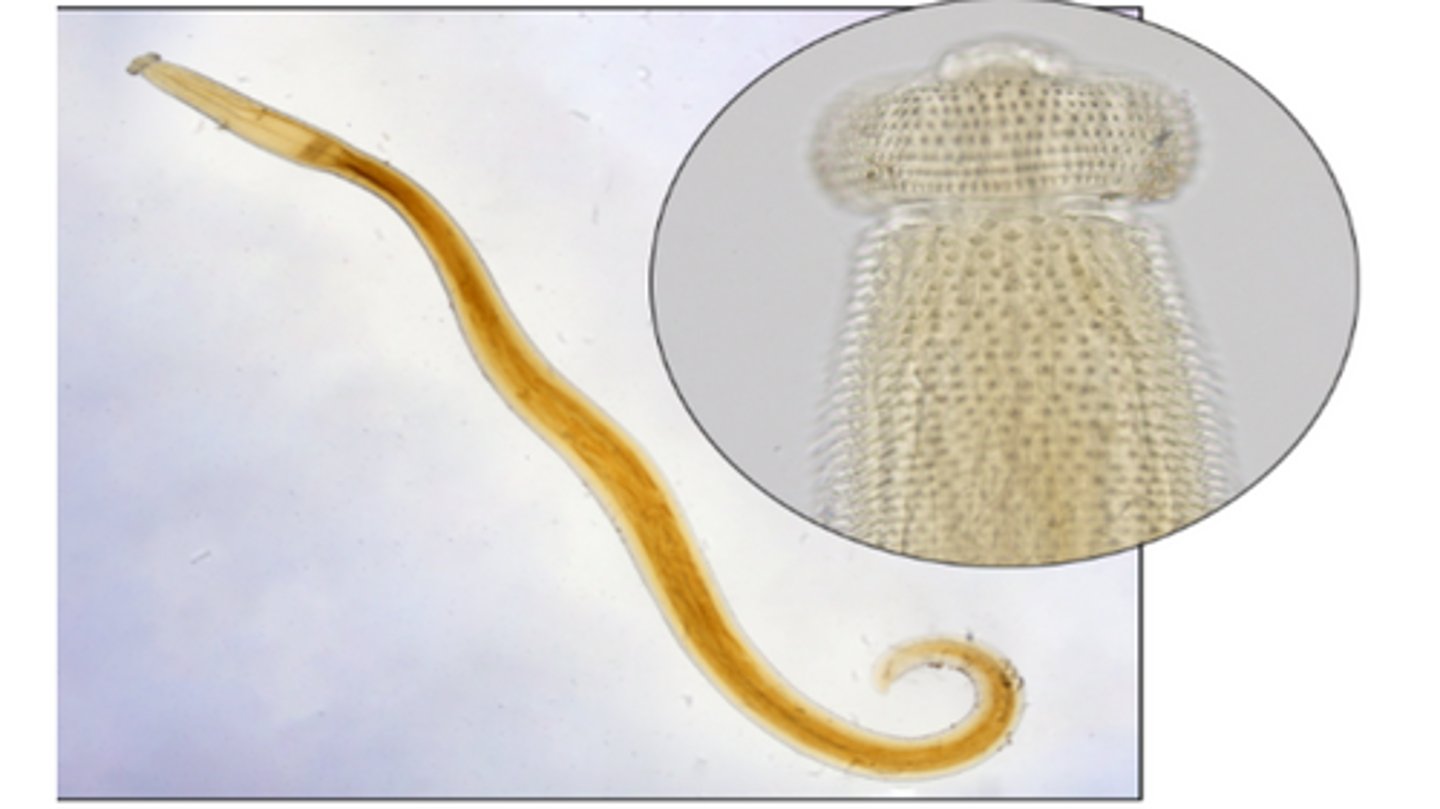
Oxyuris equi
Equine pinworms that infect the colon
-Adults females are stout with a large esophagus that has a prominent esophageal bulb; may be passed in the feces
-Eggs are laid in the perineal/anal skin area
Scotch tape method
Best method for detecting Oxyuris equi eggs (ovoid, yellowish, thick shelled, flattened, operculated)

Setaria equina
Nonpathogenic filariid that infects equids (DH) with mosquitoes (IH)
-Adults are large, long, & white that are found in the peritoneal cavity & pleural cavity
-Microfilariae are shed by adult females & found in the blood

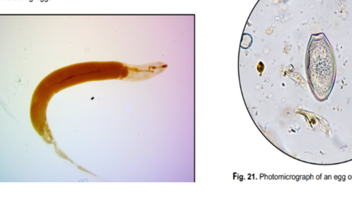
Gnathostoma
Nematodes that infects the stomach of mammals (including humans)
-1st IH: freshwater copepods
-2nd IH: Freshwater fish & amphibians
-Adults & larvae have cuticular spines along the anterior portion of the body with a prominent cephalic bulb
-Unembryonated, operculated eggs are passed in feces
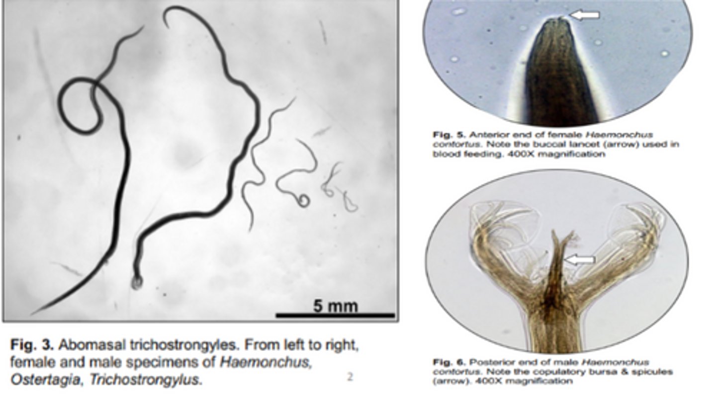
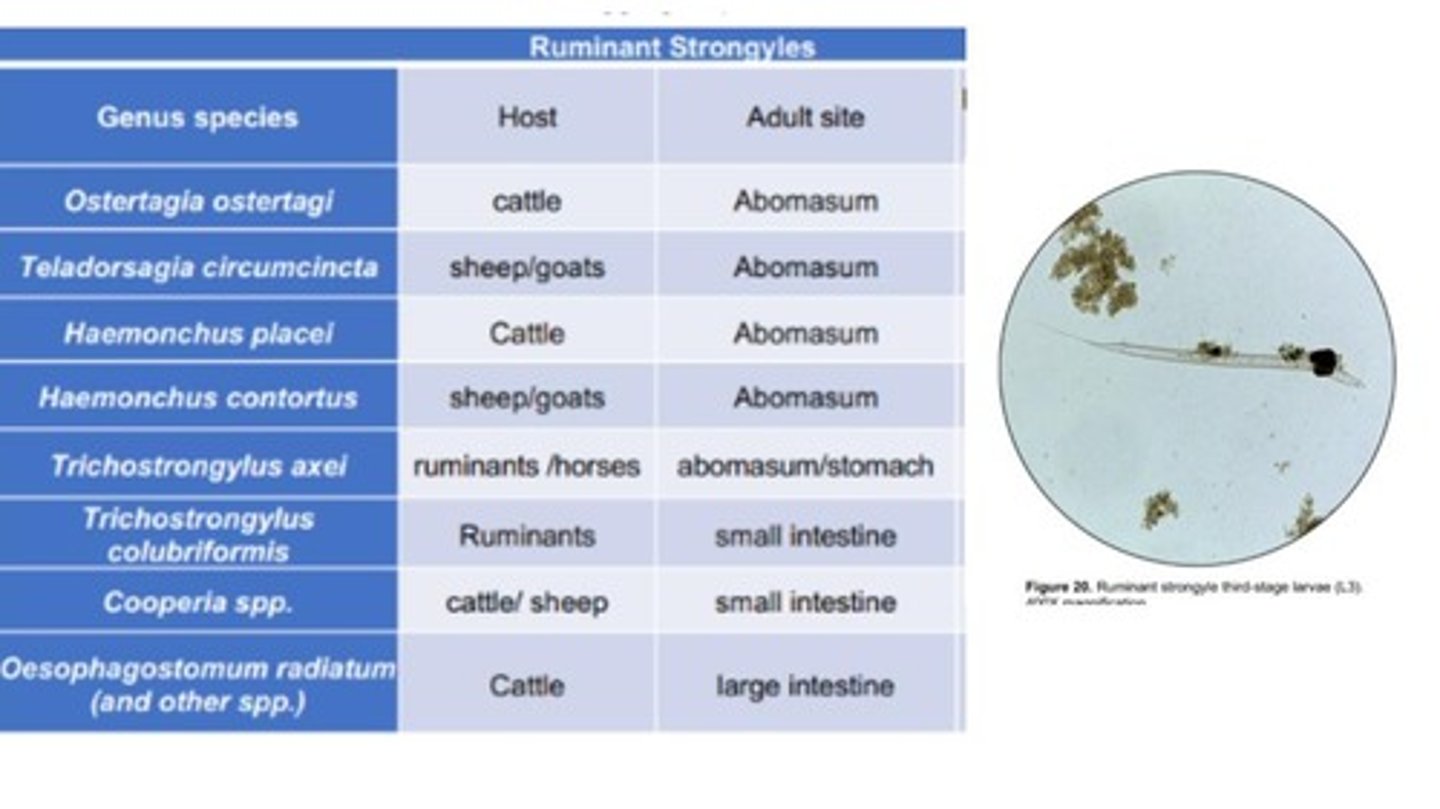
Haemonchus
Largest of the abomasal trichostrongyle
-Small buccal cavity
-Common name: Barber pole (female worms coil/wrap their uterus/ovaries around their intestine)
Contortus, Placei
Haemonchus Species:
-_____ = infects small ruminants
-_____ = infects cattle

FAMACHA
Eye chart is used only for assessing the severity of H. contortus-induced anemia in small ruminants & camelids
Adults, L4
What stages of Haemonchus contortus are aggressive blood feeders that can cause anemia in small ruminants?

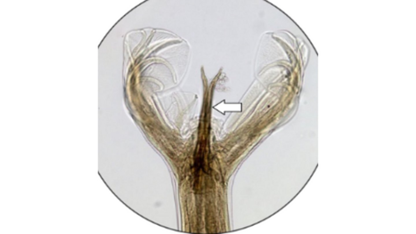
Haemonchus contortus
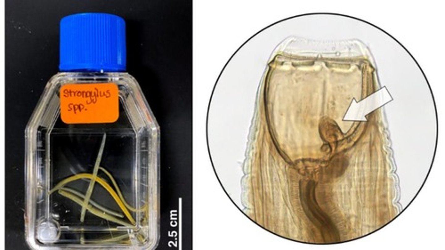
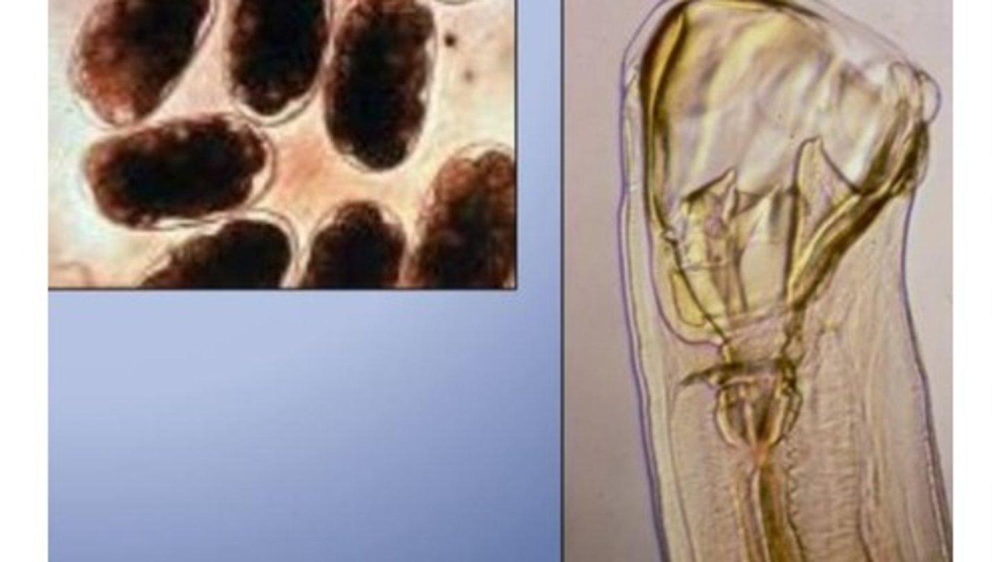
What abomasal, male nematode is this based on its copulatory bursa and spicules (arrow)?
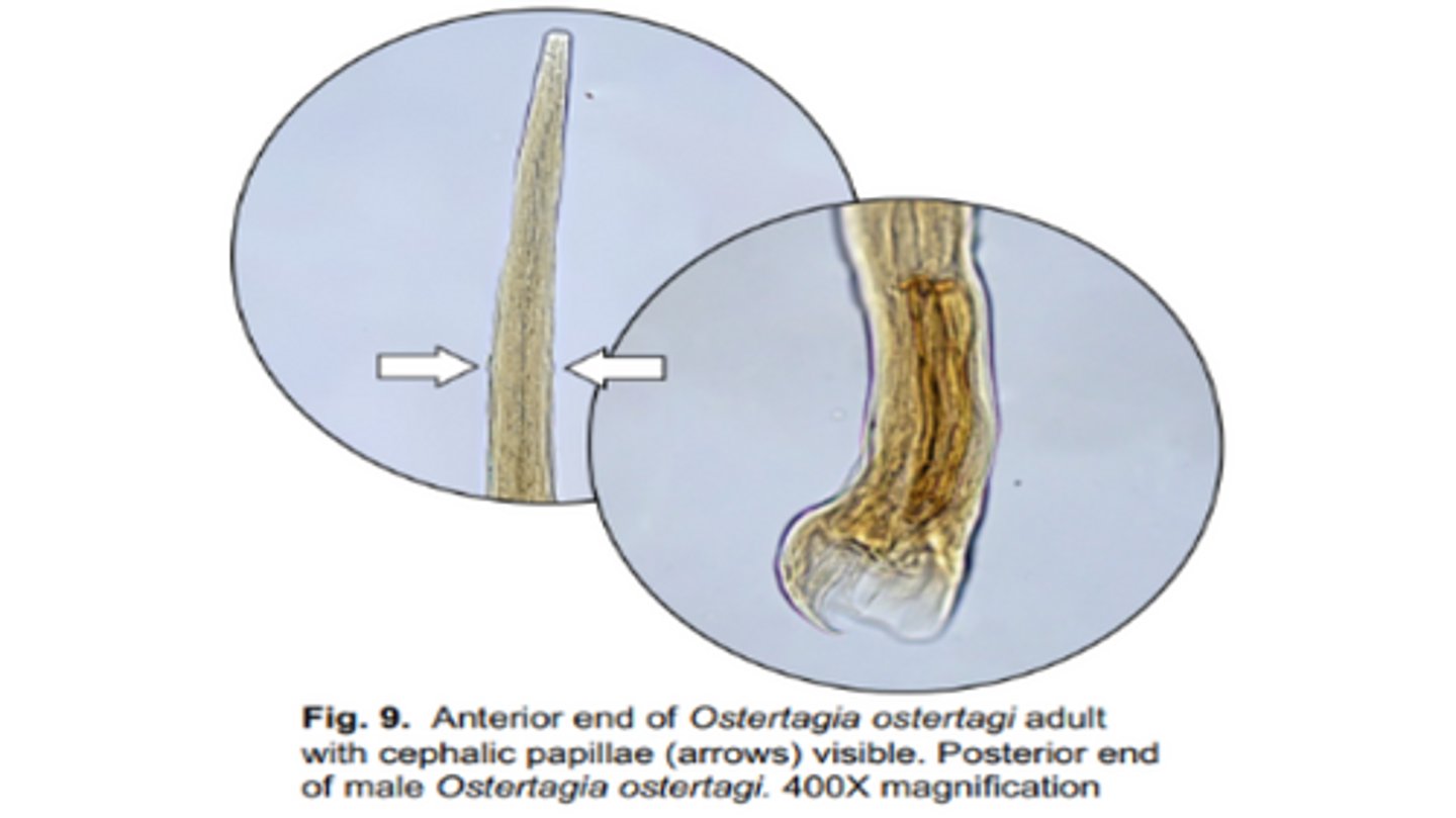
Ostertagia ostertagi
Causes cobblestone like lesions on the mucosal surface of the abomasum in cattle (DH)
-Tiny buccal cavity with cephalic papillae
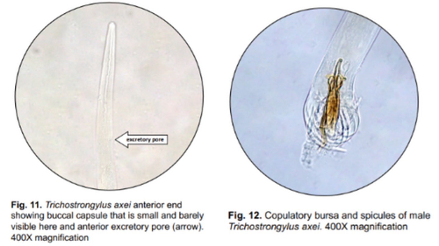
Trichostrongylus
Smallest abomasal nematode that infects large/small ruminants & horses
*Have a small buccal capsule

Colubriformis, Axei
Trichostrongylus Species:
-T. _____ = major species found in the SI of small ruminants
-T. ____ = infects abomasum of cattle & stomach of horses
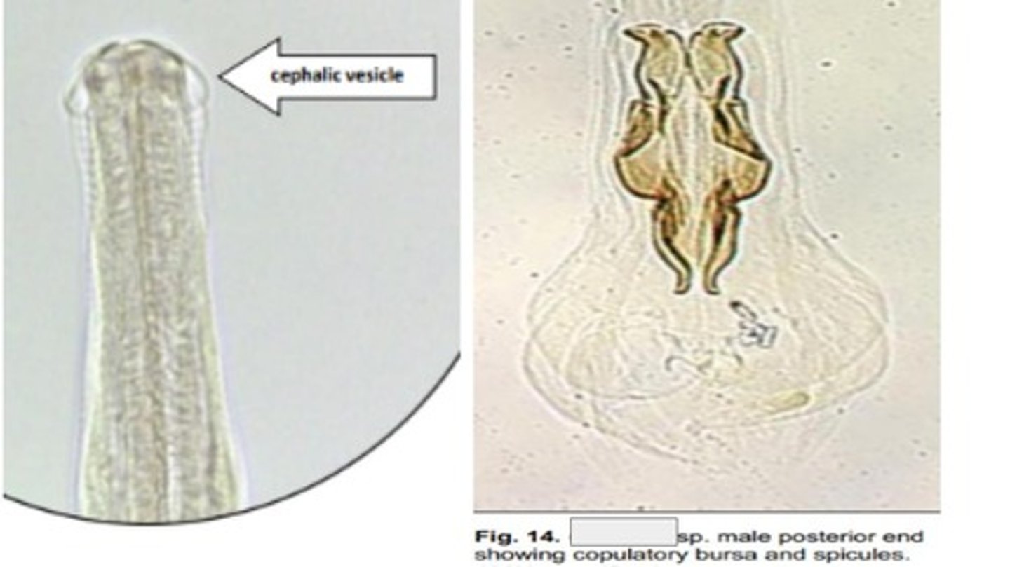
Cooperia
Ruminant strongyle with a characteristic cephalic vesicle/swelling at its anterior end
-Found in the SI of large & small ruminants
-Similar in size to Trichostrongylus spp.
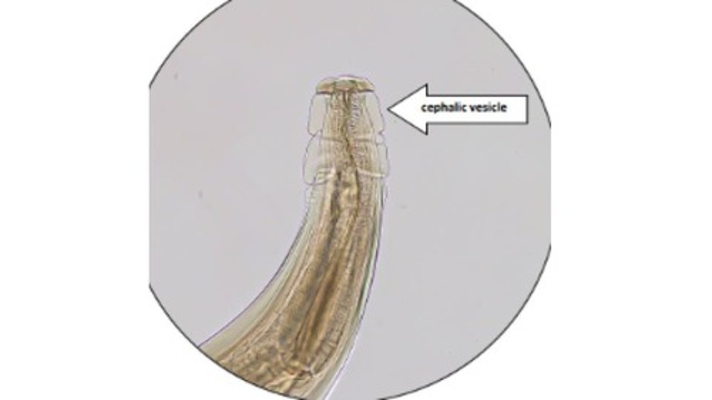
Oesophagostomum
Large bursate ruminant strongyle that forms nodules in large intestine of small & large ruminants
-AKA nodular worm
*Characteristic cephalic vesicle surrounding a cylindrical buccal capsule

Columbianum, venulosum
Oesophagostomum Species:
-O. _____= occurs in sheep & goats
-O. ______ = occurs in cattle, sheep, & goats
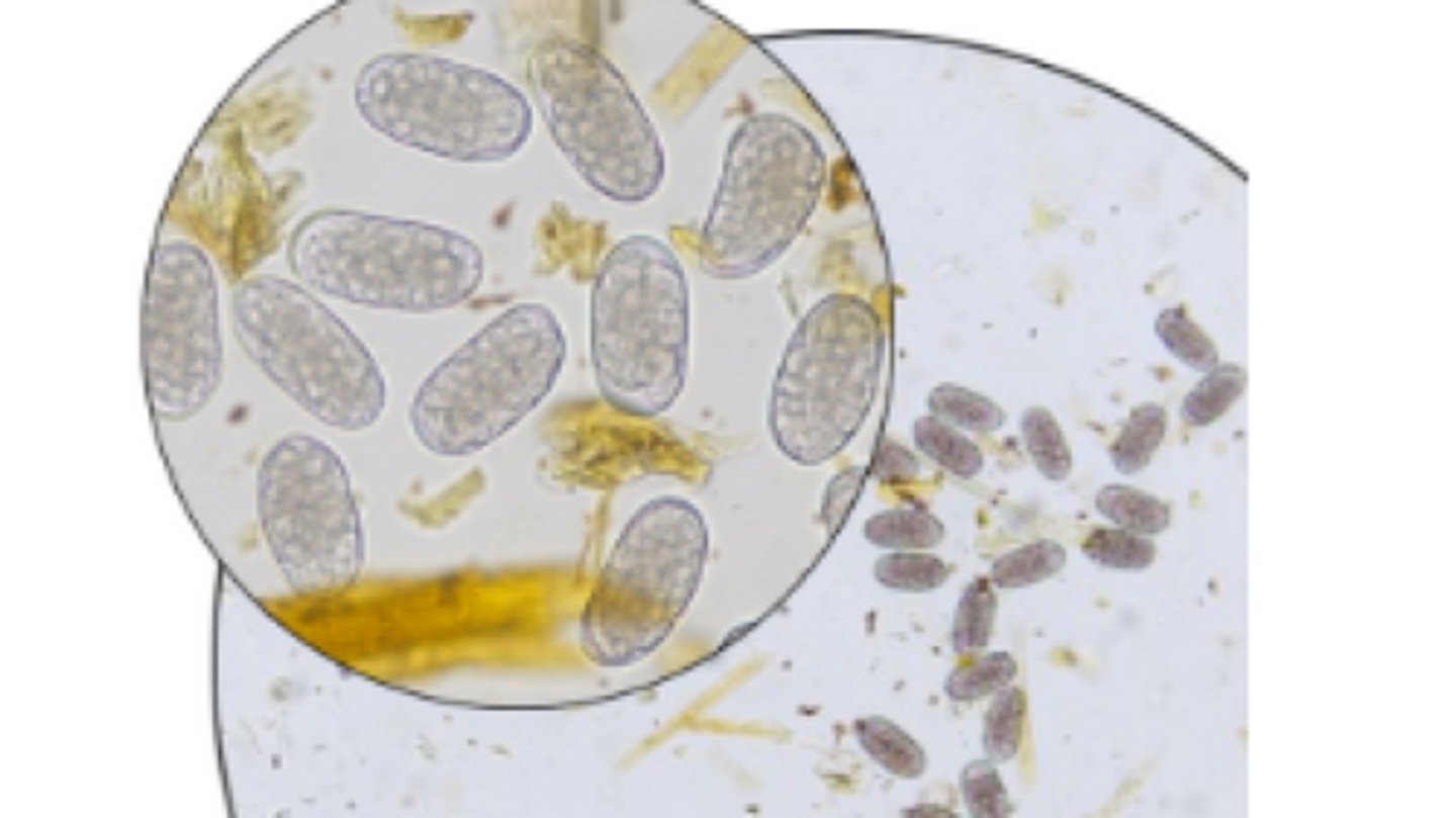
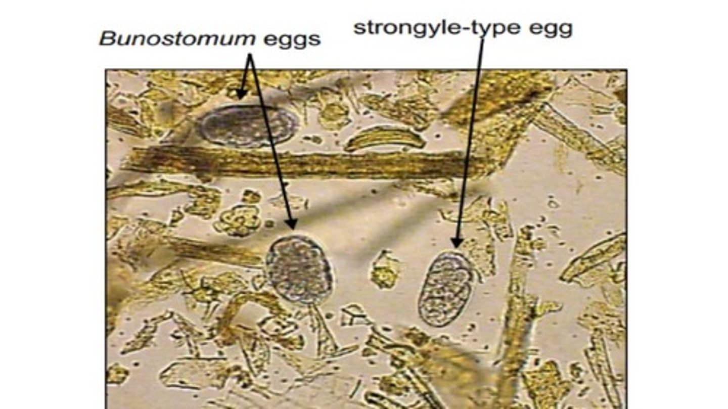
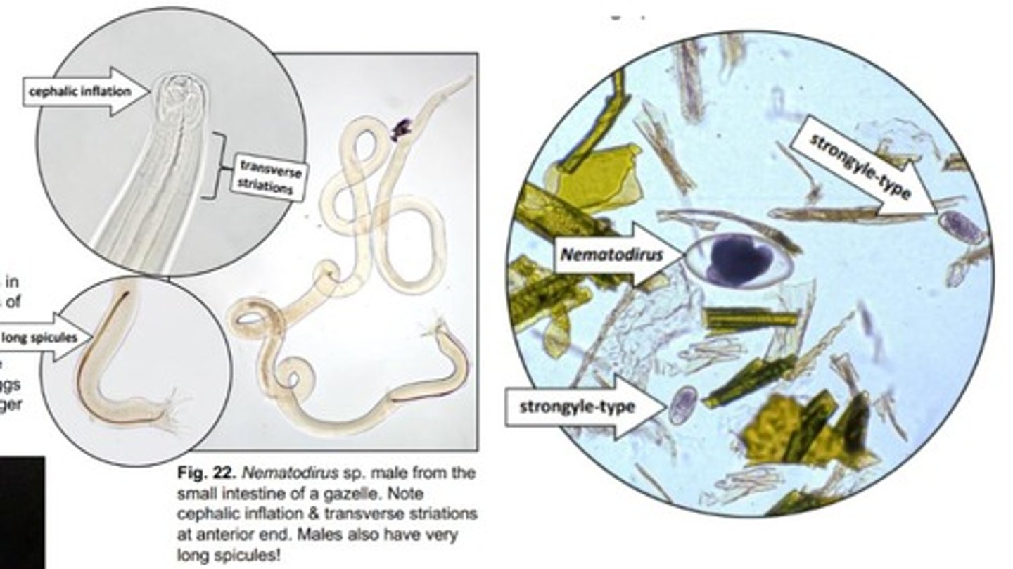
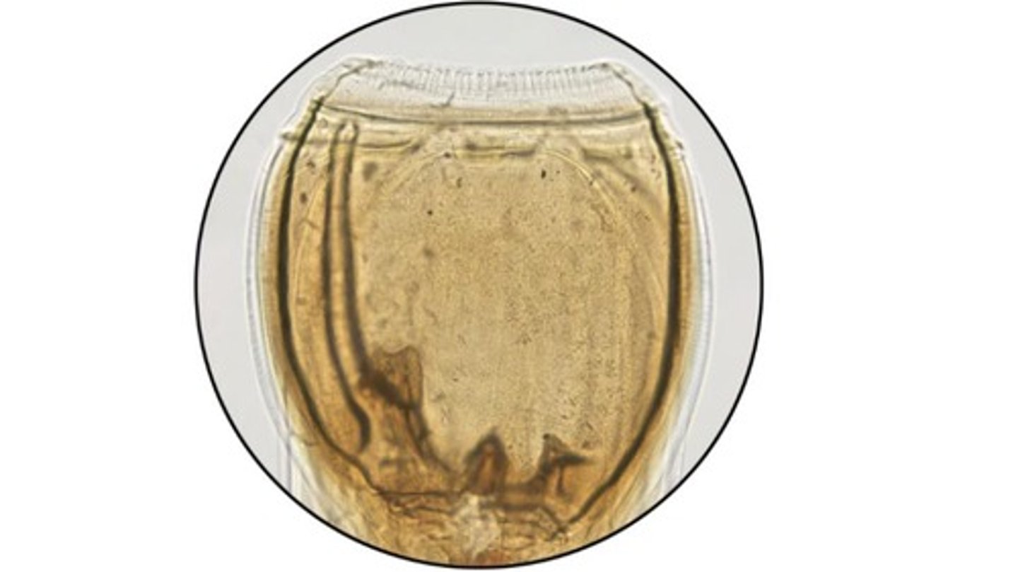
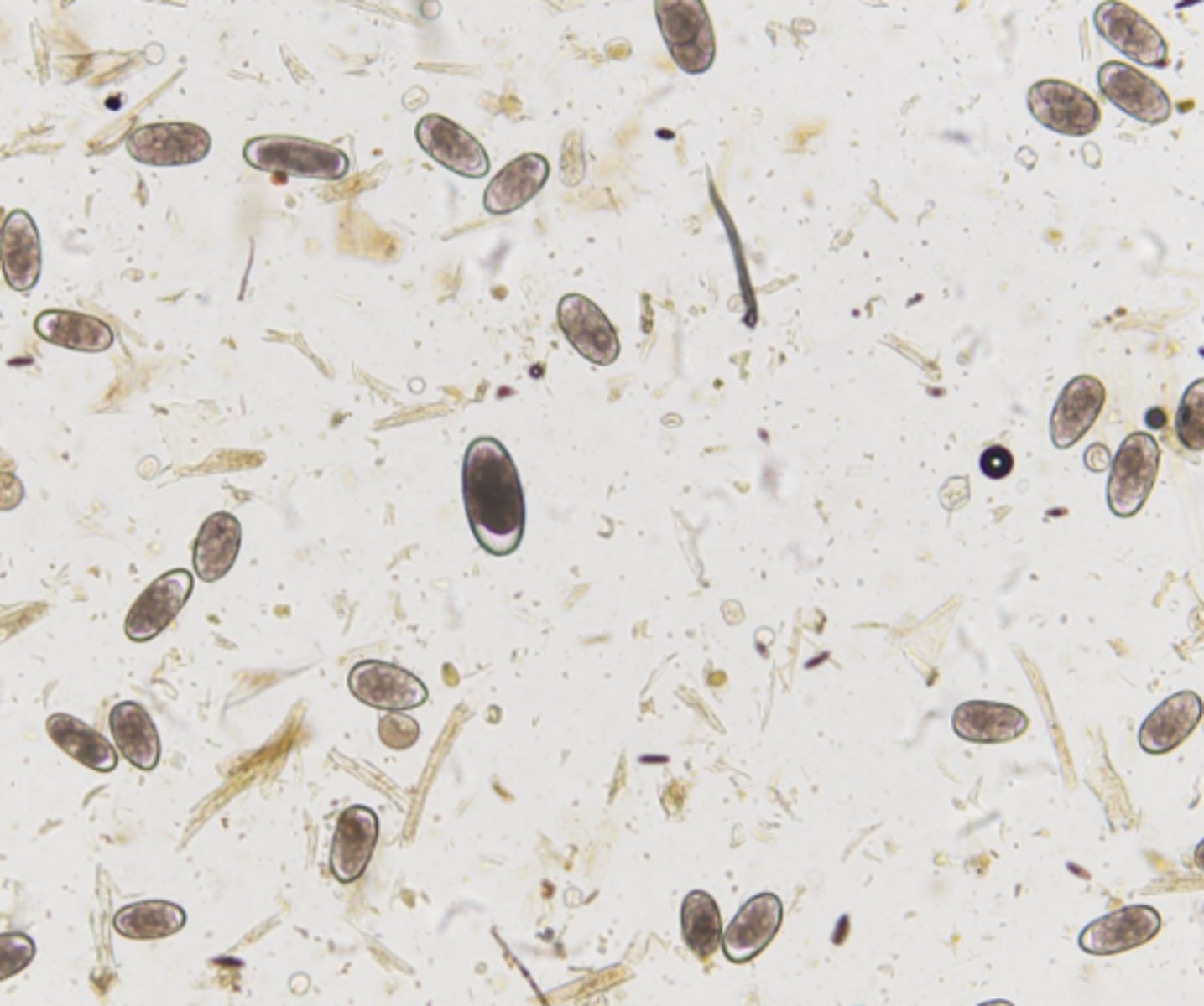
Ruminant strongyle type eggs
Identify these thin shelled, ellipsoidal, greyish eggs found in large & small ruminants
Bunostomum
Hookworm species that infects the small intestine of ruminants with chitinous cutting plates
-Eggs have thickened shells, more bluntly rounded ends, & more darkly pigmented cells compared to strongyle-type eggs
L3
The Baermann technique (following Coproculture of eggs) identifies what larval stage of Ruminant & Equine strongyles based on morphology?
*Retained cuticle gives this larvae stage a crinkled appearance
Nematodirus
Ruminant strongyle that infects the SI of small/large ruminants & passes large non-"strongyle type eggs"
-Adult characteristics: inflated cuticle at anterior end, long/slender spicules in males
Strongylus vulgaris
Large equine strongyle that is smaller than other strongylus species but the most pathogenic
-Has 2 rounded ear-shaped teeth
Strongylus equinus
Large equine strongyle that has 3 teeth (1 large dorsal, 2 small subventral)
-Ovoid buccal cavity with external & internal leaf-crowns
Strongylus edentatus
Large equine strongyle that has no teeth
-Ovoid buccal cavity with external & internal leaf-crowns
Cyathostomes
Small equine strongyle that infects the cecum & colon
-Has a well-developed buccal cavity that's more cylindrical in shape than the ovoid buccal cavity of the large strongyles
-Have external & internal leaf-crowns
*Larvae can be recovered using a Baermann technique
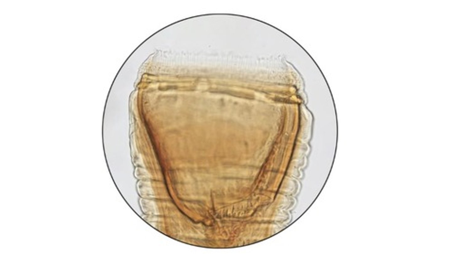
Equine strongyle type eggs (includes large & small)
Identify these thin shelled, ellipsoidal, a grey colored eggs in equine
Phlebotomum
Bunostomum species that infects the SI of wild & domestic ruminants
-Has chitinous cutting plates & multiple sub-ventral lancets
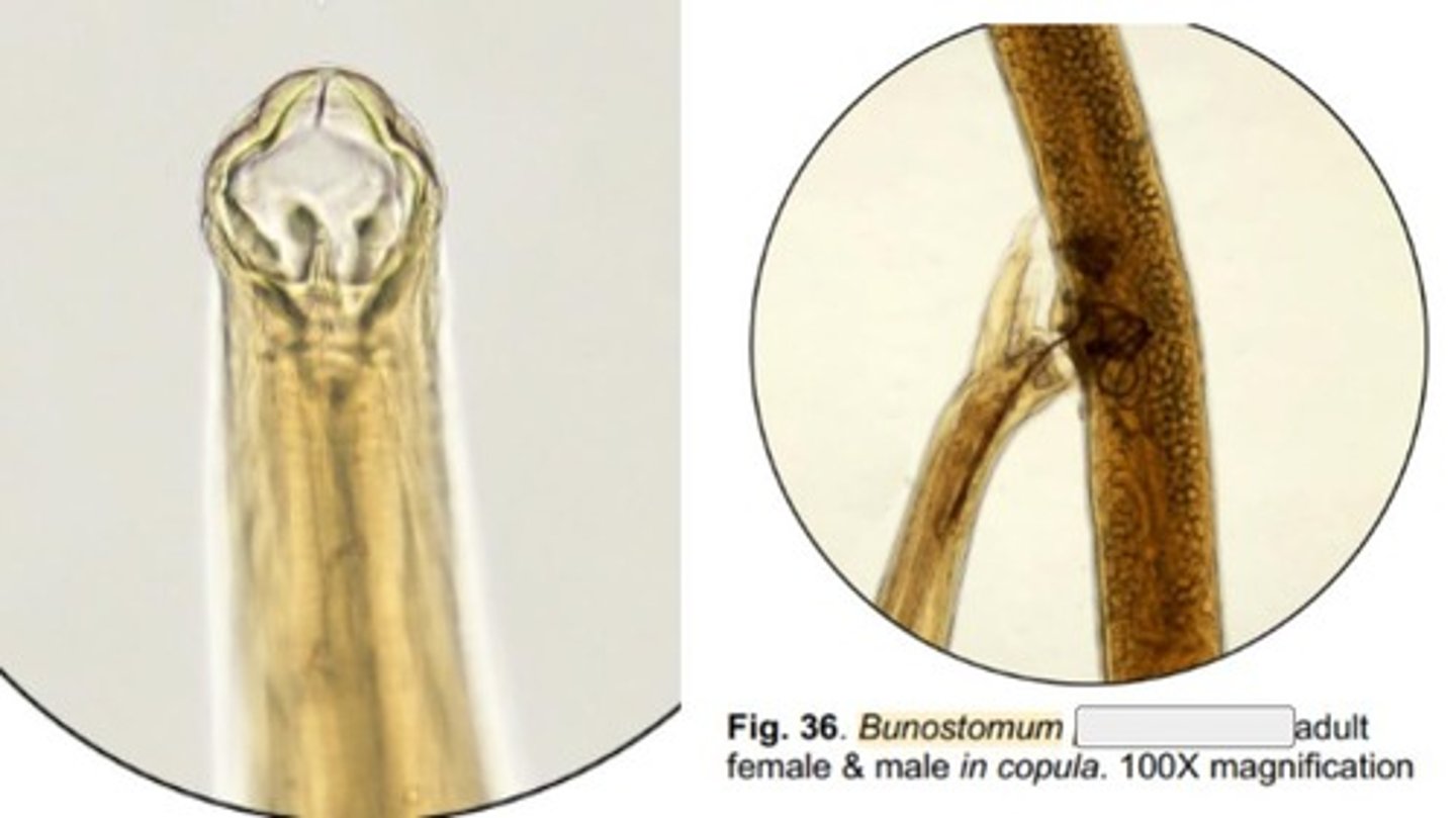
Trigonocephalum
Bunostomum species that infects the SI of sheep, goats, alpaca, & llamas
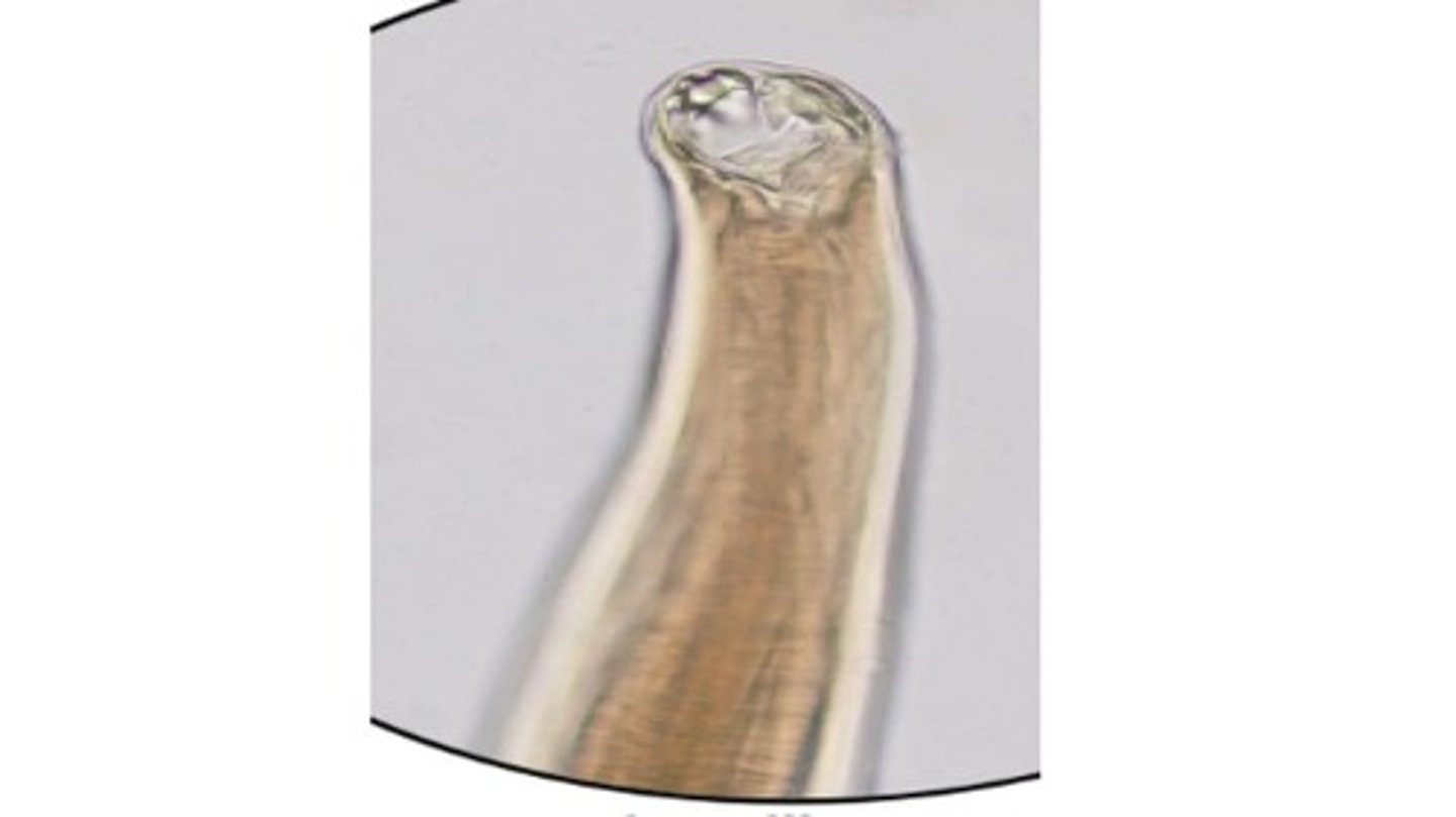
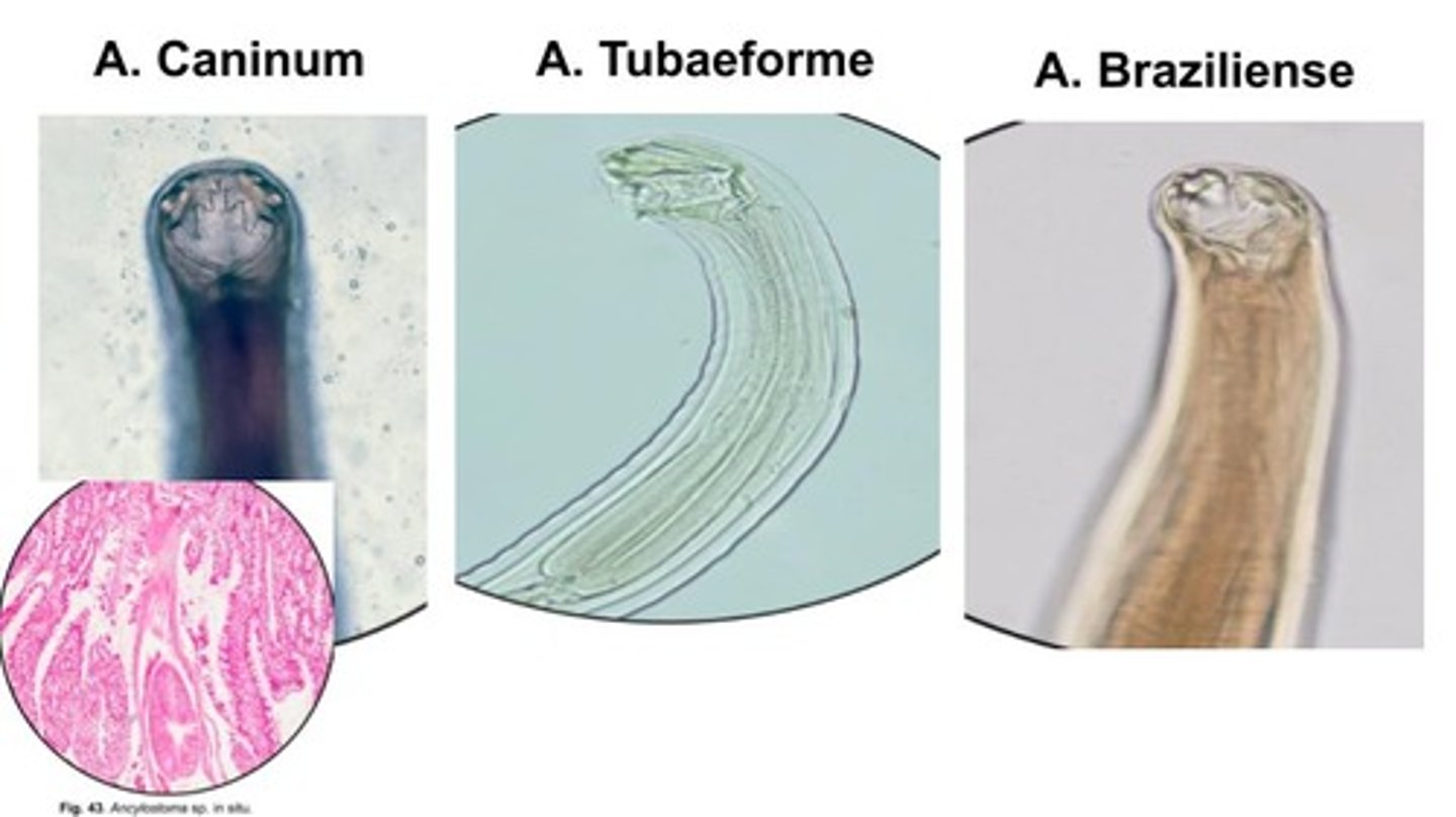
Ancylostomum caninum
Most pathogenic hookworm species in dogs
-Very aggressive blood feeders with 3 pairs of teeth that infect the SI
-Transmammary transmission = most significant route of infection
Ancylostomum tubaeforme
Hookworm species with 3 pairs of teeth that infects the SI of felids only
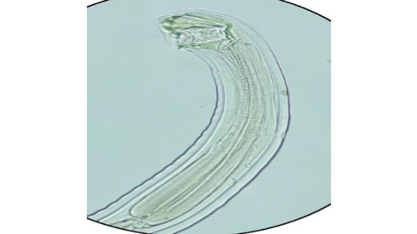
Ancylostomum braziliense
Hookworm species with 1 pair of teeth that infects the SI of dogs & cats
-Percutaneous route of transmission = most significant
*Associated with Cutaneous larva migrans in humans
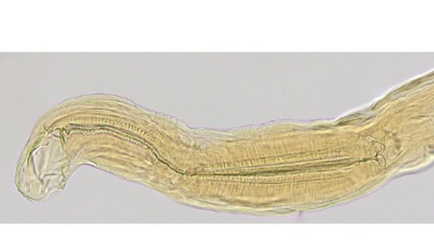
Small intestine
What part of the DH do Anyclostomum spp. infect?
*Bloodfeeders that attach to the mucosa
Uncinaria stenocephala
Hookworm with chitinous cutting plates that infects the SI of canids & felids
-more likely to be found in cooler regions of the US
Hookworm eggs
Identify these thin shelled, ovoid, containing morula with 2-8 cells
-Found in dogs & cats
Strongyloides
AKA threadworms, infect SI of multiple DH (depending on spp)
-Parthenogenetic females with long filariform esophagus, no parasitic males
*Females seen in mucosal scrapings of intestines
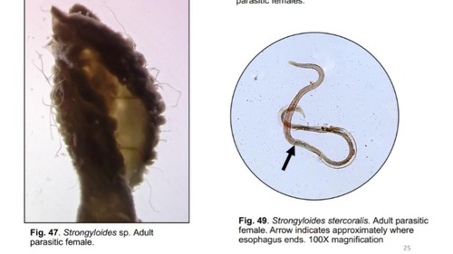
Stercoralis
Strongyloides species that infects the SI of dogs, cats, & humans
-Has L1 stages (rhabditiform larvae) with a club shaped esophagus & posterior bulb that hatch within the DH & are passed directly in feces
-L1s also have a characteristic genital primoridum
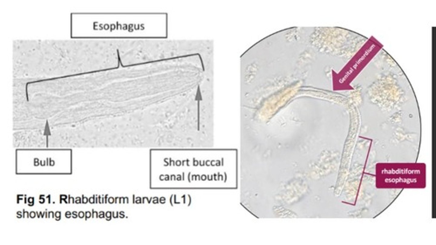
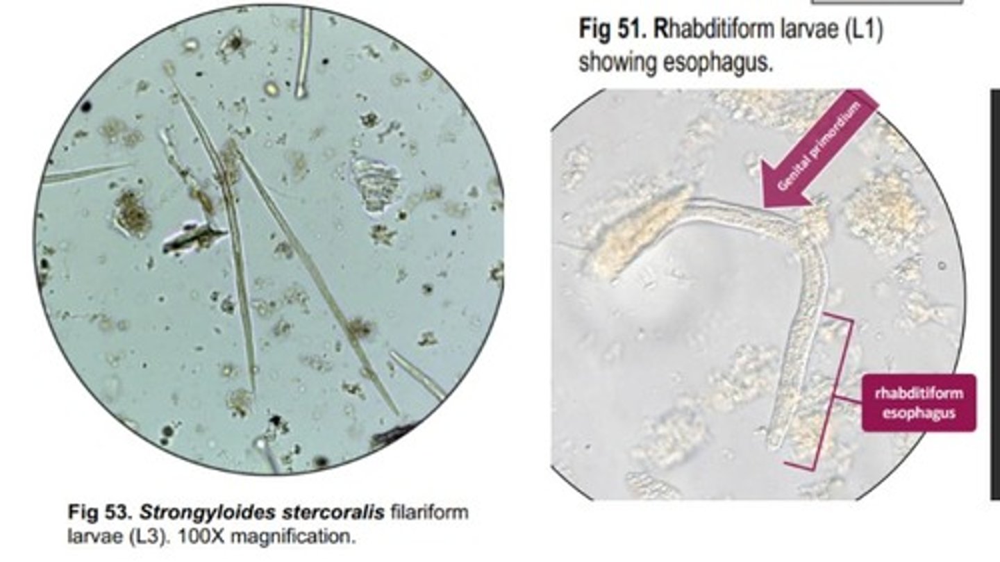
L3
What is the infective stage in all Strongyloides spp?
-Has a filariform esophagus (longer & thinner than rhabditiform esophagus)
*Does not have a narrow section or a bulb
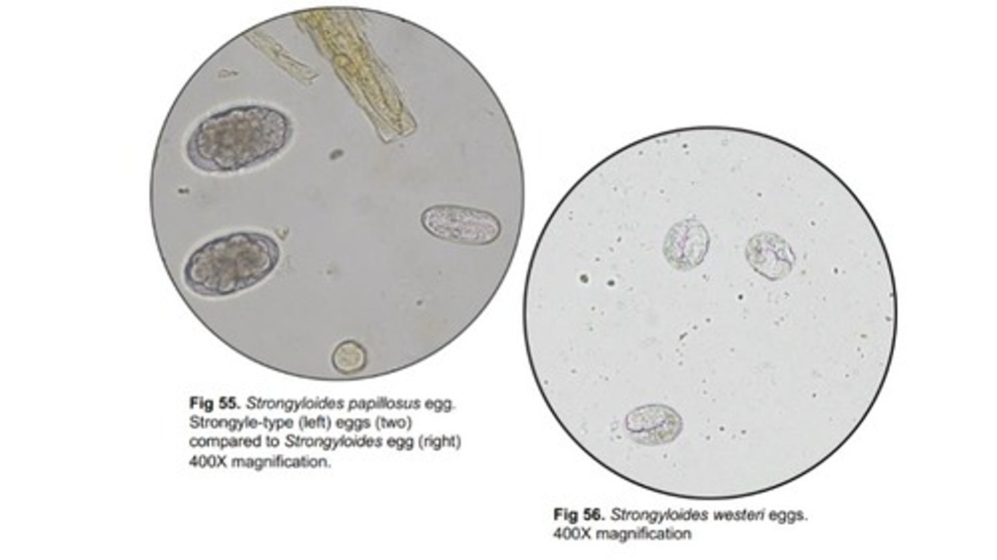
Papillosus, Westeri
Strongyloides Species:
-S. ____ = ruminants
-S. ____ = equids
*Strongyloides eggs are smaller than strongyle-type eggs
Gordian worms
AKA Horsehair worms
-Larval stages develop in arthropods (crickets, grasshoppers)
-Adult stages are free-living
Angiostrongylus cantonensis
AKA rat lungworm
-Causes eosinophilic meningitis in humans & is prevalent in Southeast Asia and tropical Pacific islands
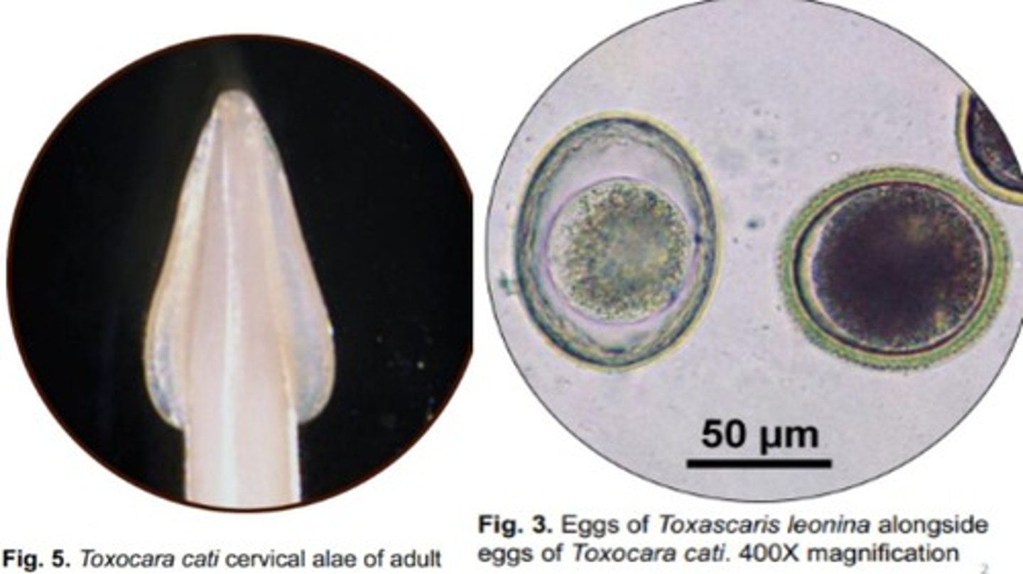
Toxascaris leonina
Roundworm that infects the SI of domestic/wild felids & canids
-Cervical alae is similar to that of Toxocara canis
-Eggs are ovoid with a smooth outer shell & more spacious interior compared to eggs of Toxocara cati & canis
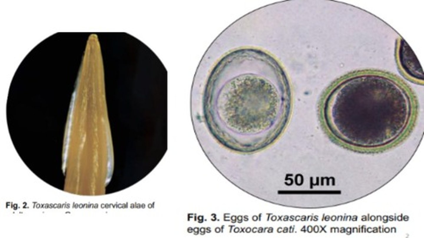
Toxocara cati
Roundworm that infects the SI of wild/domestic felids
-Prominent, arrowhead-like cervical alae
-Eggs are ovoid with a bumpy outer surface with a less spacious interior compared to Toxascaris leonina
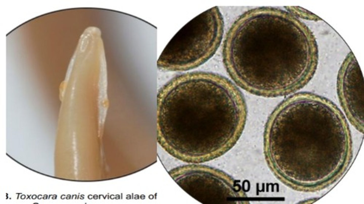
Toxocara canis
Roundworm that infects the SI of domestic/wild canids
-Cervical alae are similar to Toxascaris leonina
*Ovoid eggs with a bumpy outer surface & less spacious interior than to Toxascaris leonina
*Transplacental transmission is a significant transmission route
Baylisascaris procyonis
Roundworm that infects SI of racoons & rarely domestic canids
-Eggs are subspherical & brown with a finely pitted outer shell, & contain a single cell that doesn't fill the egg
*Can lead to neural larva migrans & ocular larva migrans in humans
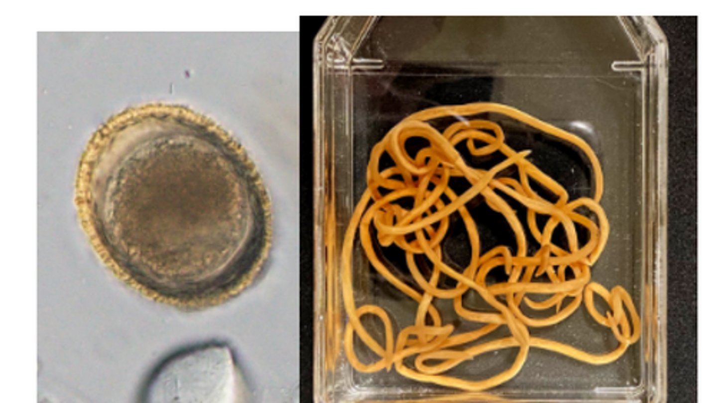
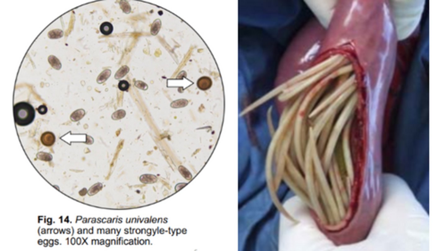
Parascaris univalens
Roundworms that infect the SI of equids
-Eggs are almost round with a golden brown, thick pitted shell
-Sometimes, eggs lack dark rough protein coat
Ascaris suum
Roundworm that infects the SI of swine
-Eggs are ovoid, golden brown, thick shelled with bumpy exterior
*Eggs are sticky, very resistant, & can remain infective for many years; zoonotic risk
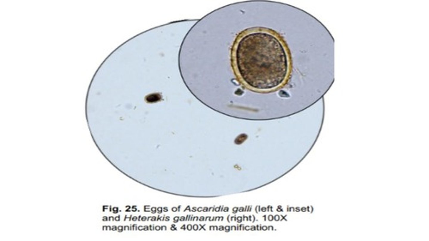
Heterakis gallinarum
Roundworm that infects the ceca of galliform birds
-Adults are small white worms, while eggs are ellipsoid with a thick smooth shell containing a single cell that fills the egg
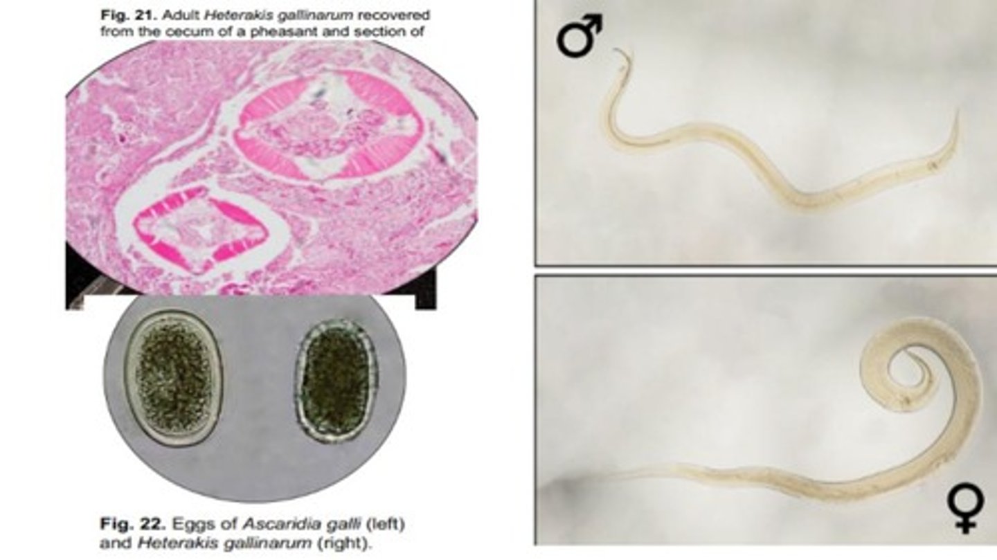
Ascaridia galli
Roundworm that infects the SI of Galliform birds
-Adults are large white worms, while eggs are ovoid with a thick smooth shell
-Eggs are Larger & more ovoid than Heterakis gallinarum
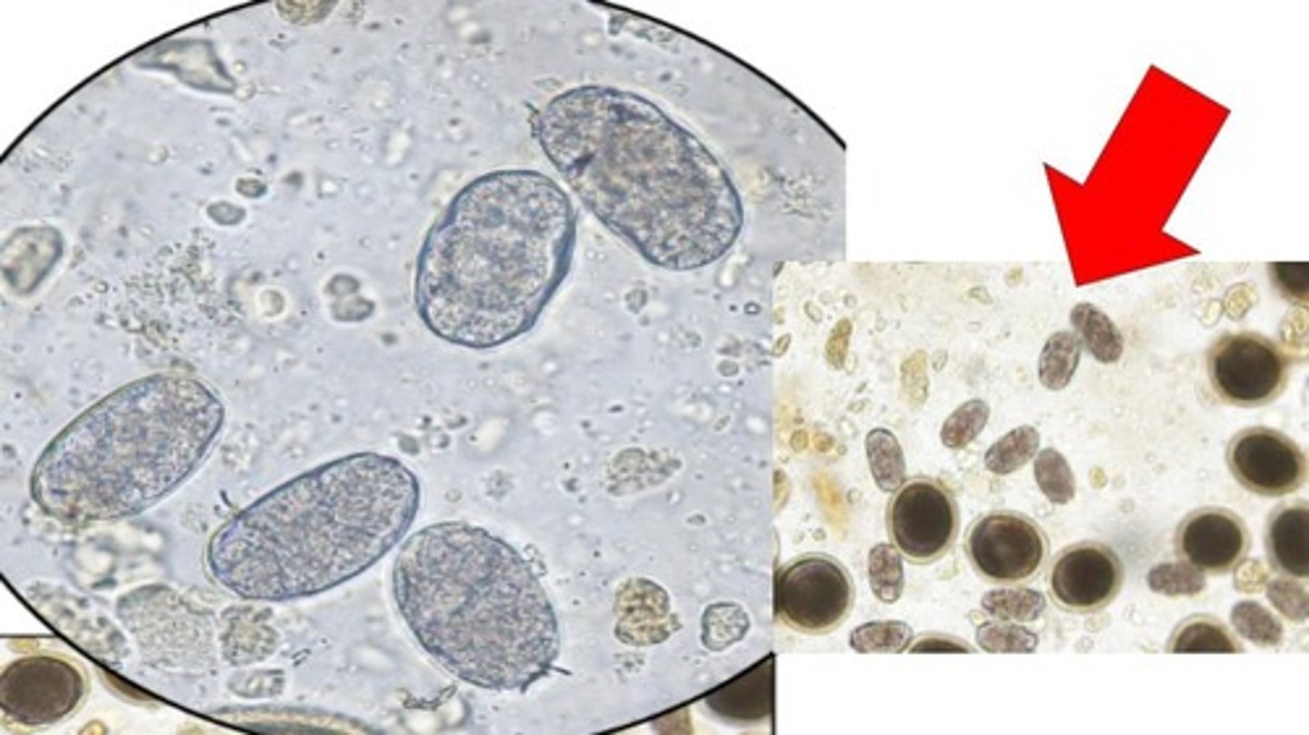
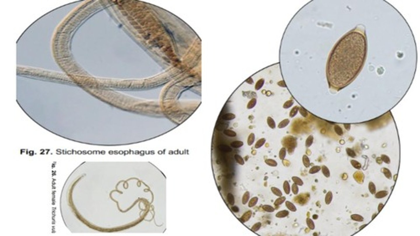
Trichuris vulpis
Whipworms that infect the cecum & colon of domestic/wild canids
-Adults have a long stichosome esophagus composed of stichocytes
-Eggs are football-shaped with bipolar plugs & a smooth surface
Trichuris ovis
Whipworm that infects the cecum & colon of domestic/wild ruminants
-Adult males have a spined spicule
-Eggs have a football shape & bipolar plugs
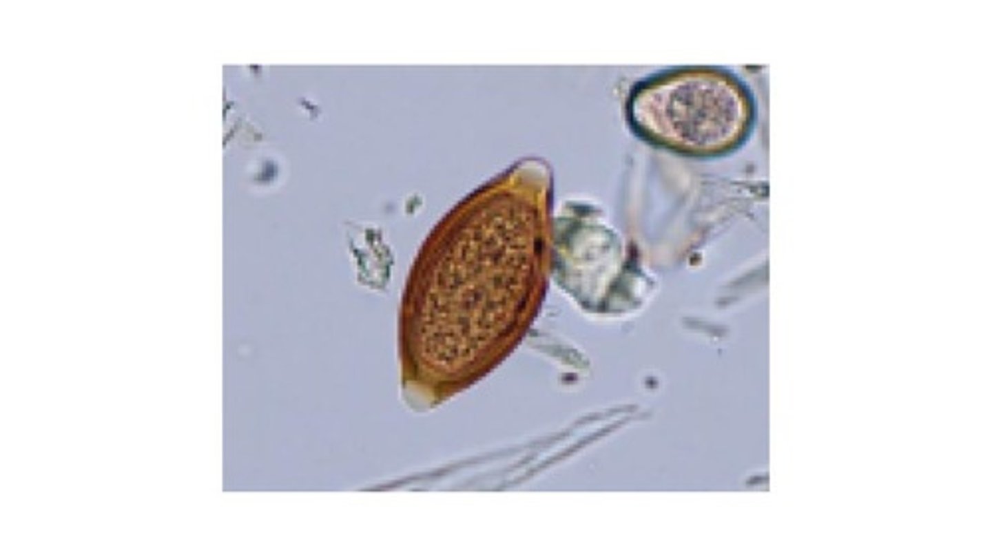
Capillarids
Very small, thin nematodes with a stichosome esophagus typical of Enoplida
-Difficult to see grossly at necropsy
-Eggs are more barrel shaped (sides are more parallel) with a rough surface compared to Trichuris eggs
-Eggs may be in feces or urine depending on the type
*Ex: Eucoleus, Pearsonema
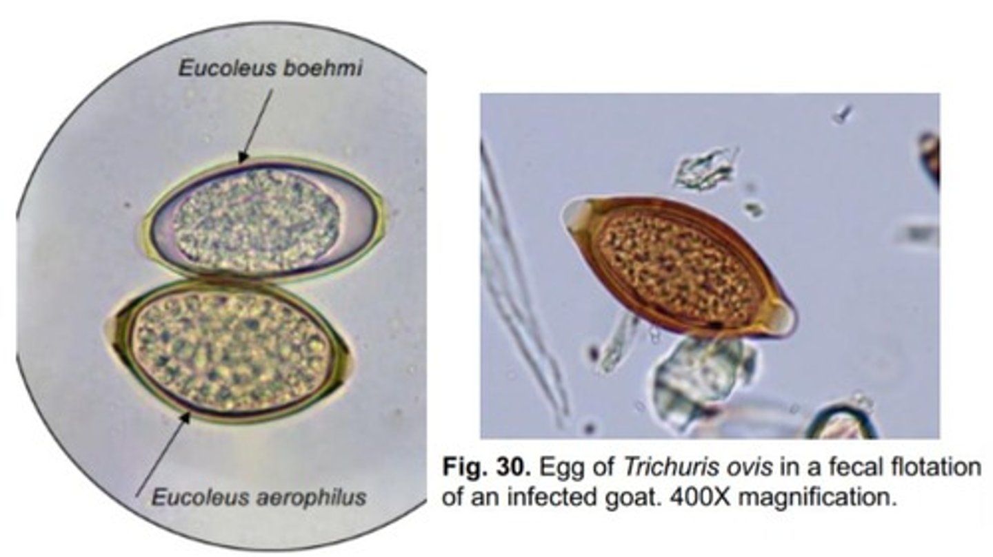
False (not expected in urine sediment because it is a respiratory parasite)
True or False: You would expect to see Eucoleus aerophilus in a urine sediment.
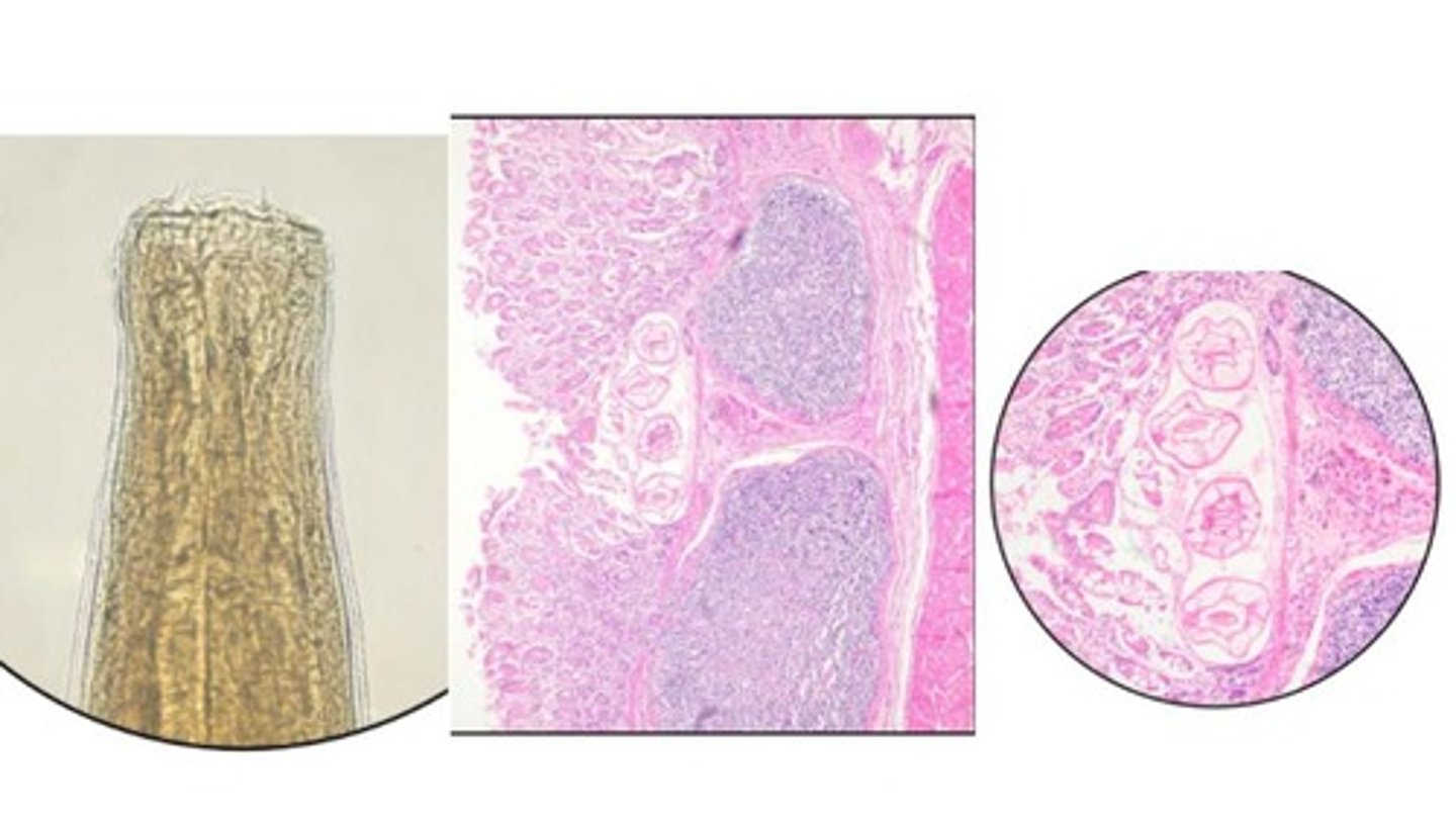
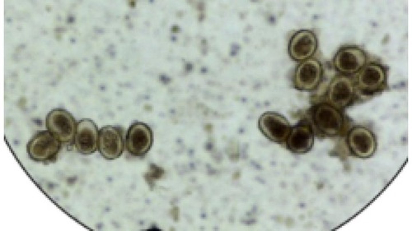
Capillarid type eggs
Identify what type of nematode eggs these are.
-Present in quail
*Bipolar plugged egg in histo sections can aid in the diagnosis of infection
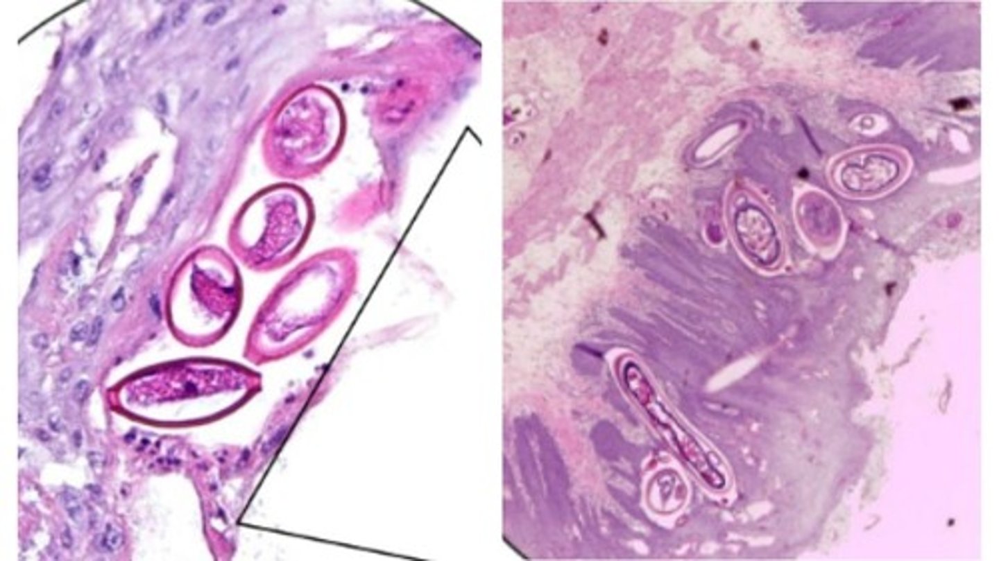
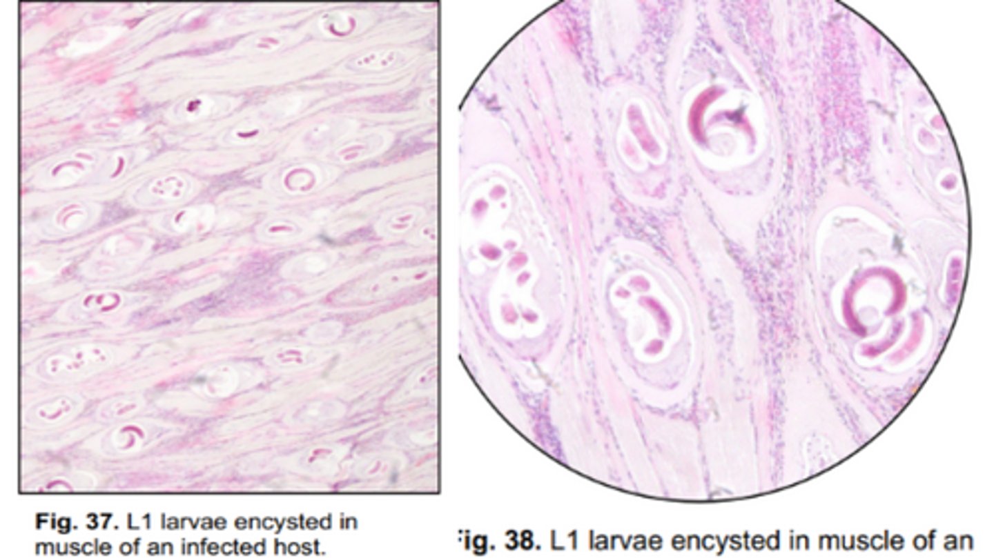
Trichinella spiralis
Trichinelloidea that infects the SI in a wide range of mammalian hosts (including humans) & some avian spp.
-Adults are very small with a stichosome esophagus
-Nurse cells encapsulate & protect the L1s in muscle
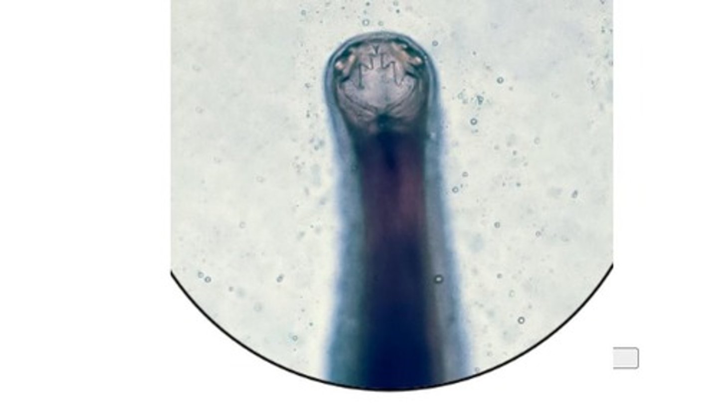
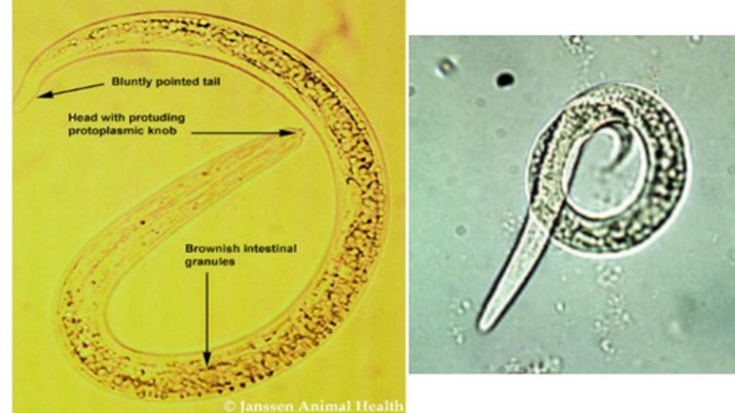
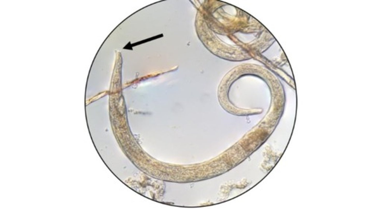
Dictyocaulus
Group of Trichostrongyloidea lungworms that infect the bronchioles, bronchi, & trachea
*Adults are plain, long, thin worms
-Baermann technique to recover L1 (blunt end, brown intestinal granules, & button at mouth)
Muellerius capillaris
Lungworm that infects the bronchioles & lung parenchyma of sheep & goats
-L4 are found in lung parenchyma & form nodules where adults develop
-L1 have a wavy, pointed tail with dorsal spine & are found in feces --> Baermann technique
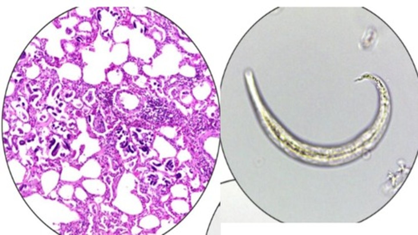
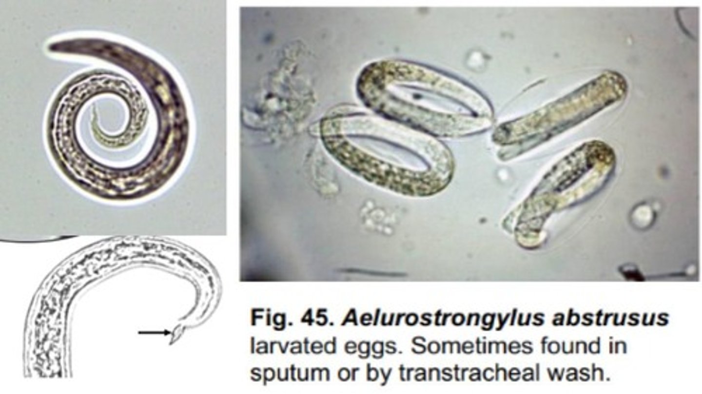
Aelurostrongylus abstrusus
Lungworm that infects the bronchioles, lung parenchyma, & alveoli of domestic/wild felids
-Eggs can be found in sputum or transtracheal washes
-Characteristic tail of L1 with an S-shaped bend & dorsal spine
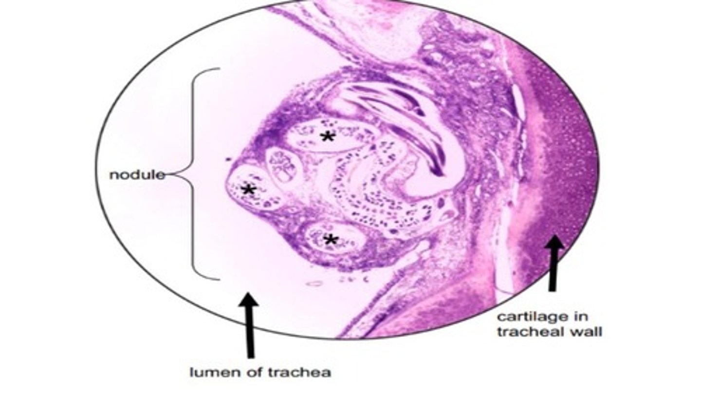
Oslerus (Filaroides) osleri
Metastrongyloidea (lungworm) that forms nodules in the trachea & bronchi of domestic/wild canids
-Not often found in the lungs but can be confirmed by bronchoscopy as lesions observed by bronchoscope are pathognomonic
Metastrongylus
Are Trichostrongyloidea or Metastrongylus the lungworm superfamily that infect the bronchi & bronchioles in swine?
-Larvated eggs passed in feces; earthworms are IH & harbor the infective L3 stage (indirect life cycle)
Ollulanus triscuspis
Trichostrongylid nematode that infects the gastric mucosa of the stomach in domestic/wild felids
-Associated with chronic cases of vomiting in the absence of other nematodes (i.e. Physaloptera)
*Life cycle is direct with the transmission of L3s are expelled in the vomitus that are then ingested
-Females are ovoviviparous, autoinfection is possible
-Adults have 3 unique projections called tubercles
Dictyocaulus viviparous
This lungworm of cattle has a direct LC and sheds L1s in fresh feces that are isolated on a Baermann
Ascaridia galli, heterakis gallinarum, ascaris suum
Earthworms serve as PH for these 3 roundworms by bioaccumulating infectious larvae/eggs within soil
Strongyloides ransomi and westerni
What 2 Strongyloides species is transmammary transmission is important in their epidemiology?
Trichinella spiralis
L1s of this nematode infect striated muscle cells of warm blooded animals & the adults reside in their SI
Strongyloides stercoralis, Aelurostrongylus abstrusus, Dictyocaulus filaria
Baermann technique is used to isolate the L1s of which of these nematodes:
-Strongyloides papillosus
-Strongyloides stercoralis
-Aelurostrongylus abstrusus
-Dictyocaulus filaria
Baylisascaris procyonis
Racoons should be handled with caution because of the zoonotic risk of this nematode & associated with neural larval migrans
False
True or False; Domestic dogs can't be a DH of Baylisascaris procyonis & wouldn't be a source of zoonotic transmission.
Dioctophyme renale
Giant kidney worm with adults found in the kidneys
Strongyloides stercoralis
In addition to Ancylostoma braziliense (hookworm), this nematode is also responsible for cutaneous larval migrans in people
*Walking around barefoot
Bear
Zoonoses with Trichinella spiralis occur primarily in the US from consuming undercooked meat of this host
Sheep, goats
What are the DH of these lungworms (Infect respiratory tract):
-Dictylocaulus filaria
-Muellerius capillaris
Strongyloides stercoralis
Autoinfection with this nematode in canids can be associated with severe disease
*Can recolonize in the SI
Toxocara canis
A puppy with these symptoms likely is infected with what parasite:
-Ovoid spherical eggs with a bumpy outer edge & dark single cell within
-Potbellied appearance
-Ascarid worms expelled in vomitus or feces
-Small intestinal obstruction
Toxocara cati
A kitten with these symptoms likely is infected with what parasite:
-Ovoid spherical eggs with a bumpy outer edge & dark single cell within
-Potbellied appearance
-Ascarid worms expelled in vomitus or feces
-Small intestinal obstruction
Ascaris suum
What pig nematode is associated with fibrotic areas in the liver tissue known as "milk spots"?
Strongyloides tumefaciens
This nematode infects the SI of most hosts but in cats infections consist of large intestinal nodules
True
True or False: Rodents harboring L3s of Toxocara spp & Toxocaris Leonina can serve as PH if they're ingested by a hsot.
Oslerus (Filaroides) osleri
Tracheal nodules observed by bronchoscopy in a canid are pathognomonic for infection with ...
Pilobolus
This fungus is important in the dispersal larvae of Dictylocaulus viviparus from a cow paddy
Toxocara canis, toxocara cati
What 2 ascarids are associated with visceral larval migrans or ocular larva migrans
True
True or False: Peroral transmission is a route of entry for Trichuris into a new host.
False
True or False: Trichuris spp. infect the esophagus of their host.
True
True or False: Trichuris is commonly called whipworms.
True
True or False: Trichuris eggs are football-shaped with 2 polar plugs at either ends.
False (only find parasitic females)
True or False: At necropsy, male & female adults of Strongyloides ransomi could be recovered from mucosal scrapings of the duodenum of a pig.
Toxocara canis, toxascaris leonina, Baylisascaris procyonis
Which of these nematodes have eggs that can be found in a fecal flotation of an infected canine host:
-Strongyloides westeri
-Toxocara canis
-Toxascaris leonina
-Baylisascaris procyonis
Cattle, sheep
Nematodirus by Species:
-N. helvetianus = infects cattle
-N. battus = infects sheep