Quizzes lecture 1-8
1/23
There's no tags or description
Looks like no tags are added yet.
Name | Mastery | Learn | Test | Matching | Spaced | Call with Kai |
|---|
No analytics yet
Send a link to your students to track their progress
24 Terms
A necropsy of a 6 y/o, male neutered, DSH reveals edematous kidneys and pulmonary edema. Histopathology of the kidney shows numerous intratubular oxalate crystals. You recognize that this cat suffered from ethylene glycol toxicity and likely died due to acute renal failure resulting in cardiac arrest. In this case the cause of death is ______ and the mechanism of death is _________.
Ethylene glycol toxicity, acute renal failure and cardiac arrest
Acute renal failure and cardiac arrest, ethylene glycol toxicity
Acute renal failure and cardiac arrest, ethylene glycol toxicity
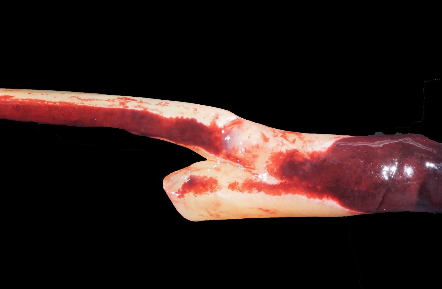
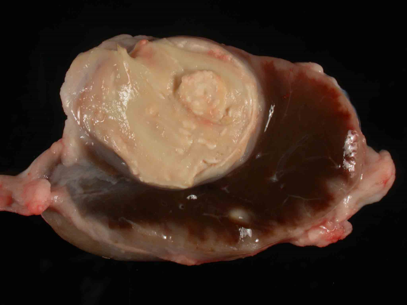
The image shows a clot that would be described as elastic, smooth, and gelatinous and was easily removed from the vessel lumen (not adhered to the wall). This is an example of a _________ and represents a/an ________.
Antemortem thrombus (before death); incidental lesion
Postmortem clot (after death); incidental lesion
Postmortem clot (after death); postmortem artifact
Postmortem clot (after death); postmortem artifact
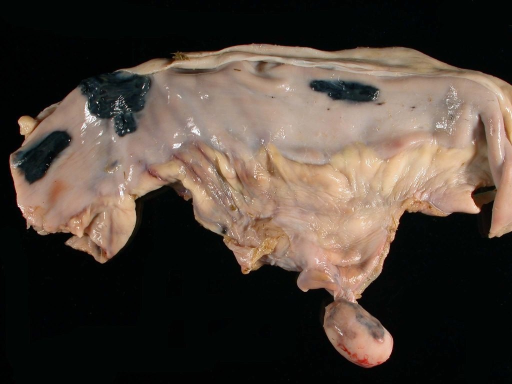
This is small intestine from a horse that shows multifocal black foci representative of a condition referred to as hemomelasma ilei as well as a mesenteric, pedunculated lipoma that was NOT wrapped around the intestine. These lesions both are best defined as:
Incidental lesions
Clinically significant lesions
Agonal lesions
incidental lesions
In the “3D ATP” paradigm used to formulate a morphologic diagnosis, the three “Ds” are:
disorder, degree, distribution
degree, duration, distribution
disease, degree, duration
degree, duration, distribution
You perform a necropsy on a 3 y/o female spayed cat and notice multifocal white to tan, pinpoint to 4mm diameter foci scattered throughout the spleen. You quickly move the cat to a biosafety cabinet as you recognize that this lesion (along with multiple enlarged lymph nodes) is consistent with the zoonotic disease, Tularemia caused by the bacterial agent Francisella tularensis. Under the microscope, the white foci observed on the spleen are areas of necrosis surrounded by neutrophilic inflammation.
Which of the following represents the etiologic diagnosis:
Francisella tularensis splenitis (splenic francisellosis)
Spleen, necrotizing and suppurative splenitis, multifocal, severe, acute
Viral splentitis
Francisella tularensis splenitis (splenic francisellosis)
You perform a necropsy on a 3 y/o female spayed cat and notice multifocal white to tan, pinpoint to 4mm diameter foci scattered throughout the spleen. You quickly move the cat to a biosafety cabinet as you recognize that this lesion (along with multiple enlarged lymph nodes) is consistent with the zoonotic disease, Tularemia caused by the bacterial agent Francisella tularensis. Under the microscope, the white foci observed on the spleen are areas of necrosis surrounded by neutrophilic inflammation.
Which of the following represents the morphologic diagnosis:
Francisella tularensis splenitis
Spleen, necrotizing and suppurative splenitis, multifocal, severe, acute
Viral splentitis
Spleen, necrotizing and suppurative splenitis, multifocal, severe, acute
Which of the following is an example of an exogenous cause of disease?
Type I Hypersensitivity (immunologic condition)
Type I Diabetes (genetic condition)
Rodenticide poisoning
Rodenticide poisoning
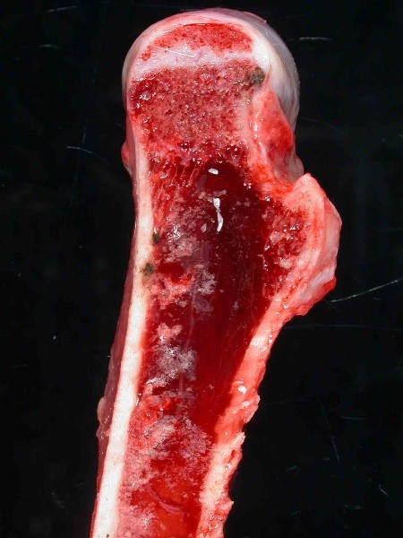
This image depicts the bone marrow of the femur taken from an emaciated gazelle. There is complete loss of adipose (fat) from marrow cavity. The term used to describe this lesion is:
Fat necrosis
Serous atrophy of fat
Bone marrow hemorrhage
Serous atrophy of fat
Which of the following represents the pathogenesis (mechanism of disease) of primary photosensitivity:
Inherited defect in porphyrin metabolism → aberrant porphyrin metabolites deposited in skin → photosensitization
Chlorophyll from plants → metabolized to phylloerythrin by bacteria in rumen/colon → phylloerythrin persists in circulation in animals with liver disease (e.g. due to pyrrolizidine alkaloid intoxication) → phylloerythrin deposited in skin → photosensitization
Uptake/Ingestion of photodynamic compounds with food (e.g. St. John's wort) → deposition in skin → photosensitization
Uptake/Ingestion of photodynamic compounds with food (e.g. St. John's wort) → deposition in skin → photosensitization
Which of the following tissues/cell types is most sensitive to hypoxia?
Neurons
Cardiomyocytes
Keratinocytes
neurons
Which of the following is considered a hallmark of reversible cell injury?
Cellular swelling
Cellular shrinking
Cellular necrosis
Cellular swelling
The image shows a thrombus at the aortic bifurcation (“saddle thrombus”) in a cat with hypertrophic cardiomyopathy. What type of hypoxia would result from this thrombus?
Hypoglycemic hypoxia
Anemic hypoxia
Ischemic hypoxia
Ischemic hypoxia
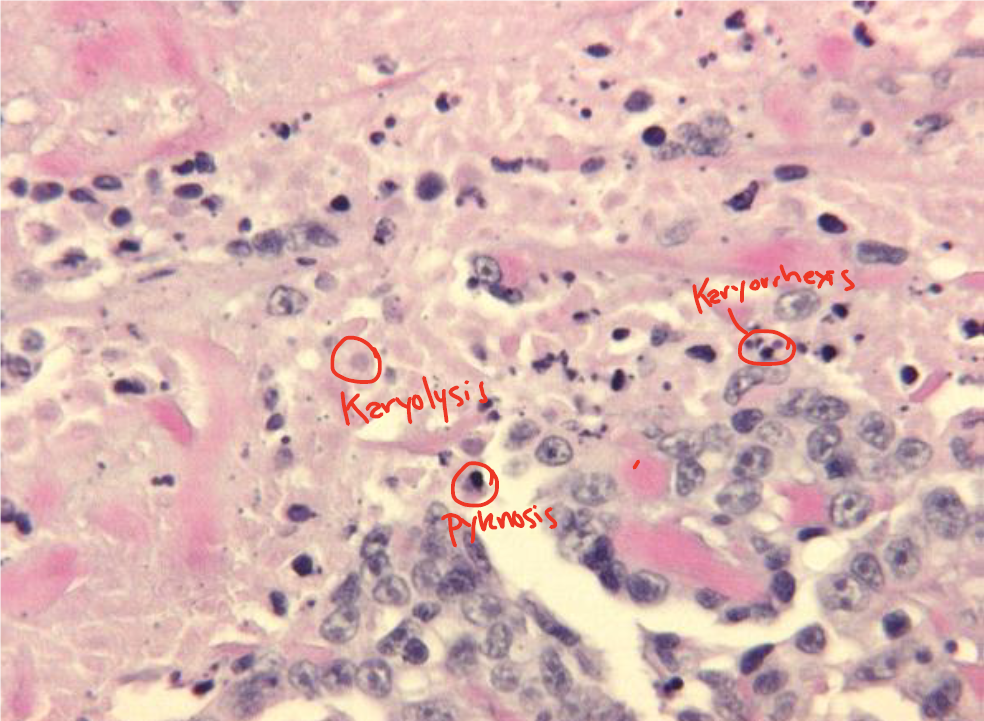
The image depicts the various nuclear morphologies associated with ______________ cell injury, which include karyolysis, karyorrhexis, and pyknosis. Pyknosis is the term used to describe ____________.
Reversible; fragmented nucleus
Irreversible; shrunken nucleus
Irreversible; fragmented nucleus
Irreversible; shrunken nucleus
Oxidative Stress refers to:
Free radical induced injury
Oxygen induced injury
Hypoxia induced injury
Free radical induced injury
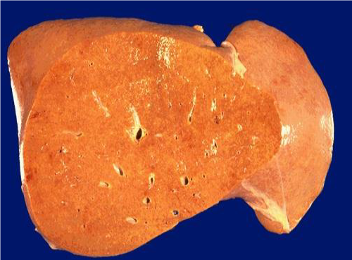
The image depicts a liver with fatty change, which is the result of inadequate apoprotein production (the enzyme needed to transport lipid out of cells) as a result of cell injury and damage to the rough endoplasmic reticulum. A way to confirm that this liver is affected by fatty change is:
Place in formalin and if it floats, this confirms fatty change
Place in formalin and if it sinks, this confirms fatty change
No need to do anything – nothing else can make a liver look this enlarged or pale yellow/orange.
Place in formalin and if it floats, this confirms fatty change
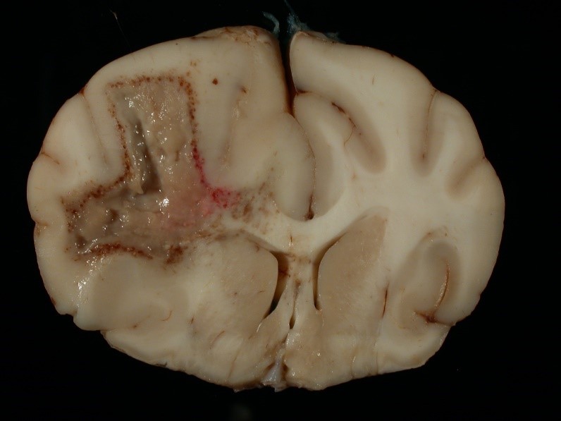
The image depicts a brain from a dog. In this type of necrosis, enzymatic digestion of cells predominates over protein denaturation and grossly (macroscopically), there is accumulation of soft, viscous, fluid. This is an example of what type of necrosis?
Coagulation (coagulative) necrosis
Caseous necrosis
Liquefactive (lytic) necrosis
Liquefactive (lytic) necrosis
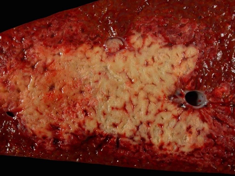
The image depicts liver from a horse. In this type of necrosis, protein denaturation predominates over enzymatic digestion and grossly (macroscopically), the tissue is paler in color, is often firm, and may be swollen or shrunken. This is an example of what type of necrosis?
Coagulation (coagulative) necrosis
Caseous necrosis
Liquefactive (lytic) necrosis
Coagulation (coagulative) necrosis
The image depicts a lymph node from a cow. In this type of necrosis, is typically seen with specific bacterial diseases, such as tuberculosis, and grossly (macroscopically), the tissue is grey to white to tan, dry, and friable to pasty. This is an example of what type of necrosis?
Coagulation (coagulative) necrosis
Caseous necrosis
Liquefactive (lytic) necrosis
Caseous necrosis
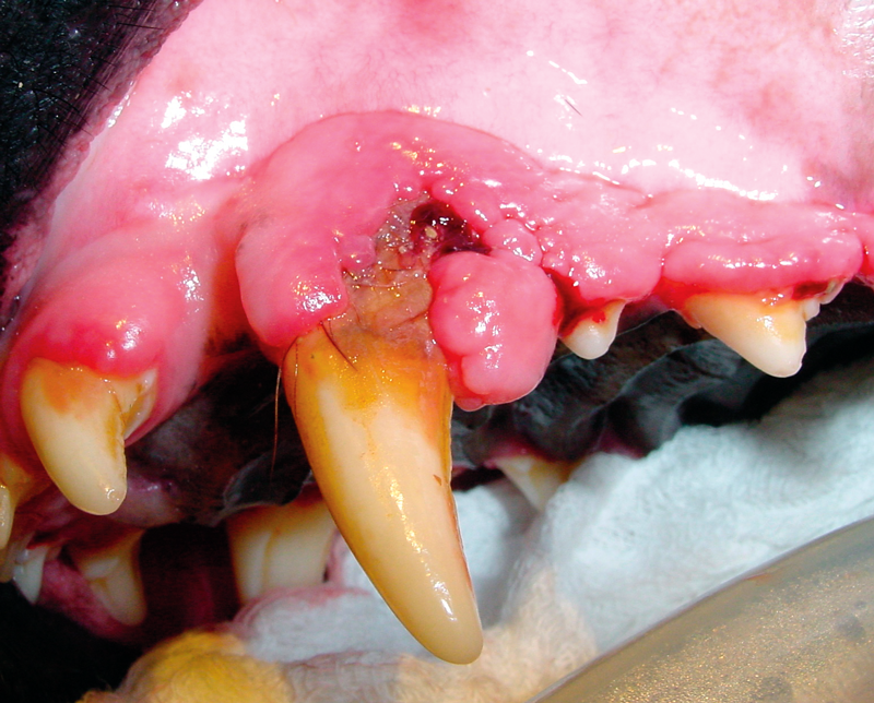
The image depicts gingival hyperplasia in a dog. In the case of hyperplasia, the tissue/organ is enlarged due to:
Increased size of the cells
Increased number of the cells
Replacement of one cell type by a different cell type
Increased number of the cells
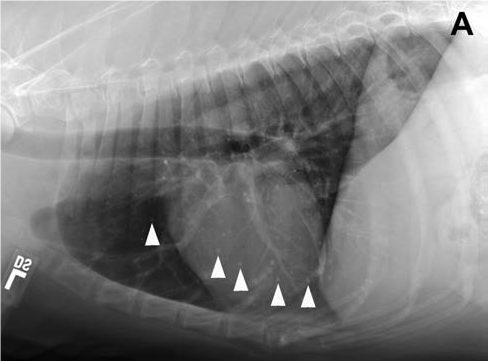
This radiograph is from a 15 year old, male neutered, mixed-breed dog. The arrows are pointing to multifocal, well-defined, 1- to 3.5-mm-diameter mineral opacities. These represent pulmonary osteomas, a common finding in clinically normal older dogs. The development of these nodules involves a change in differentiation from one cell type to another. This is an example of _______ and these would be considered a(n) _____________.
Metaplasia, postmortem artifact
Metaplasia, incidental lesion
Dysplasia, postmortem artifact
Metaplasia, incidental lesion
You receive a necropsy report that describes hepatocellular vacuolar degeneration within the liver of a dog and the cytoplasmic vacuoles are described as clear, indistinct, and irregular and the nuclei of the hepatocytes are centrally located. This represents accumulation of _________ within hepatocytes and is also referred to as ____________.
Lipid; steroid-induced hepatopathy
Lipid; hepatic lipidosis
Glycogen; steroid-induced hepatopathy
Glycogen; steroid-induced hepatopathy
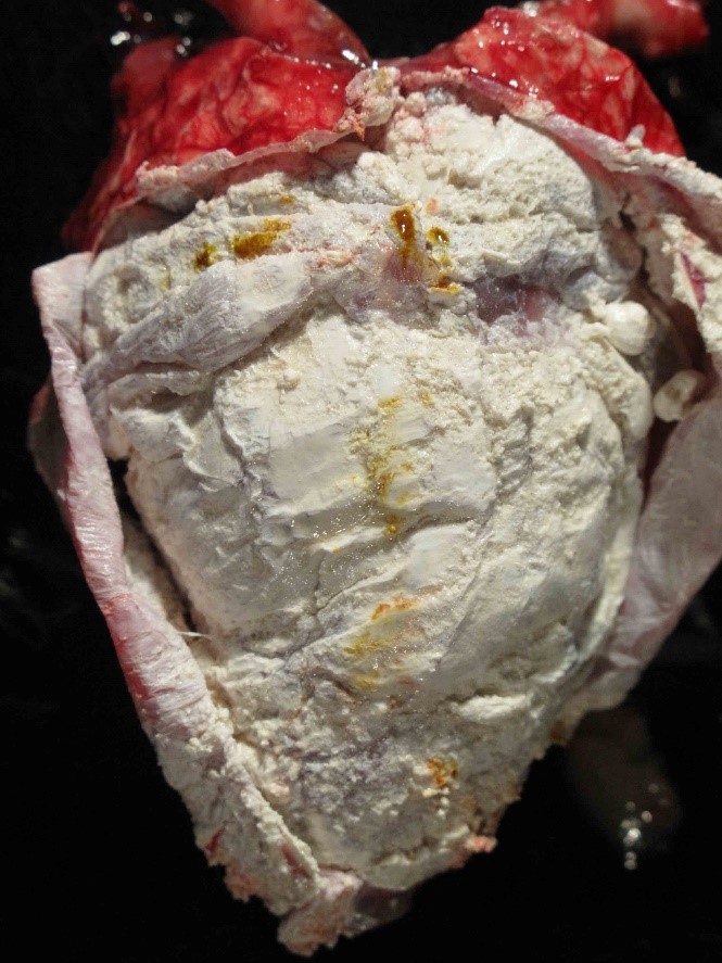
The image shows the heart and pericardial sac from a bald eagle. The white, chalky material represents the deposition of urate salts. Name the condition.
Metastatic calcification
Dystrophic calcification
Gout
gout
You are examining a 1 year old, male neutered, Saint Bernard and notice that the pad of the 3rd
digit of the left forelimb is enlarged by a hard nodule. The owner mentions that the dog had a grass awn (foxtail) embedded in that pad approximately 6 months prior. You suspect this may be an area of calcium deposition secondary to the previous trauma. This is an example of:
Metastatic calcification
Dystrophic calcification
Pseudogout
Dystrophic calcification
A goat digests Cestrum diurnum, a calcium accumulating plant that results in hypervitaminosis D. On necropsy examination, you note mineralization of the abomasal mucosa (as evidenced by hard texture and white/chalky, pinpoint lesions widely disseminated over the mucosal surface) and mineralization of the lungs (as noted by non-collapsed lungs upon opening of the thoracic cavity, and a firm to hard, crackly texture). This is an example of:
Metastatic calcification
Dystrophic calcification
Hypercalcemia of malignancy
Metastatic calcification