A&P Nervous/Endocrine/Cardiovascular/Lymph System Lab Practical
1/44
There's no tags or description
Looks like no tags are added yet.
Name | Mastery | Learn | Test | Matching | Spaced | Call with Kai |
|---|
No analytics yet
Send a link to your students to track their progress
45 Terms
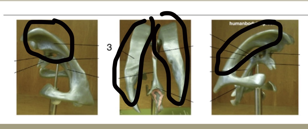
Lateral ventricle
Location: 2 largest ventricles; Function: Produce & hold CSF (cerebrospinal fluid)
Third ventricle
Location: Midline (more at the top); Function: Receives CSF from lateral ventricles
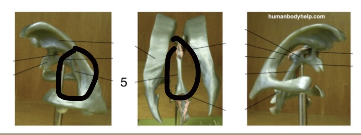
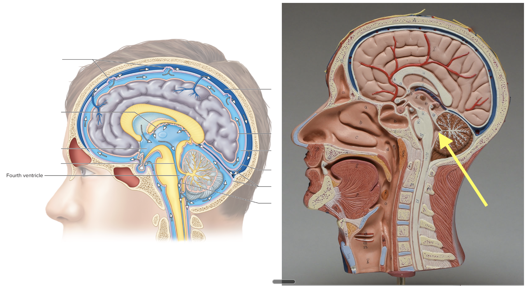
Fourth ventricle
Location: Midline, more towards the bottom; Function: Sends CSF to spinal cord
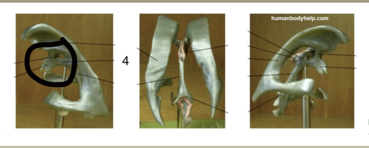
Cerebral aqueduct
Location: Midbrain connecting the 3rd & 4th ventricles; Function: CSF flow
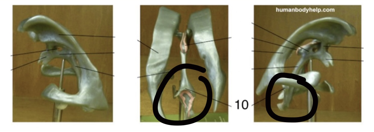
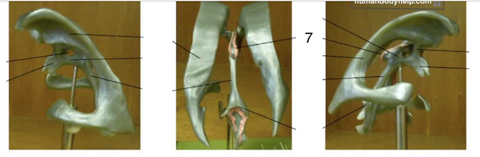
Choroid plexus
Location: Pink/red areas; Function: Produce CSF (cerebrospinal fluid)
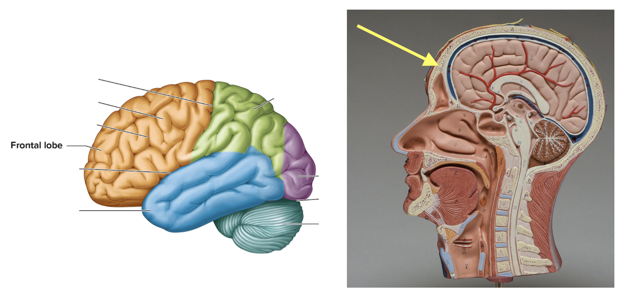
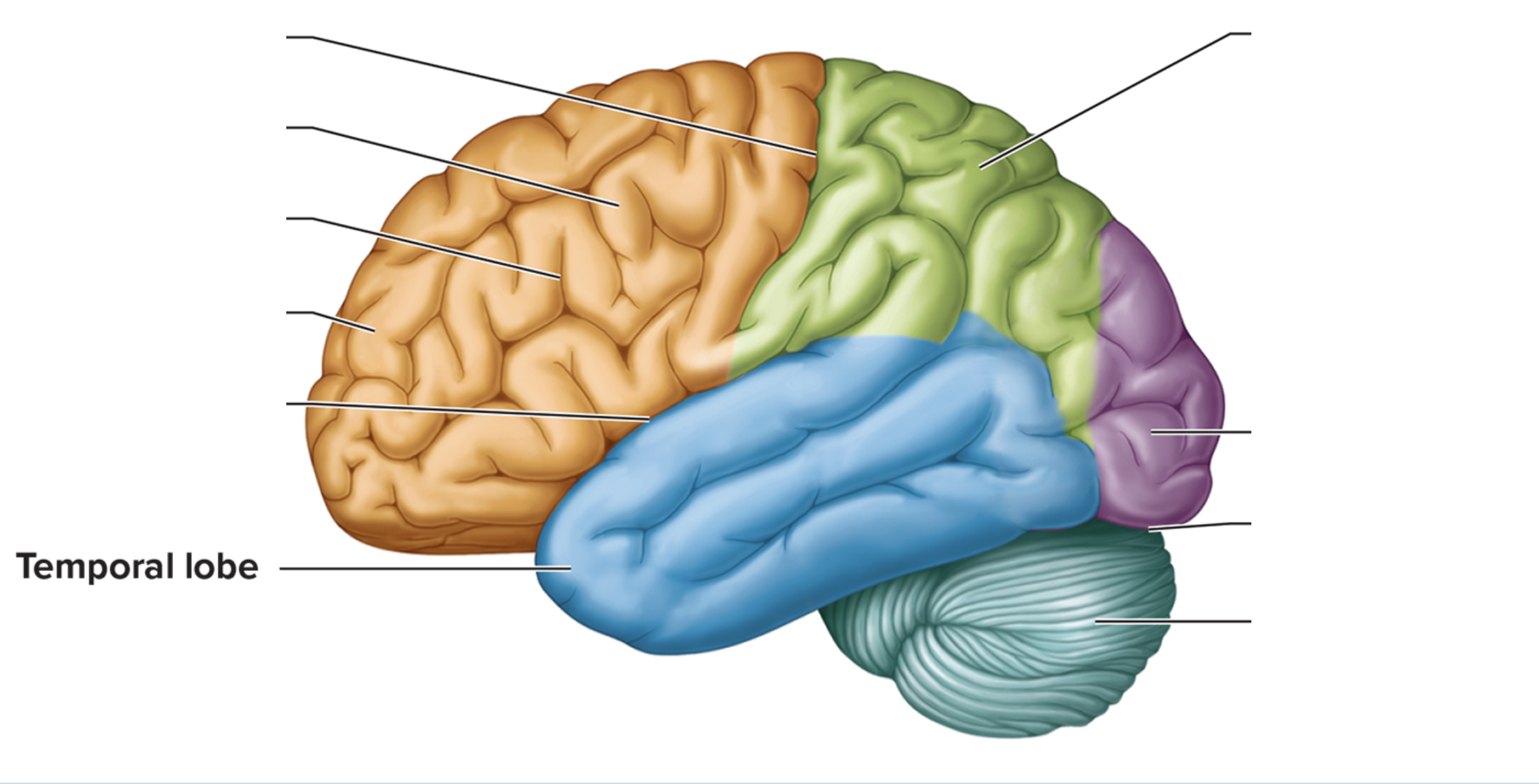
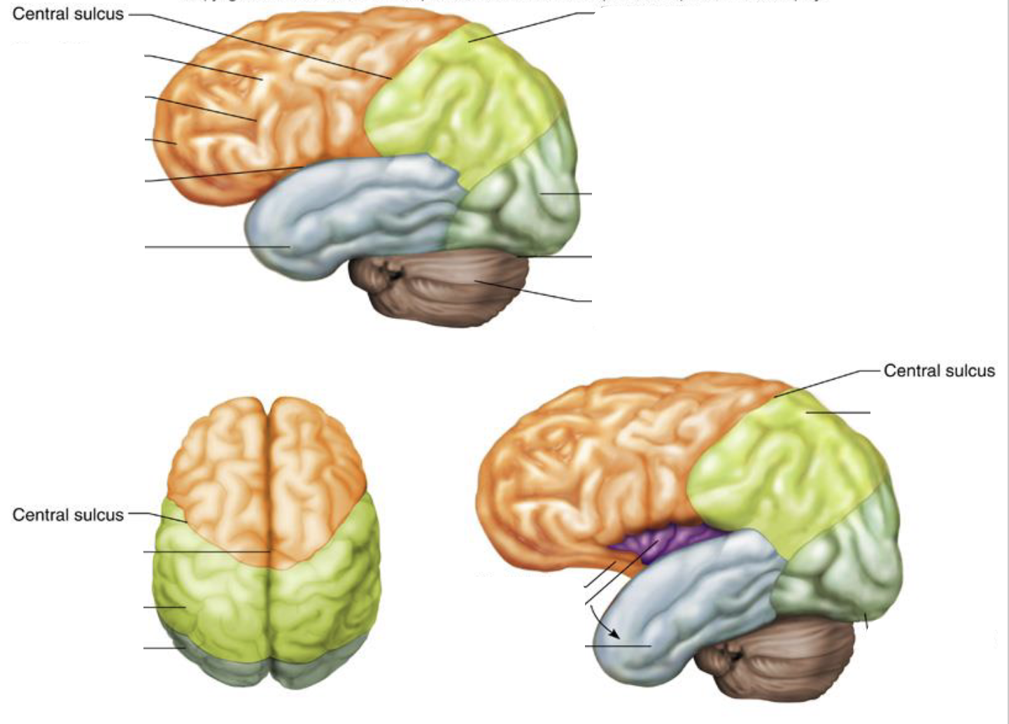
Frontrol lobe
Location: Front of cerebrum; Function: Voluntary motor movement
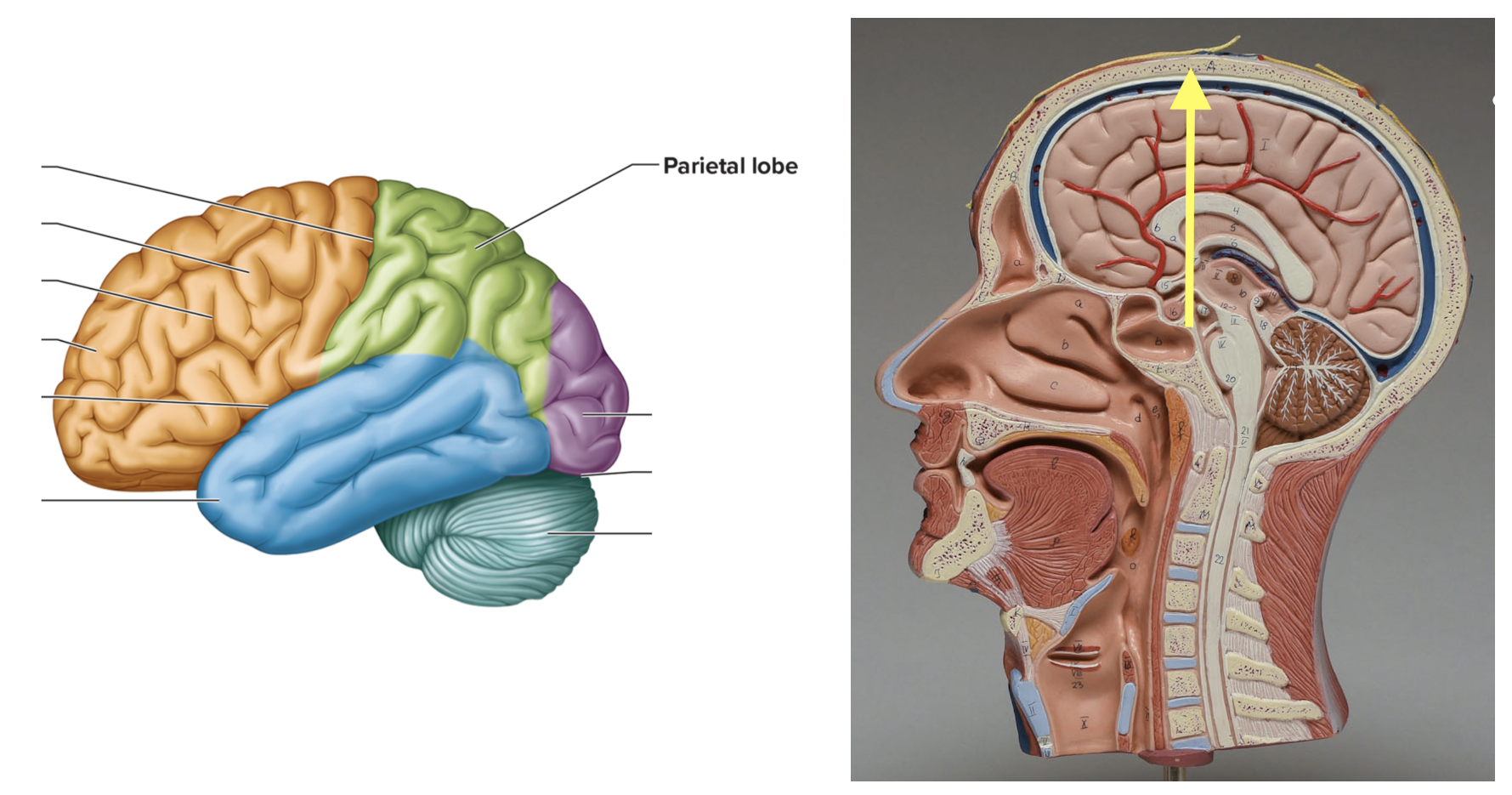
Parietal lobe
Location: Upper middle of cerebrum (skull); Function: Interprets sensory info
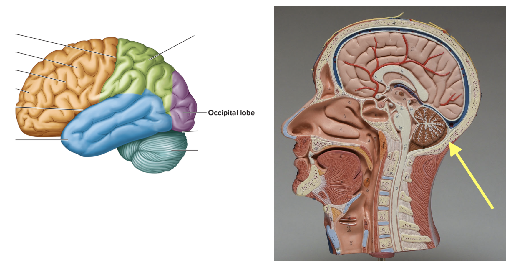
Occipital lobe
Location: Back of the brain; Function: Vision
Temporal lobe
Location: Sides of brain near ears; Function: Hearing
Corpus callosum
Location: Large C-shaped, middle of brain; Function: Connects left & right hemispheres
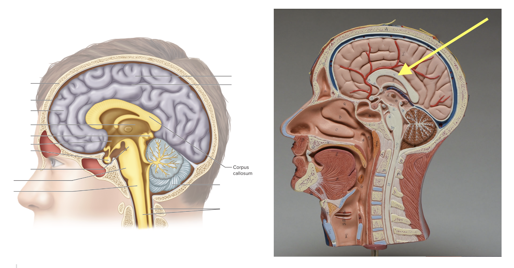
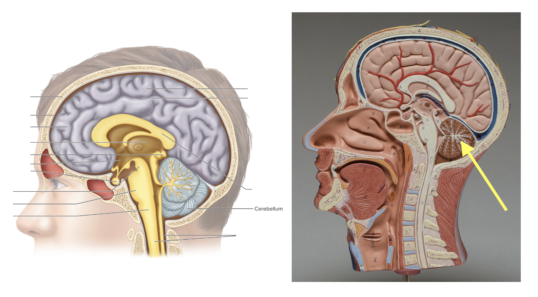
Cerebellum
Location: Rounded tree-like pattern structure in back of brain; Function: Balance & coordination
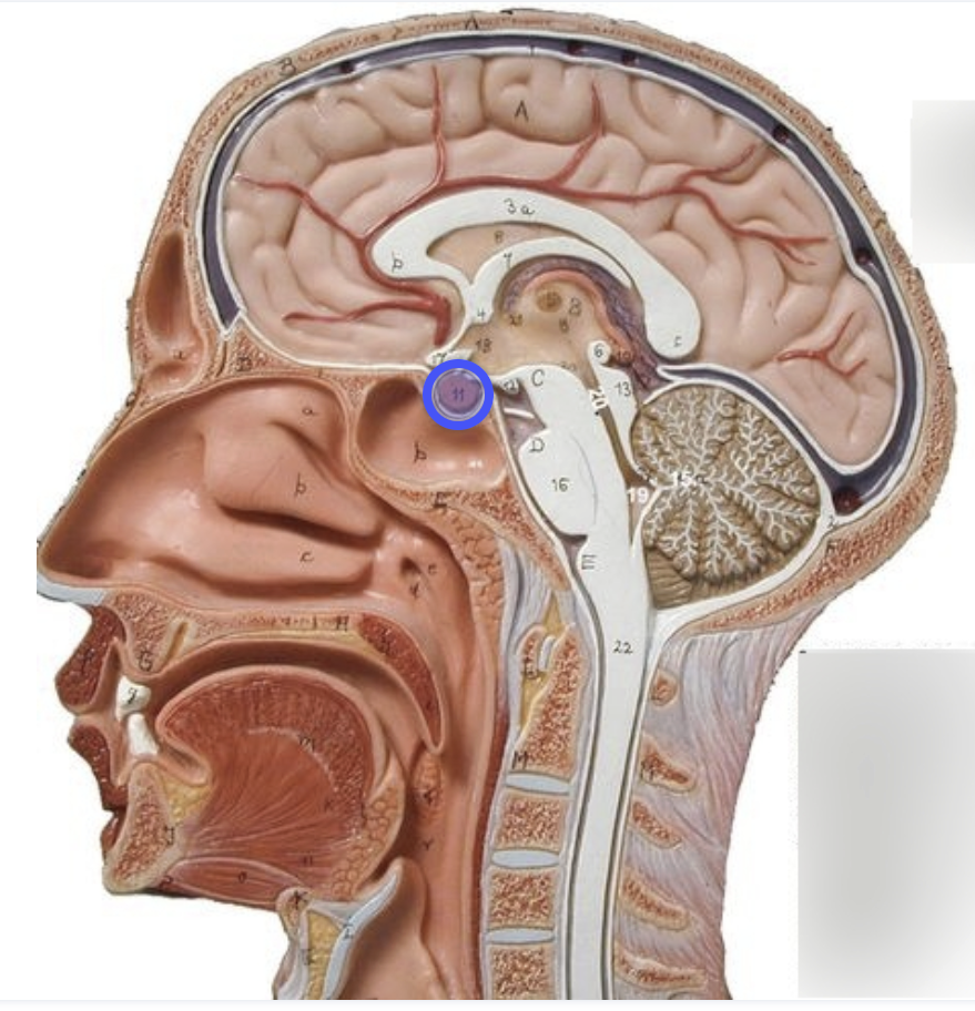
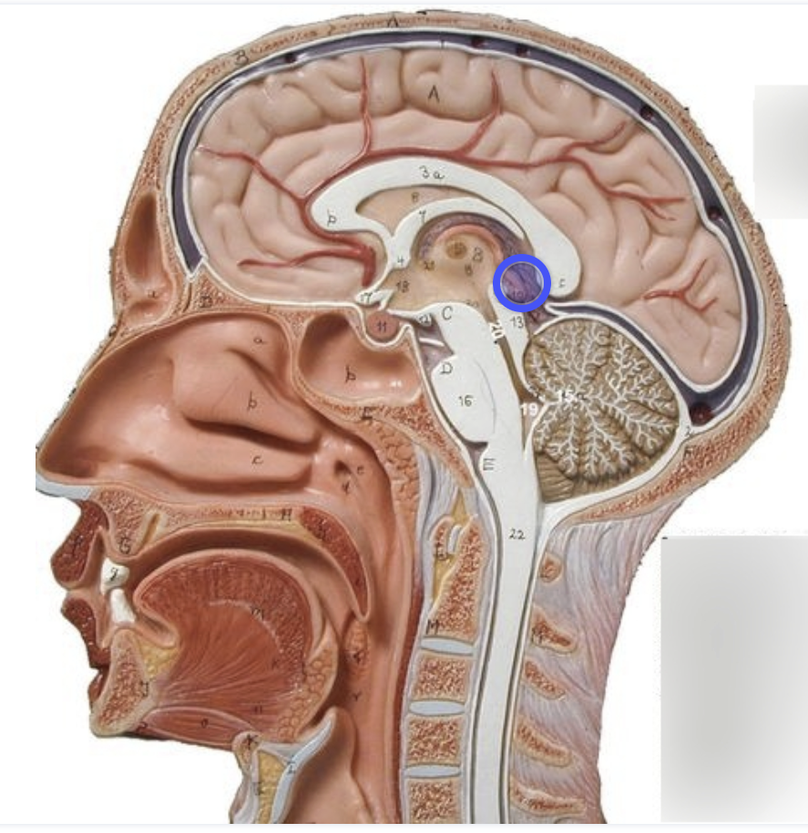
Pituitary gland
Location: Below hypothalmus, attached by infundibulum; Function: Release hormones
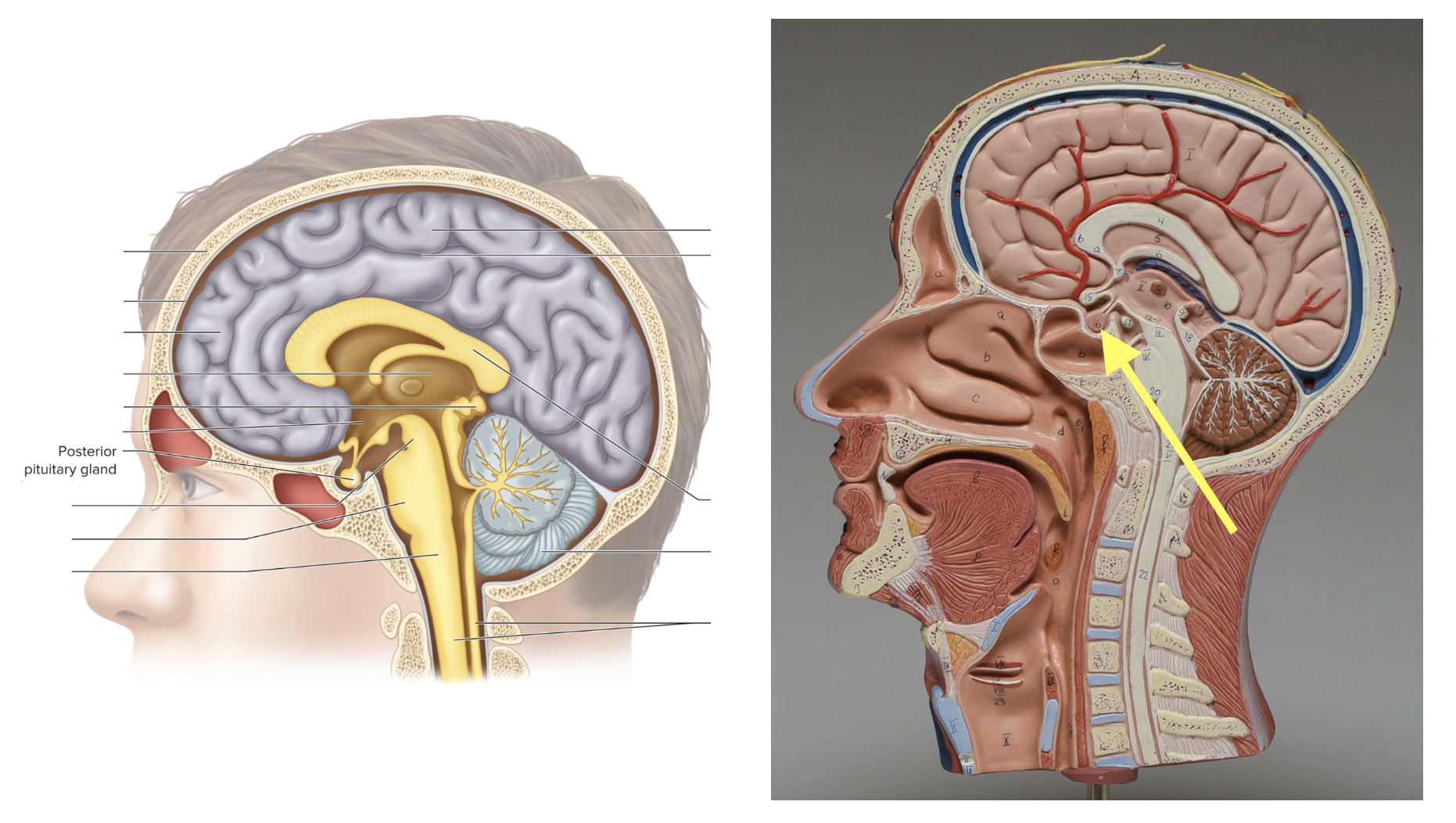
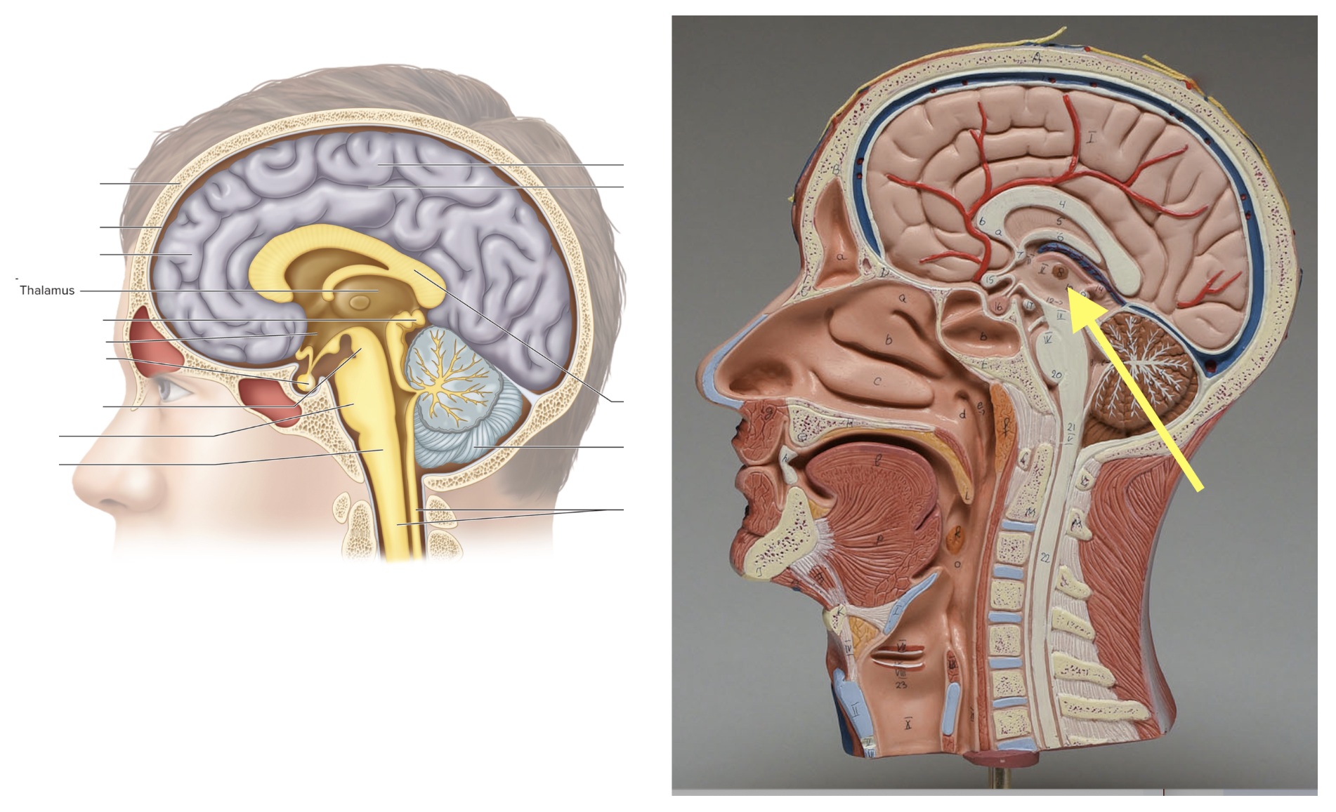
Thalamus
Location: Center of brain; Function: Sensory relay station
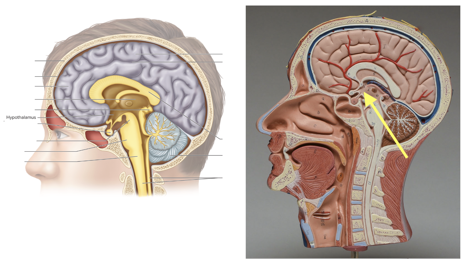
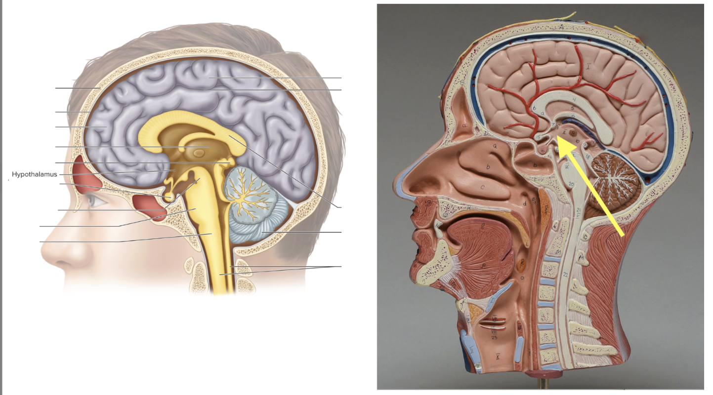
Hypothalamus
Location: Below thalamus; Function: Controls homeostasis
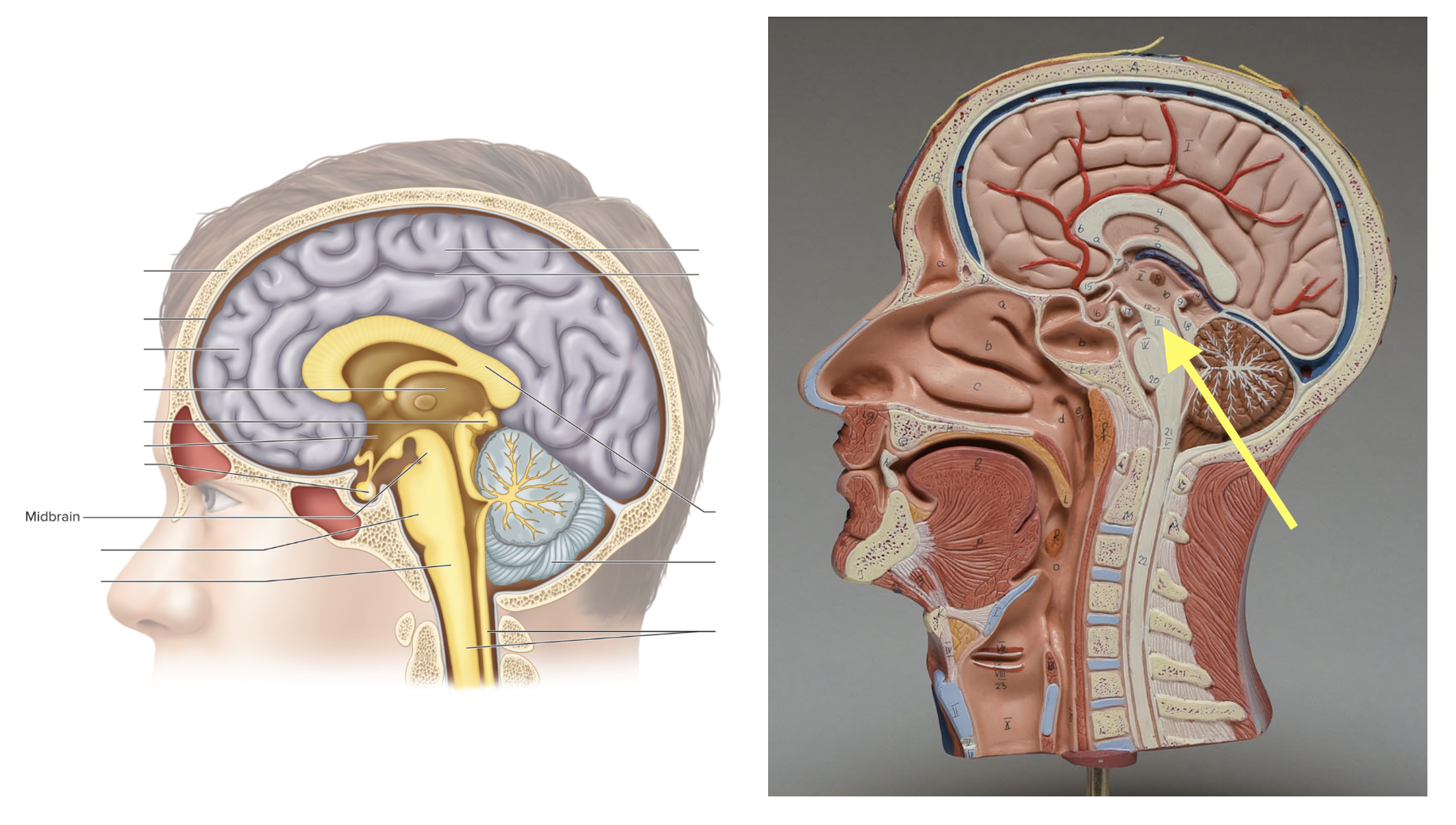
Midbrain
Location: Top of brainstem; Function: Visual & auditory reflexes
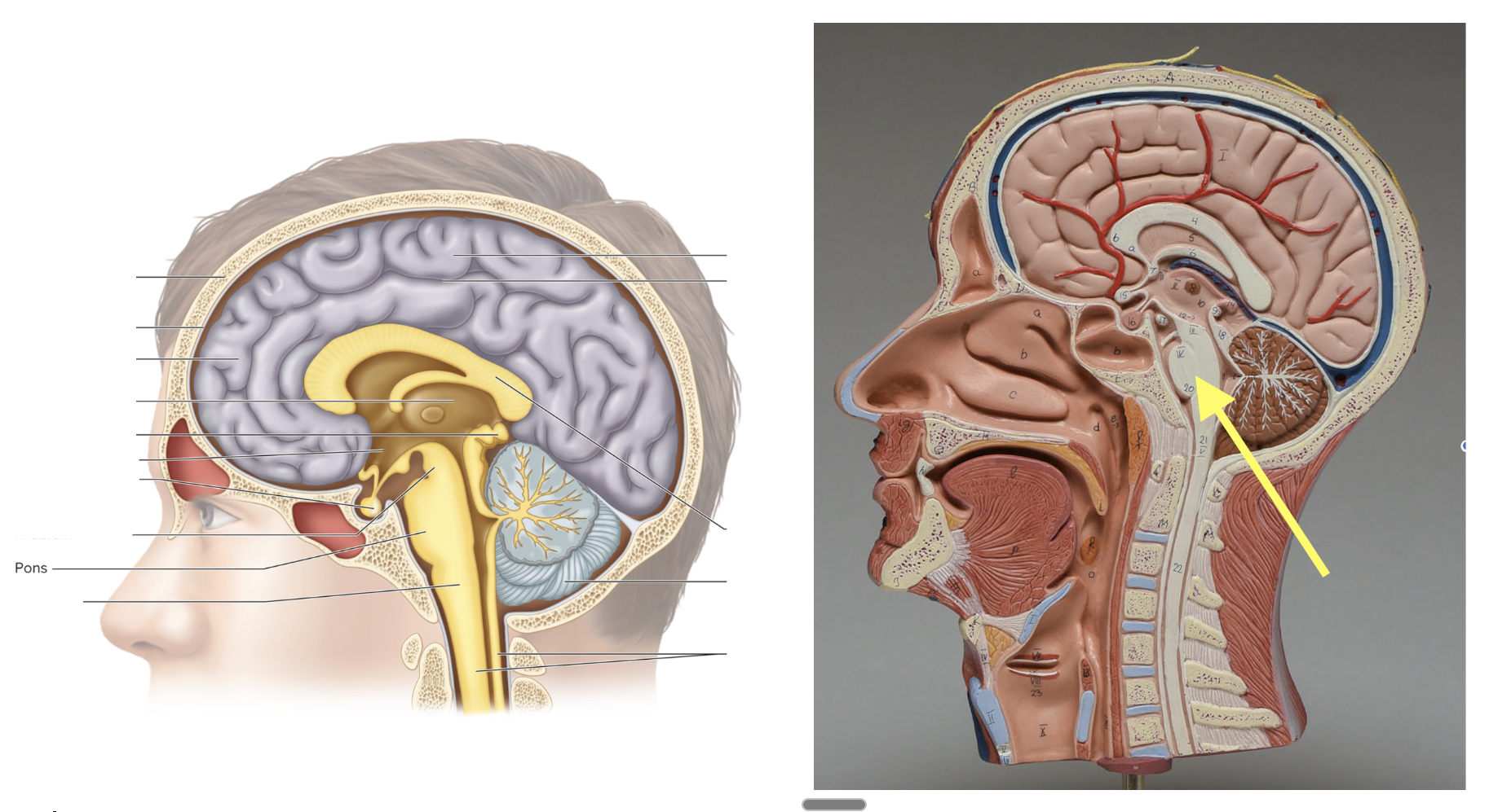
Pons
Location: Rounded bulge on the brainstem (middle); Function: Controls breathing rate
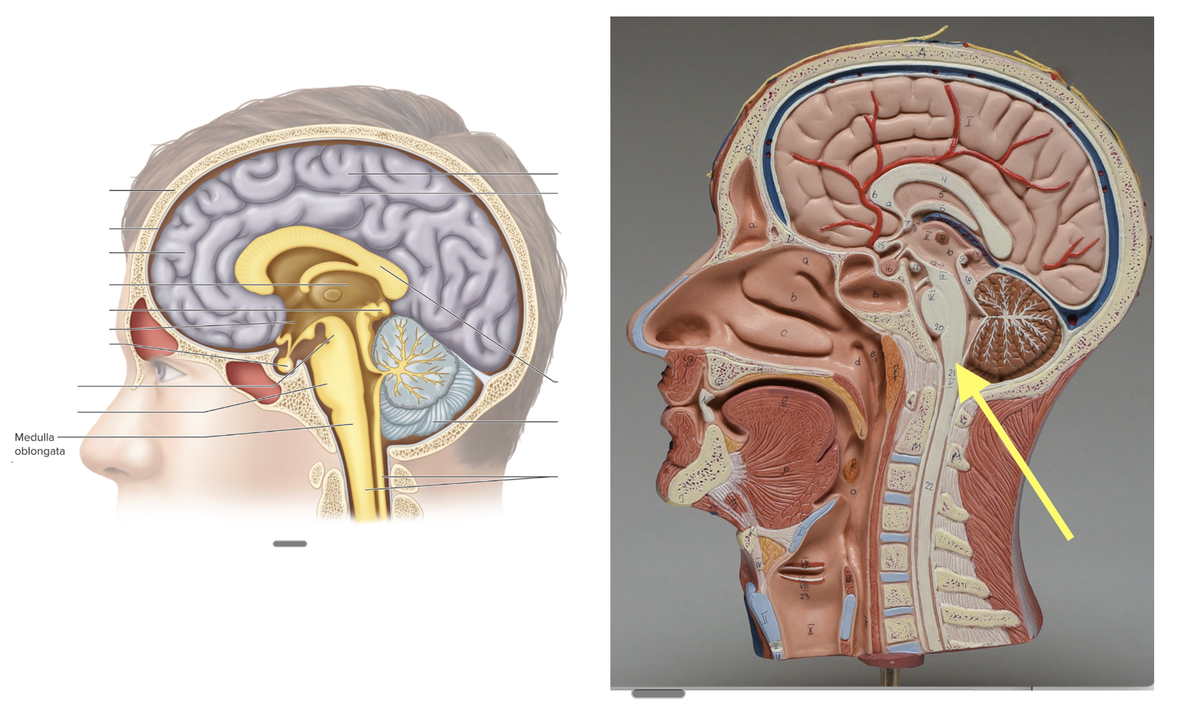
Medulla oblongata
Location: Lowest part of brainstem; Function: Controls heart rate & breathing
Central sulcus
Location: Grove between the frontal and parietal lobes; Function: separates motor and sensory areas
4th ventricle
Location: Between cerebellum & brainstem; Function: Allows CSF to flow
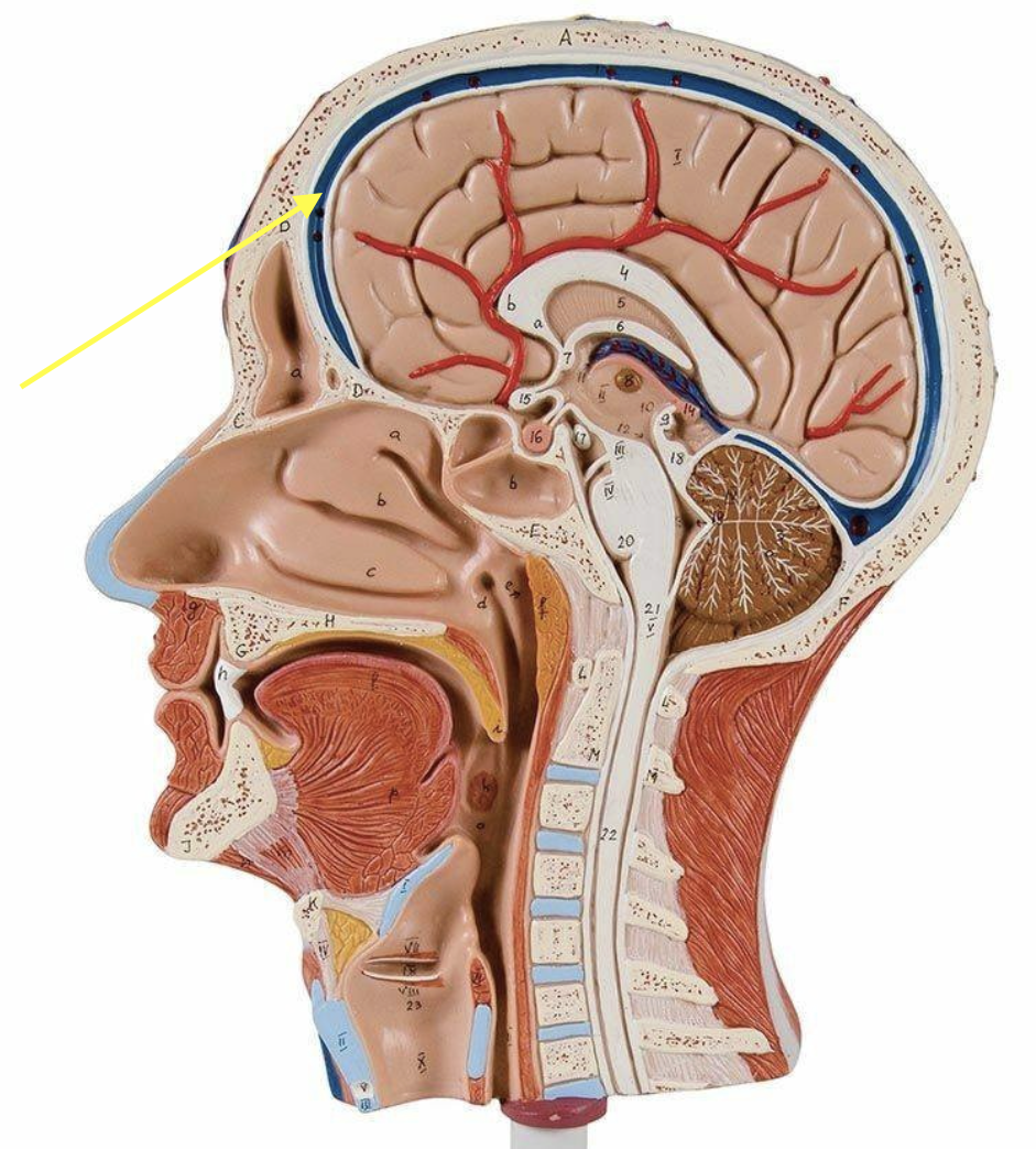
Dural sinus
Location: Within dura mater (between layers); Function: Drains venous blood from brain
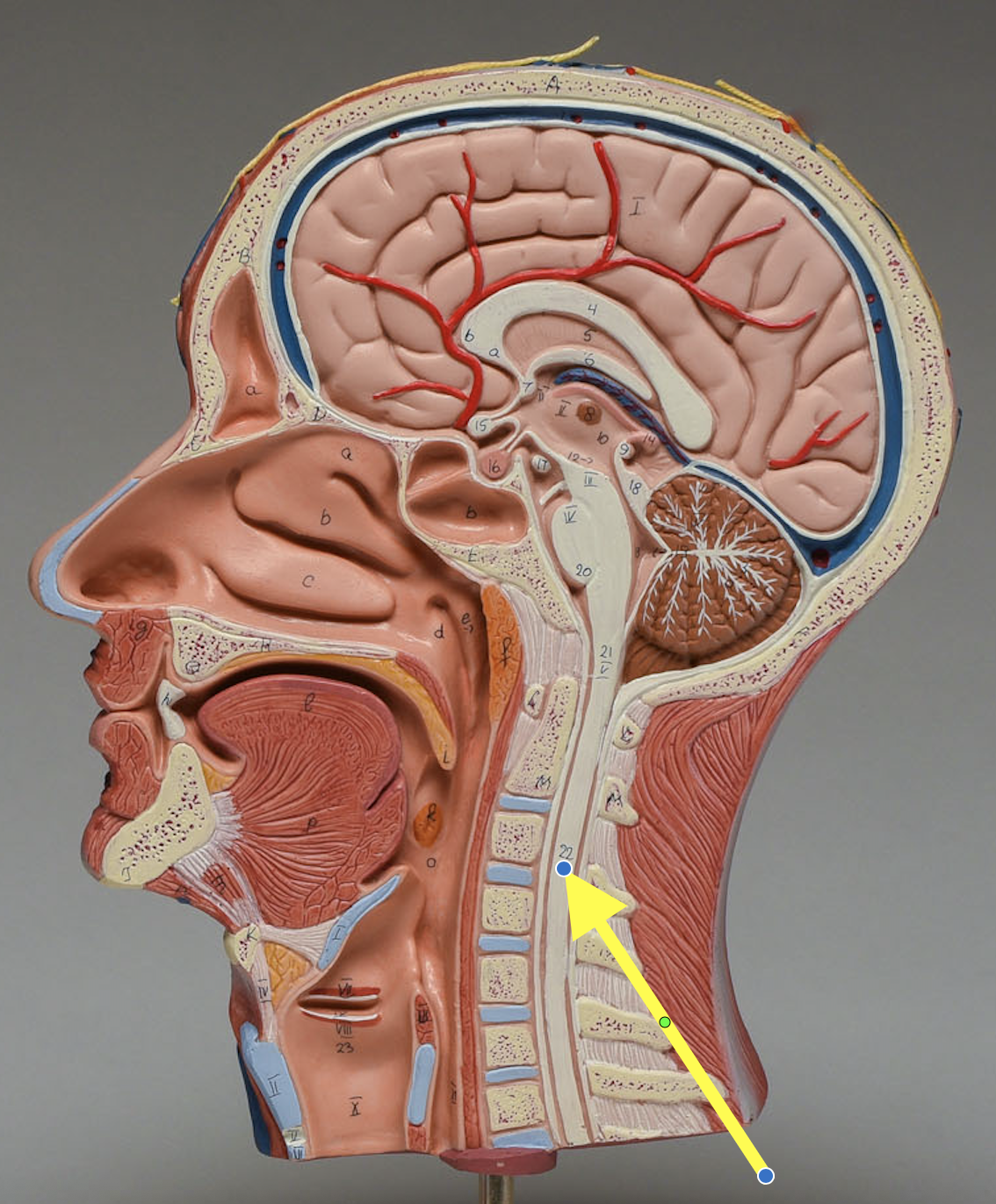
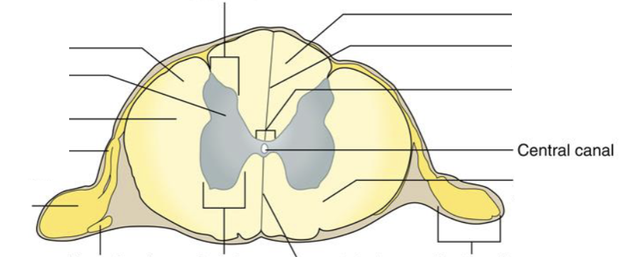
Spinal cord
Location: Inside the spine; Function: Pathway for impulses to/from brain/reflexes
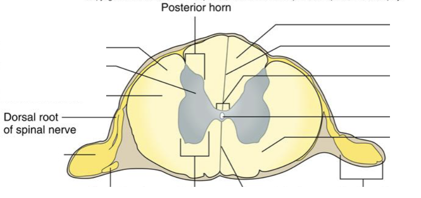
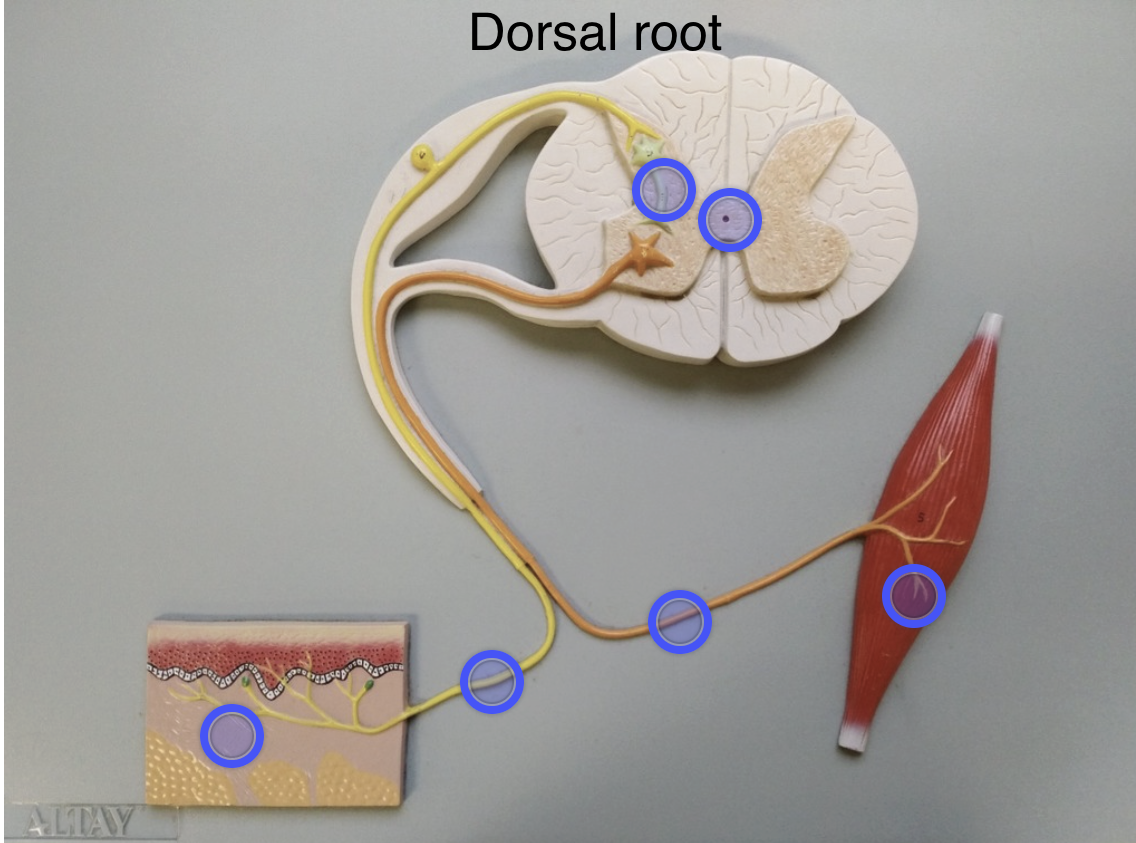
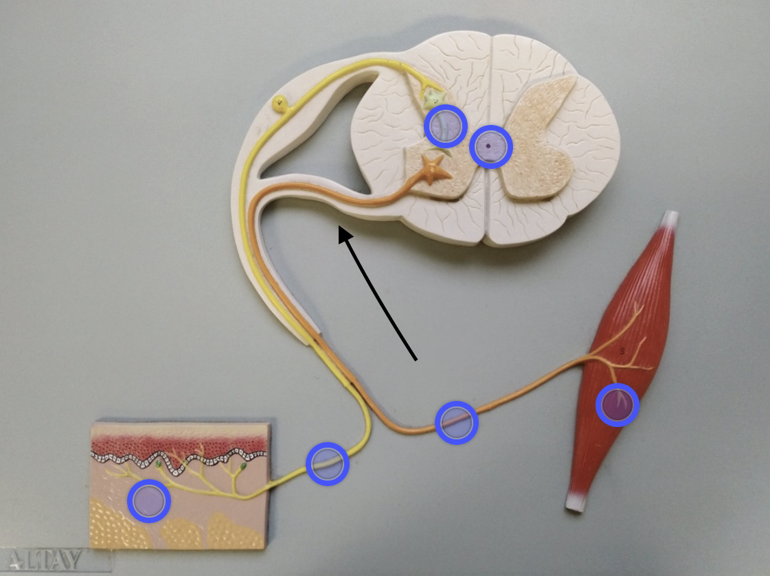
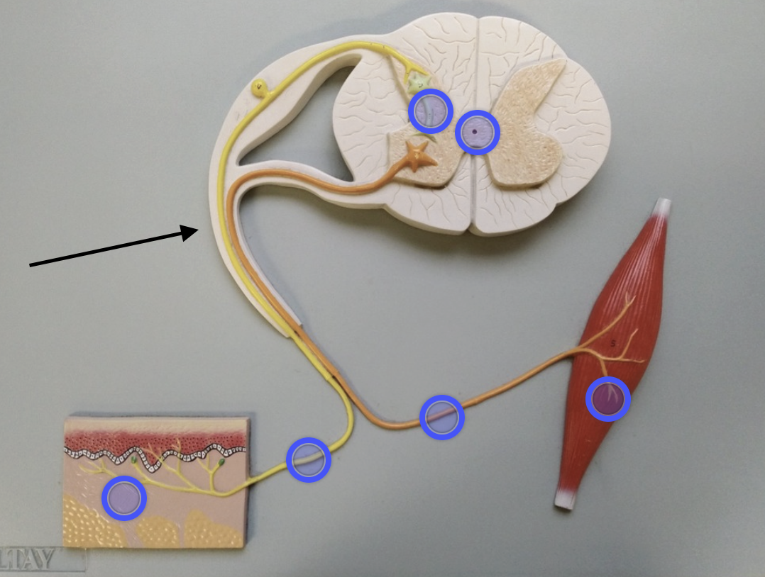
Dorsal roots of spinal nerves
Location: Back side of spinal cord; Function: Carries sensory info TO spinal cord
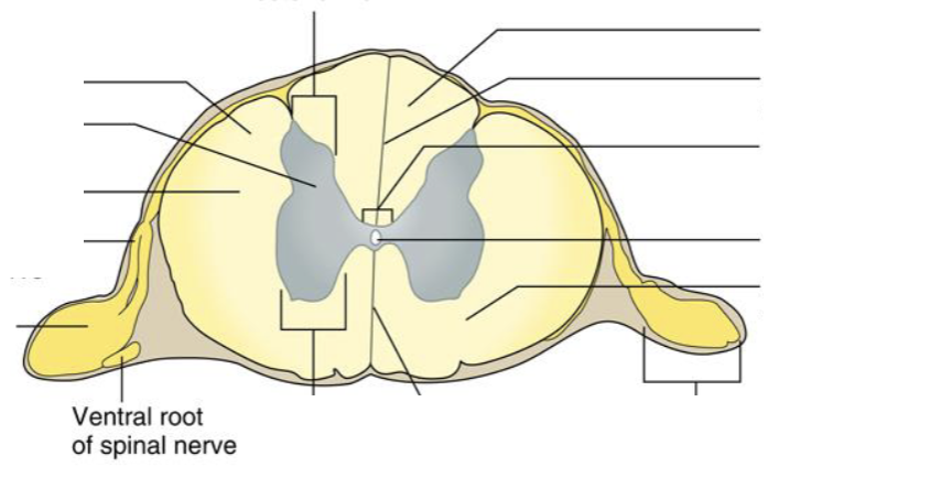
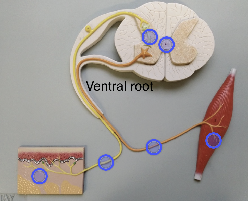
Ventral roots of spinal nerves
Location: Front side of spinal cord; Function: Carries motor signals AWAY from spinal cord
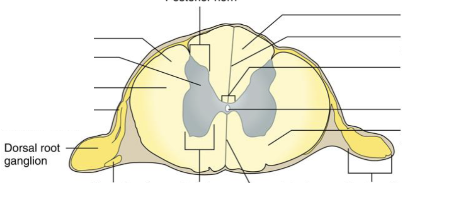
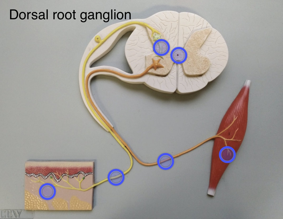
Dorsal root ganglion
Location: Bulge on dorsal root; Function: Contains cell bodies of sensory neurons
Central canal
Location: Center of spinal cord; Function: Contains CSF
Intervertebraal foramen/foramina
Function: Passageway for spinal nerves to exit the spinal cord
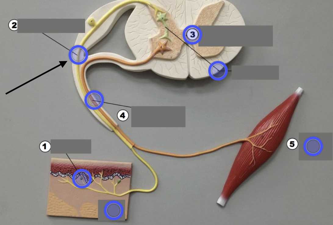
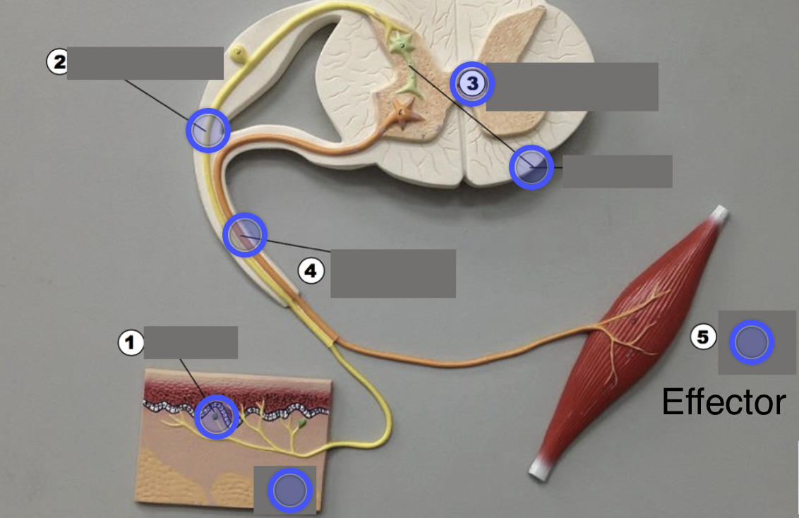
Sensory neuron
(Unipolar) Sends signal to CNS
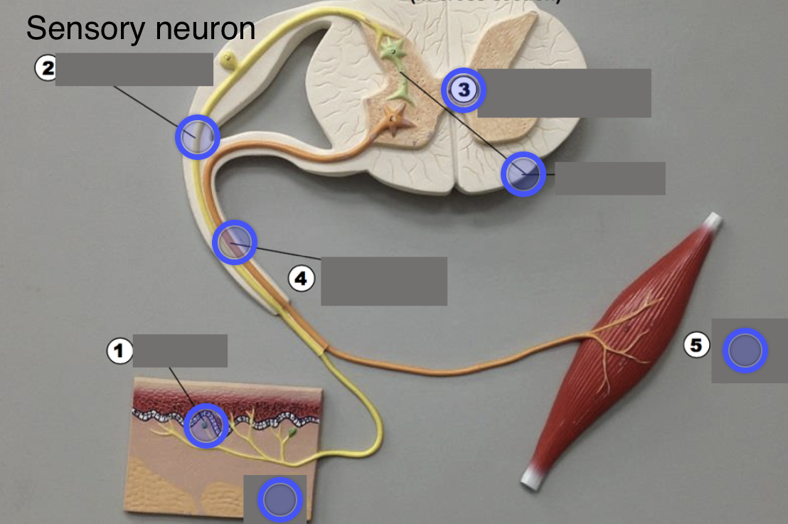
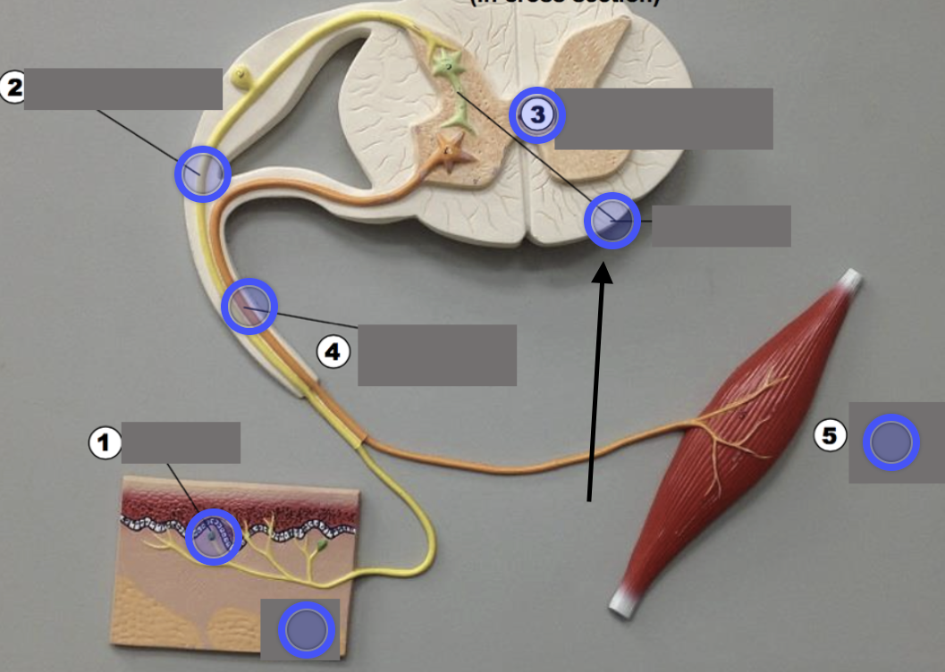
Interneuron
(Multipolar) Processes signal & passes signal
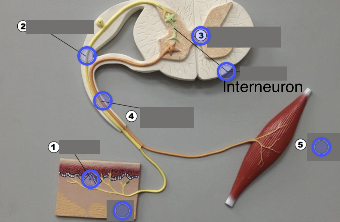
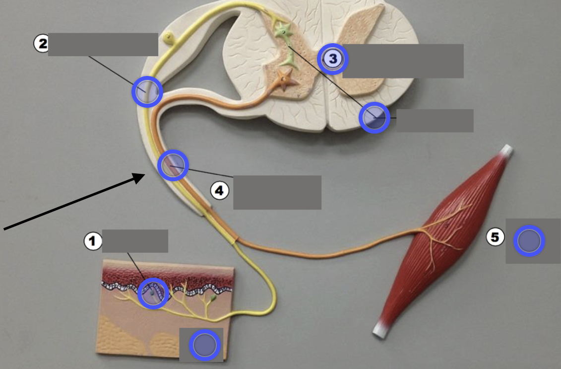
Motor neuron
(Multipolar) Sends signal to effector
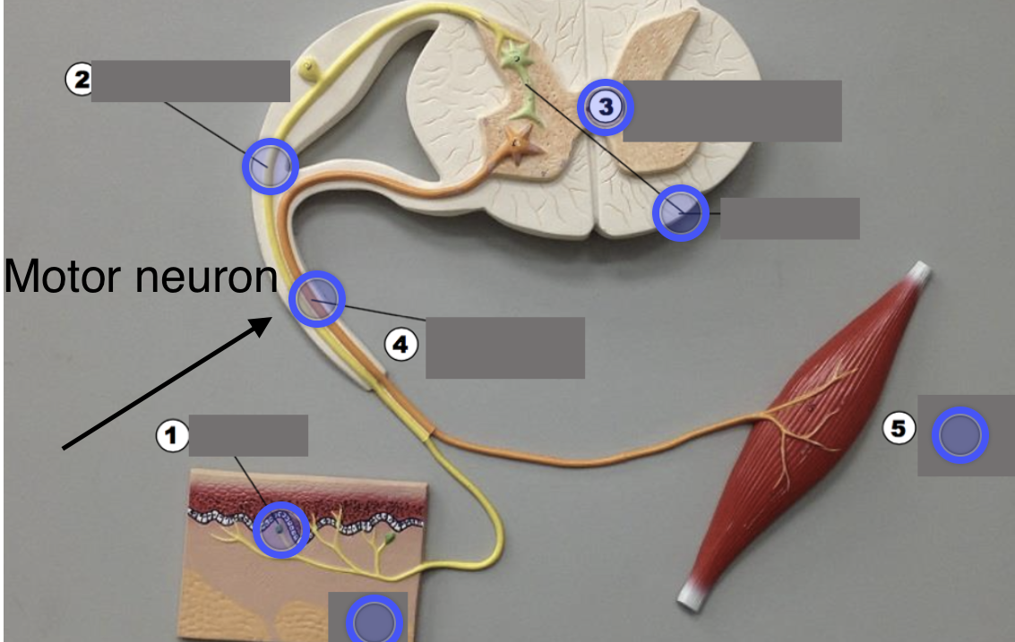
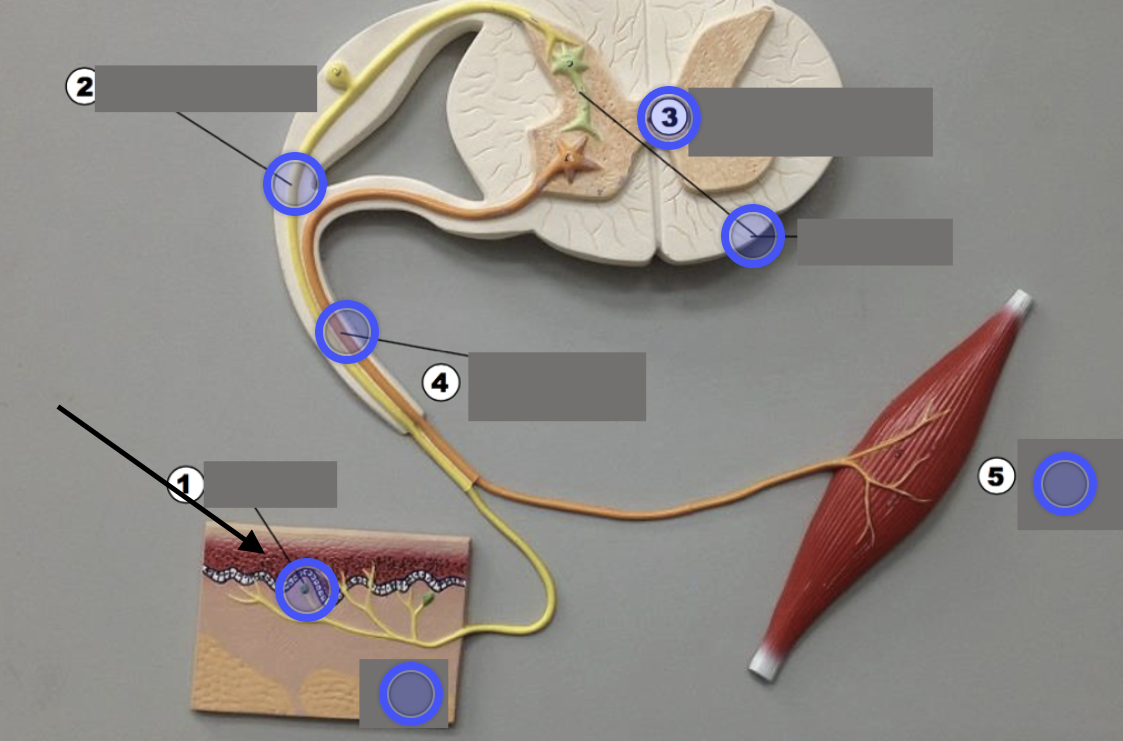
Receptor
Detects stimulus
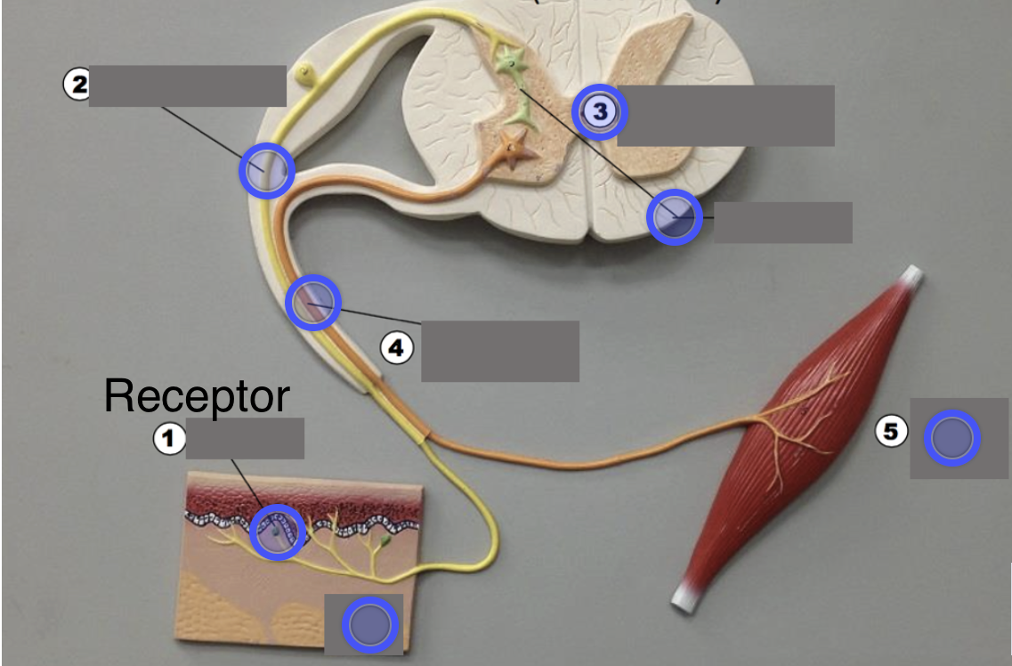
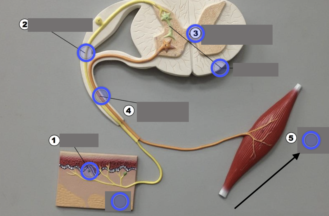
Effector
Carries out response
What is a unipolar neuron?
One process; used for sensory input
What is a multipolar neuron?
Many dendrites and one axon; used for processing and motor output
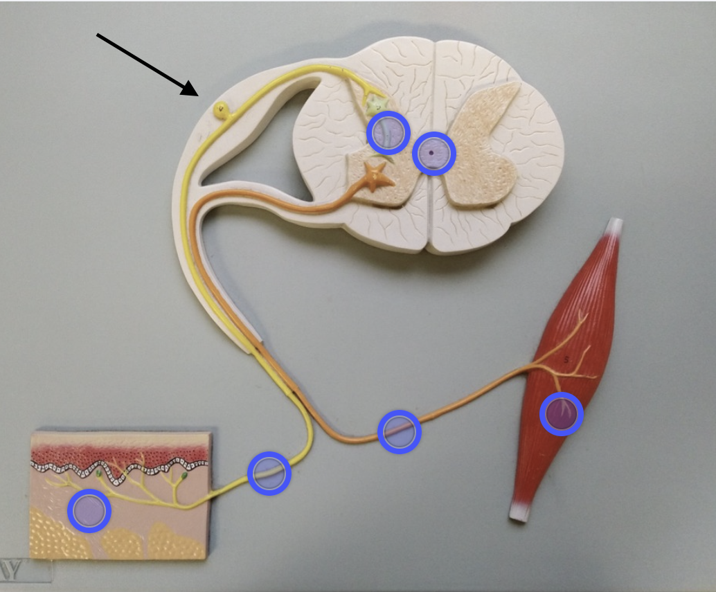
Dorsal root ganglion
Houses sensory neuron cell bodies

Dorsal root
Carries sensory signals INTO the spinal cord
Ventral root
Sends signals from the motor neuron to the effector
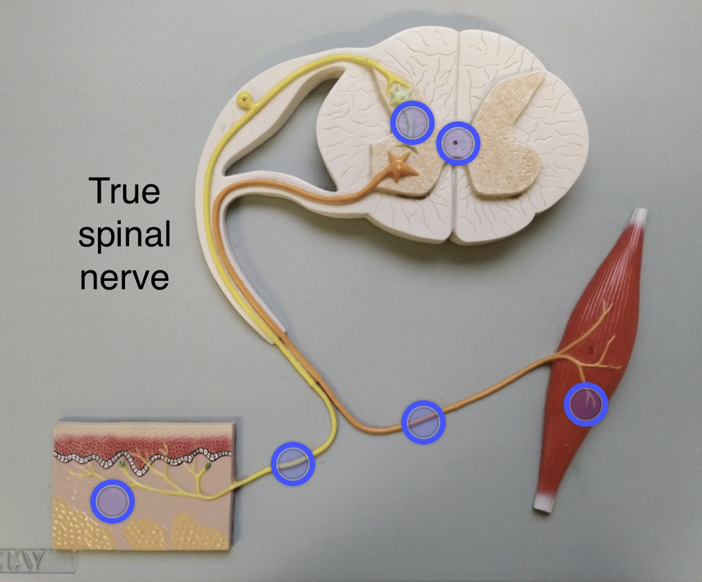
True spinal nerve
Carries both sensory and motor signals
Hypothalamus
Function: Controls pituitary (releasing hormones)
Pitutary gland
Function: “Master gland” (controls other glands)
Pineal gland
Function: Secretes melatonin (sleep cycle)
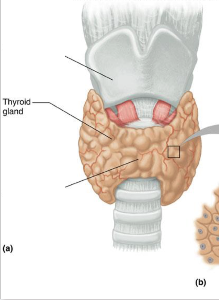
Thyroid gland
Function: Regulates metabolism
Adrenal gland
Function: Stress response (fight-or-flight)
Pancreas
Function: Regulates blood sugar
Testis
Function: Produces testosterone
Ovary
Function: Produces estrogen & progesterone