Unit IV - The Nervous System I
1/80
There's no tags or description
Looks like no tags are added yet.
Name | Mastery | Learn | Test | Matching | Spaced | Call with Kai |
|---|
No analytics yet
Send a link to your students to track their progress
81 Terms
The Nervous System
The endocrine and nervous systems maintain internal coordination
Endocrine: chemical messengers (hormones) delivered to the bloodstream
Nervous: three basic steps
Sense organs receive information
The brain and spinal cord determine responses
The brain and spinal cord issue commands to glands and muscles
Subdivisions of the Nervous System
Two major anatomical subdivisions
Central Nervous System (CNS)
The brain and spinal cord are enclosed in bony coverings
Peripheral Nervous System (PNS)
Nerve: bundle of axons in connective tissue
Ganglion: swelling of cell bodies in a nerve
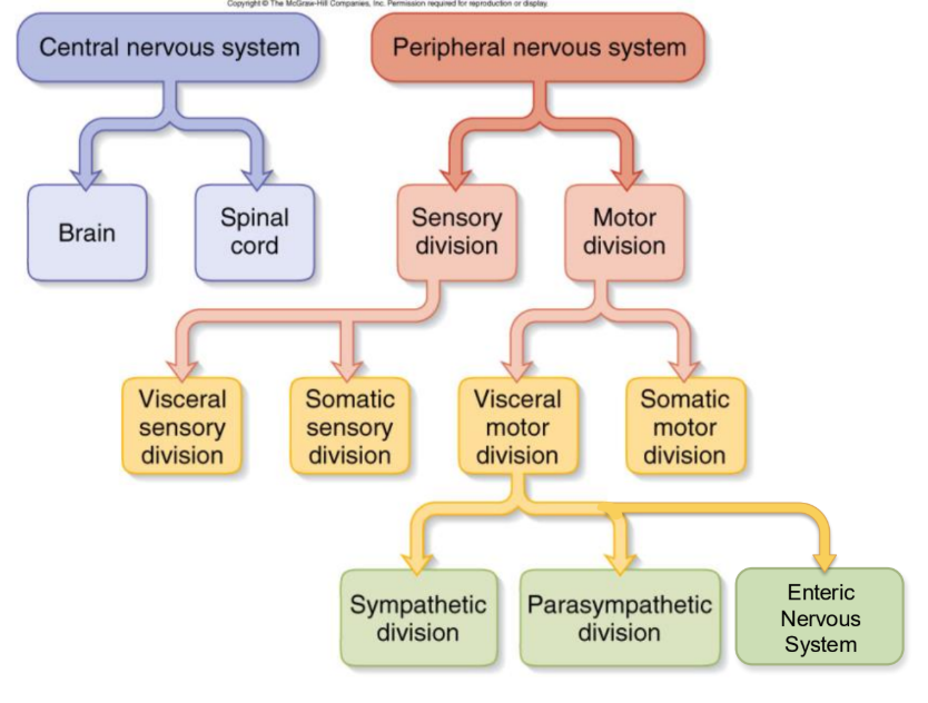
Functional Divisions of the PNS
Sensory (afferent) divisions (receptors to CNS)
Visceral sensory and somatic sensory division
Motor (efferent) division (CNS to effectors)
Visceral motor division (ANS)
Effectors: cardiac, smooth muscle, glands
Sympathetic division (action)
Parasympathetic division (digestion)
Somatic motor division effectors: skeletal muscle
Effectors: skeletal muscle
Sensory (Afferent) Neurons
Afferent Neurons
Detect changes in the body and the external environment
Information transmitted into brain or spinal cord
Interneurons
Association Neurons
Lie between the sensory and motor pathways in the CNS
90% of our neurons are interneurons
Process, store, and retrieve information
Motor Neuron
Efferent Neuron
Send signals out to muscles and gland cells
Organs that carry out responses called effectors
Properties of Neurons
Excitability (irritability)
Ability to respond to internal and external environmental stimuli
Conductivity
Produce traveling electrical signals
Secretion
Secretion of a chemical neurotransmitters from the nerve termini in response to an electrical signal
Structure of a Neuron
Cell Body - Perikaryon Soma
Single, central nucleus with a large nucleolus
Cytoskeleton of microtubules and neurofibrils (bundles of actin filaments)
Lipofuscin is a product of the breakdown of worn-out organelles -- more with age
A vast number of short dendrites
For receiving signals
A single axon (nerve fiber) arising from the axon hillock for rapid conduction
Axoplasm, axolemma, and synaptic vesicles
Multipolar Neuron
Most common
Many dendrites / one axon
Very common in the cerebellum
Bipolar Neuron
One dendrite / one axon
Olfactory, retina, ear
Unipolar Neuron
Sensory from the skin and organs to the spinal cord
Anaxonic Neuron
Many dendrites / no axon
Help in visual processes
Axonal Transport
Proteins made in soma must be transported to axon and axon terminal
Repairs axolemma, for gated ion channel proteins, as enzymes or neurotransmitters
Fast anterograde axonal transport
Either direction up to 400 mm / day for organelles, enzymes, vesicles and small molecules
Fast retrograde for recycled materials and pathogens
Slow axonal transport or axoplasmic flow
Moves cytoskeletal and new axoplasm at 10 mm / day during repair and regeneration in damaged axons
Can take anywhere from a few months to a few years
Axonal Transportation is Bidirectional
Substances travel continuously along the axon in both directions
Anterograde movement: movement AWAY from the cell body
Move substances needed to make neurotransmitters
Some neurotransmitters are made in the cell body, packaged into vesicles, then transported to axon terminals
Retrograde movement: movement TOWARD the cell body
Return substances to be degraded or recycled by the cell body
Move molecules like nerve growth factor that activates certain genes that promote growth
Neurons
Excitable nerve cells
Respond to stimuli by changing their action potential
Transmit electrical signals
Neuroglia
Glial cells, Glia
Far outnumber neurons
Surround / wrap neurons
Support, insulate, protect neurons
Structure
Central cell body with branching “processes” (extensions)
Smaller in size and nuclei stain darker than neurons
Cells of the CNS
Astrocytes - MOST IMPORTANT!
Microglial cells
Ependymal cells
Oligodendrocytes
Cells of the PNS
Satellite cells
Schwann cells
CNS Neuroglia - Astrocytes
Abundant, star-shaped cells
Barrier between capillaries and neurons
Control brain environment
MOST IMPORTANT
CNS Neuroglia - Microglia
Spiderlike phagocytes
Dispose of debris
SECOND MOST IMPORTANT
CNS Neuroglia - Ependymal Cells
Line cavities of the brain and spinal cord
Cilia assist with circulation of cerebrospinal fluid
CNS Neuroglia - Oligodendrocytes
Wrap around nerve fibers in the central nervous system
Produce myelin sheaths
Astrocytes
Most abundant and versatile glial cell
Forms the framework of the CNS
Support and brace neurons
Connect them to their nutrient supply lines
Control the chemical environment around neurons
“Mop up” leaked potassium ions
Recapture and recycle released neurotransmitters
Contribute to the BBB (blood-brain barrier)
Regulate the composition of brain tissue fluid
Convert glucose to lactate to feed neurons
Secretes nerve growth factor to promote synapse formation
Respond to and influence synaptic signaling
Sclerosis – hardened astrocyte mass replaces damaged neurons
Microglial Cells
Small, ovoid cells with long “thorny” processes
Monitor the status of nearby neurons
Migrate toward damaged neurons
Transform into specialized macrophages that phagocytose microbes and neuronal debris:
In the presence of invading microorganisms or dead neurons
e.g., in areas of infection, trauma, or stroke
Important since immune cells have limited access to CNS
Important for synapse elimination or pruning
Synaptic Pruning
Our body’s way of maintaining more efficient brain function as we get older and learn new complex information
Implicated in autism and schizophrenia
Ependymal Cells
Ranges in shape from squamous to columnar—many are ciliated
Line the central cavities of the brain and spinal cord
Form a fairly permeable barrier between:
Cerebrospinal fluid (CSF) fills the CNS cavities
Interstitial fluid (ISF) tissues bathing the cells
Beating of their cilia helps circulate CSF that cushions the brain and spinal cord
Oligodendrocytes
Wrap around and insulate axons
Branching cells with fewer processes than astrocytes
Line up along the thicker nerve fibers in the CNS
Wrap their processes tightly around the fibers
Produce an insulating covering called a myelin sheath
PNS Neuroglia - Satellite Cells
Like Microglia
Surround, protect, and cushion neuronal cell bodies
Thought to function similarly to astrocytes
PNS Neuroglia - Schwann Cells
The live area of the cell
Form myelin sheath in PNS
Functionally similar to oligodendrocytes
Vital to regeneration of damaged peripheral nerve fibers
Myelin
Insulating layer around a nerve fiber
Oligodendrocytes in the CNS and Schwann cells in the PNS
Formed from wrappings of the plasma membrane
20% protein and 80 % lipid (looks white)
All myelination is completed by late adolescence
In PNS, hundreds of layers wrap the axon
The outermost coil is schwann cell (neurilemma)
Covered by basal lamina and endoneurium
In CNS - no neurilemma or endoneurium
Oligodendrocytes myelinate several fibers
Myelination spirals inward with new layers pushed under the older ones
Gaps between myelin segments: Nodes of Ranvier
The initial segment (area before 1st schwann cell) and the axon hillock form a trigger zone where signals begin
Speed of Nerve Signals
Diameter of fiber and presence of myelin
Large fibers have more surface area for signals
Speeds
Small, unmyelinated fibers = 0.5 - 2.0 m / sec
Small, myelinated fibers = 3 - 15.0 m / sec
Large, myelinated fibers = up to 120 m / sec
Functions
Slow signals supply the stomach and dilate pupil
Fast signals supply skeletal muscles and transport sensory signals for vision and balance
Electrical Potentials and Currents
Nerve pathway is a series of separate cells
Neural communication: mechanisms for producing electrical potentials and currents
Electrical potential - different concentrations of charged particles in different parts of the cell
Electrical current - flow of charged particles from one point to another within the cell
Living cells are polarized
Resting membrane potential is -70 mV with a negative charge on the inside of membrane
The Role of Ion Channels
Types of plasma membrane ion channels:
Passive, or leakage, channels – always open
Chemically gated channels – open with the binding of a specific neurotransmitter
Voltage-gated channels – open and close in response to membrane potential
Operation of a Chemically - Gated Channel
Example: ACh Receptor channel
Closed when a neurotransmitter (ACh) is not bound to the extracellular receptor
Na+ cannot enter the cell and K+ cannot exit the cell
Operation of a Voltage - Gated Channel
Example: Voltage Gated Na+ channel
Closed when the intracellular environment has negative voltage
Na+ cannot enter the cell
Resting Membrane Potential ( I )
Unequal electrolyte distribution between ECF (Extracellular Fluid) / ICF (Intracellular Fluid)
Diffusion of ions down their concentration gradients
Selective permeability of the plasma membrane
Electrical attraction of cations and anions
The membrane is very permeable to K+
Leaks out until an electrical gradient is created, attracting it back in
Cytoplasmic anions can not escape due to size or charge (PO42-, SO42-, organic acids, proteins)
The membrane is much less permeable to Na+
Na+ / K+ pumps out 3 Na+ for every 2 K+ it brings in
Works continuously and requires a great deal of ATP
Necessitates that glucose and oxygen be supplied to nerve tissue
Resting Membrane Potential ( II )
Resting Membrane Potential: potential difference across the membrane
Basic Neuron: -70mV: negative inside, relative to outside
Two factors generate the resting membrane potential:
Concentration—differences in ionic composition of intracellular and extracellular fluids
Permeability—differences in the plasma membrane’s permeability to those ions
Cytosol has lower [Na+] and higher [K+] than the outside (extracellular fluid)
Active transport by Na+ / K+ - ATPase sets concentration gradients
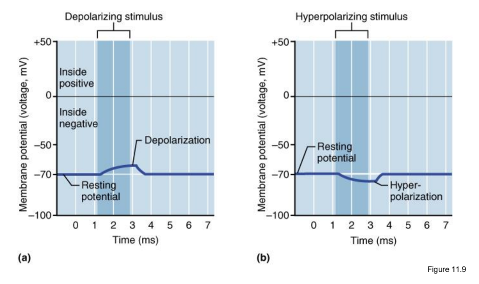
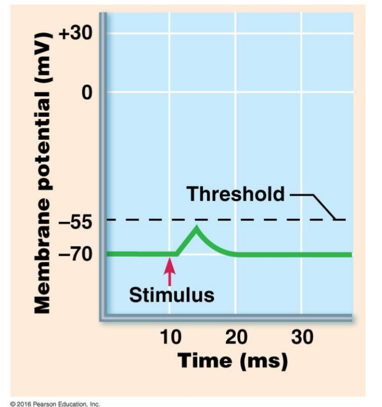
Local Potentials
Local disturbances in membrane potential
Occurs when a neuron is stimulated by chemicals, light, heat, or mechanical disturbance
Depolarization decreases the potential across the cell membrane due to the opening of gated Na+ channels
Na+ rushes in down concentration and electrical gradients
Na+ diffuses for a short distance inside the membrane, producing a change in voltage called a local potential
Differences from action potentials
Are graded (vary in magnitude with stimulus strength)
Are decremental (get weaker the farther they spread)
Are reversible as K+ diffuses out of cell
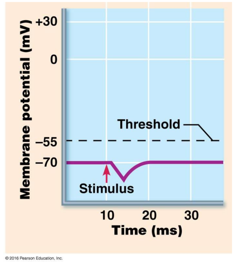
Can be either excitatory or inhibitory (hyperpolarize)
Changes in Membrane Potential
Graded Potentials
Like a Ripple - the bigger the stimulus, the bigger the response!
Voltage changes in graded potentials are decremental
Current is quickly dissipated due to the leaky plasma membrane
Can only travel over short distances
Action Potentials
More dramatic changes in areas of high density of voltage-gated channels occur
Trigger zone up to 500 channels / um2 (normal is 75)
If threshold potential (-55mV) is reached voltage-gated Na+ channels open (Na+ enters causing depolarization)
Past 0 mV, Na+ channels close: depolarization
Slow K+ gates fully open
K+ exits, repolarizing the cell
Negative overshoot produces hyperpolarization, excessive exiting of K+
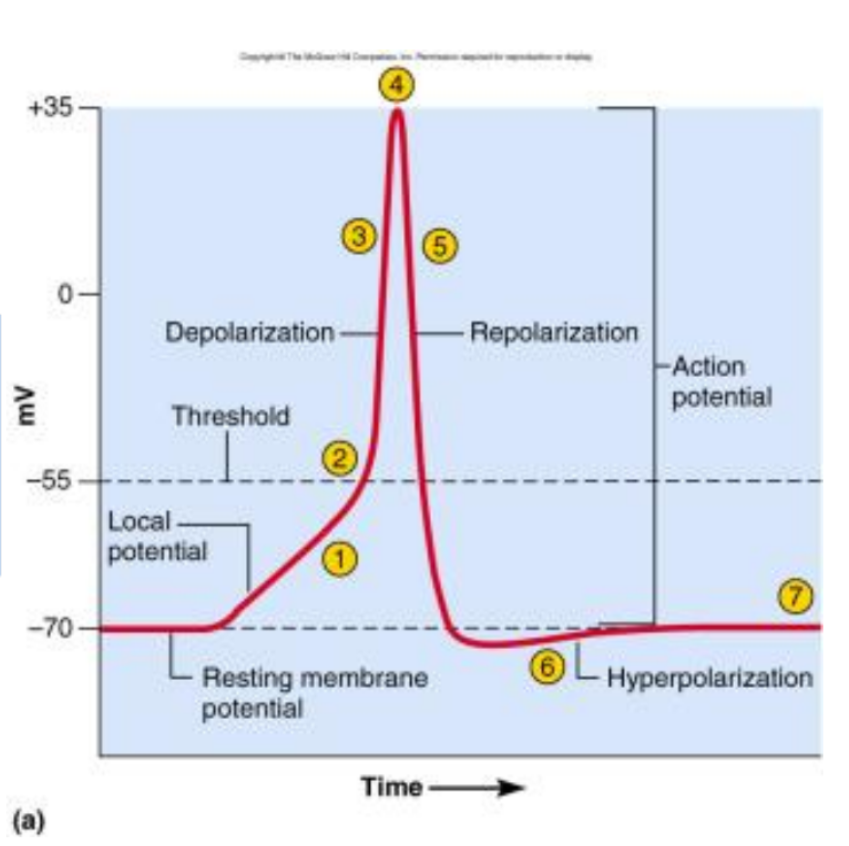
Characteristics of AP
Called a spike
Follows an all-or-none law
Voltage gates either open or don’t
Nondecremental (do not get weaker with distance)
Irreversible (once started, it goes to completion and can not be stopped)
The Refractory Period
Period of resistance to stimulation
Absolute refractory period
As long as Na+ gates are open
No stimulus will trigger AP
Relative refractory period
As long as K+ gates are open
Only especially strong stimulus will trigger new AP
Refractory period occurs only to a small patch of membrane at one time (quickly recovers)
Impulse Conduction in Unmyelinated Fibers
The threshold voltage in the trigger zone begins the impulse
Nerve signal (impulse) - a chain reaction of sequential opening of voltage-gated Na+ channels down the entire length of the axon
Nerve signal (nondecremental) travels at 2m / sec
The signal is like a wave / ripple
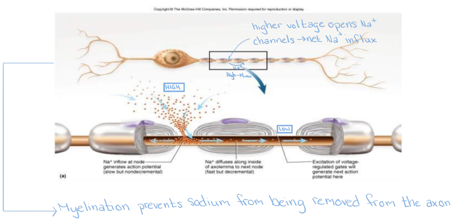
Saltatory Conduction
The rapid propagation of action potentials along myelinated axons, jumping between gaps called Nodes of Ranvier
Saltatory Conduction - Myelinated Fibers
Voltage-gated channels needed for APs
Fewer than 25 per um2 in myelin-covered regions
Up to 12,000 per um2 in nodes of Ranvier
Fast Na+ diffusion occurs between nodes
Multiple Sclerosis
An autoimmune disease that mainly affects young adults - mainly women
Symptoms include visual disturbances, weakness, loss of muscular control, and urinary incontinence
Nerve fibers are severed, and myelin sheaths in the CNS become nonfunctional scleroses
Shunting and short-circuiting of nerve impulses occur
Treatments include injections of methylprednisolone and beta interferon
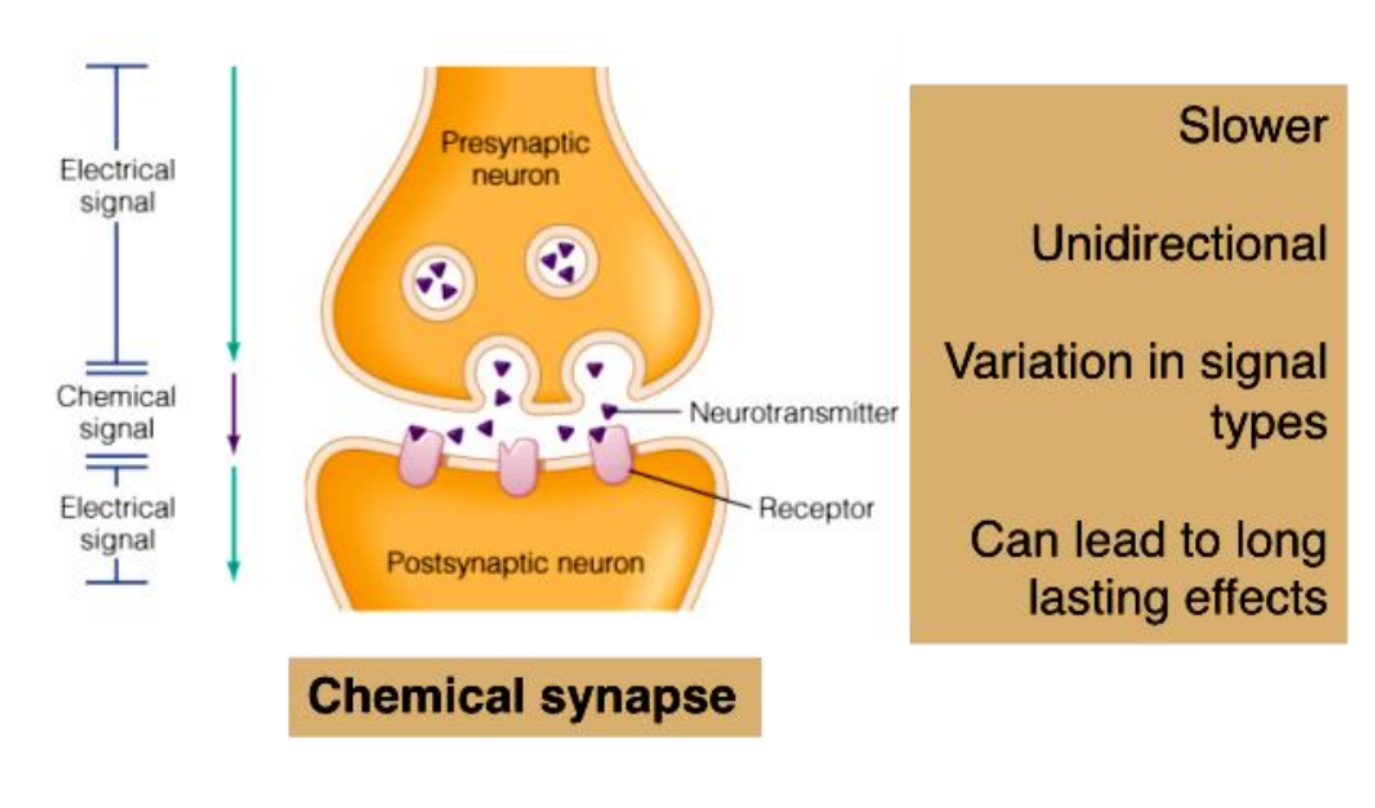
Synaptic Transmission
Action Potential (AP) arrives at the axon terminal
Calcium Influx - voltage-gated Ca2+ channels open and Ca2+ enters the axon terminal
Neurotransmitter (NT) Release - Ca2+ entry causes synaptic vesicles to release NT by exocytosis
Diffusion - NT diffuses across the synaptic cleft
Receptor Binding / Response - binding of NT opens ion channels, leading to graded postsynaptic potentials
Termination - NT effects are terminated via reuptake, degradation, or diffusion from the synapse
Synapses between Neurons
First neuron releases neurotransmitter onto second neuron that responds to it
1st neuron is presynaptic neuron
2nd neuron is postsynaptic neuron
Synapse may be axodendritic, axosomatic or axoaxonic
Number of synapses on postsynaptic cell variable
8,000 on spinal motor neuron
100,000 on neuron in cerebellum
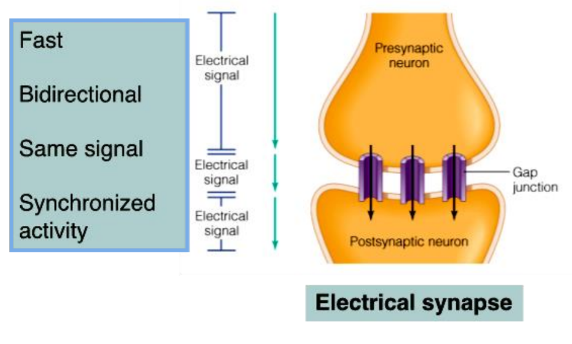
Electrical Synapses
Electrical Synapses:
Are less common than chemical synapses
Correspond to gap junctions found in other cell types
Contain intercellular protein channels
Permit ion flow from one neuron to the next
Are found in the brain and are abundant in embryonic tissue
Chemical Synapses
Specialized for the release and reception of neurotransmitters
Typically composed of two parts:
Axonal terminal of the presynaptic neuron, which contains synaptic vesicles
Receptor region on the dendrite(s) or soma of the postsynaptic neuron
Types of Neurotransmitters
Acetylcholine
Amino Acids:
GABA
Glycine
Aspartic Acid
Glutamic Acid
Monoamines:
Epinephrine
Norepinephrine
Dopamine
Serotonin
Histamine
Neuropeptides
Chains of 2 to 40 amino acids
Stored in axon terminal as larger secretory granules (called dense-core vesicles)
Act at lower concentrations
Longer-lasting effects
Some are released from non-neural tissue
Gut - Brain peptides cause food cravings
Some function as hormones
Modify actions of neurotransmitters
Ionotropic NT Receptors
Direct
Rapid signaling (ligand-gated ion channel)
Metabotropic NT Receptors
Indirect
Slower, longer-lasting changes with diverse responses (G-protein coupled receptors)
Synaptic Transmission
3 kinds of synapses with different modes of action
Excitatory cholinergic synapse = ACh
Inhibitory GABA-ergic synapse = GABA
Excitatory adrenergic synapse = NE
Synaptic delay (0.5 m / sec)
Time from the arrival of the nerve signal at a synapse to the start of an AP in the postsynaptic cell
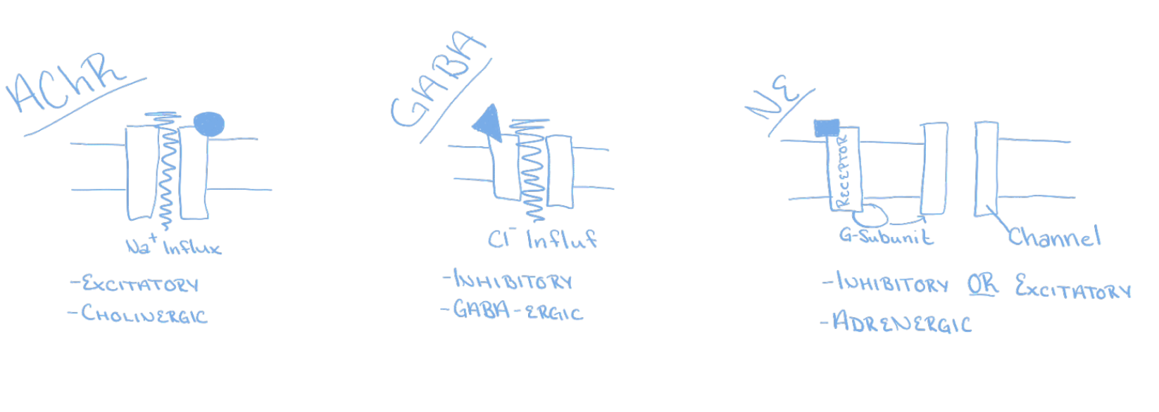
Excitatory Cholinergic Synapse
A nerve signal opens voltage-gated calcium channels in the synaptic knob
Triggers the release of ACh, which crosses synapse
ACh receptors trigger the opening of Na+ channels, producing local potential (postsynaptic potential)
When it reaches -55mV, it triggers AP in the postsynaptic neuron
Inhibitory GABA-ergic Synapse
Nerve signal triggers the release of GABA (y-aminobutyric acid), which crosses the synapse
GABA receptors trigger the opening of Cl- channels, producing hyperpolarization
The postsynaptic neuron is now less likely to reach threshold
Excitatory Adrenergic Synapse
Neurotransmitter is NE (norepinephrine)
Acts through 2nd messenger systems (cAMP)
Receptor is an integral membrane protein associated with a G protein, which activates adenylate cyclase, which converts ATP to cAMP
cAMP has multiple effects
Binds to ion gate inside of membrane (depolarizing)
Activates cytoplasmic enzymes
Induces genetic transcription and production of new enzymes
Its advantage is enzymatic amplification
Cessation and Modification of a Signal
Mechanisms to turn off stimulation
Diffusion of neurotransmitter away into the ECF
Astrocytes return it to neurons
Synaptic knob reabsorbs amino acids and monoamines by endocytosis
Acetylcholinesterase degrades ACh
Choline is reabsorbed and recycled
Neuromodulators modify transmission
Raise or lower the number of receptors
Alter neurotransmitter release, synthesis, or breakdown
Neural Integration
The more synapses a neuron has, the greater its information-processing capability
Cells in the cerebral cortex with 40,000 synapses
The cerebral cortex is estimated to contain 100 trillion synapses
Chemical synapses are decision-making components of the nervous system
The ability to process, store, and recall information is due to neural integration
Based on the types of postsynaptic potentials produced by neurotransmitters
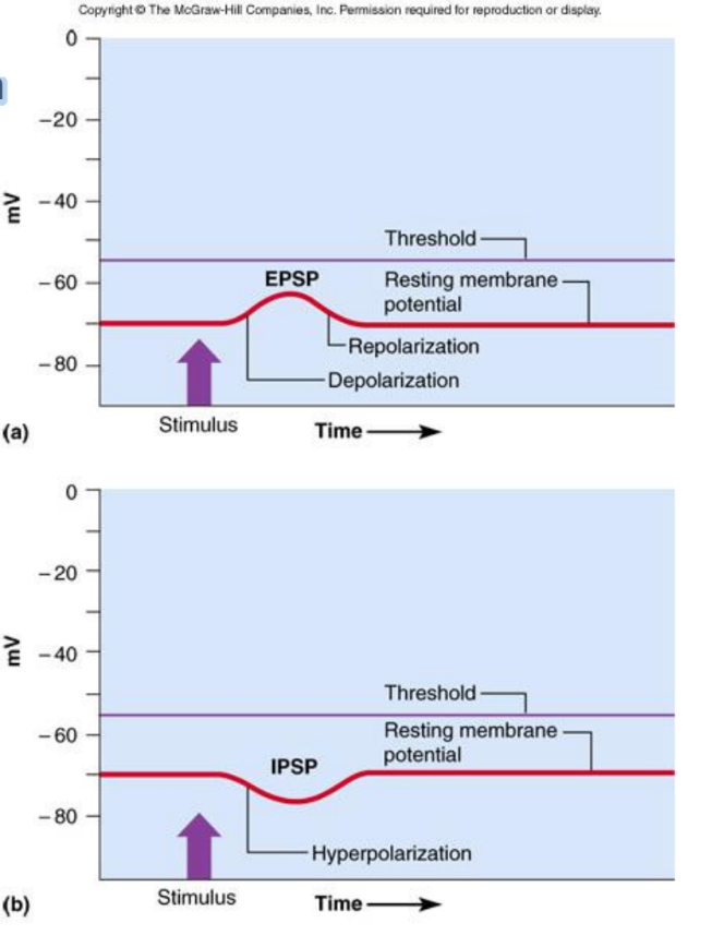
Excitatory Postsynaptic Potentials (EPSP)
A positive voltage change causes the postsynaptic cell to be more likely to fire
Result from Na+ flowing into the cell
Glutamate and aspartate are excitatory neurotransmitters
ACh and norepinephrine may excite or inhibit, depending on the cell
Inhibitory Postsynaptic Potentials (IPSP)
A negative voltage change causing postsynaptic cell to be less likely to fire (hyperpolarize)
Result of Cl- flowing into the cell or K+ leaving the cell
Glycine and GABA are inhibitory neurotransmitters
ACh and norepinephrine may excite or inhibit depending upon cell
Postsynaptic Potentials
EPSPs are graded potentials that can initiate an action potential in an axon
Use only chemically gated channels
Na+ and K+ flow in opposite directions simultaneously
Postsynaptic membranes do not generate action potentials
Neurotransmitter binding to a receptor at inhibitory synapses:
Membrane becomes more permeable to K+ and Cl-
The charge on the inner surface is negative
Reduces the postsynaptic neuron’s ability to produce an action potential
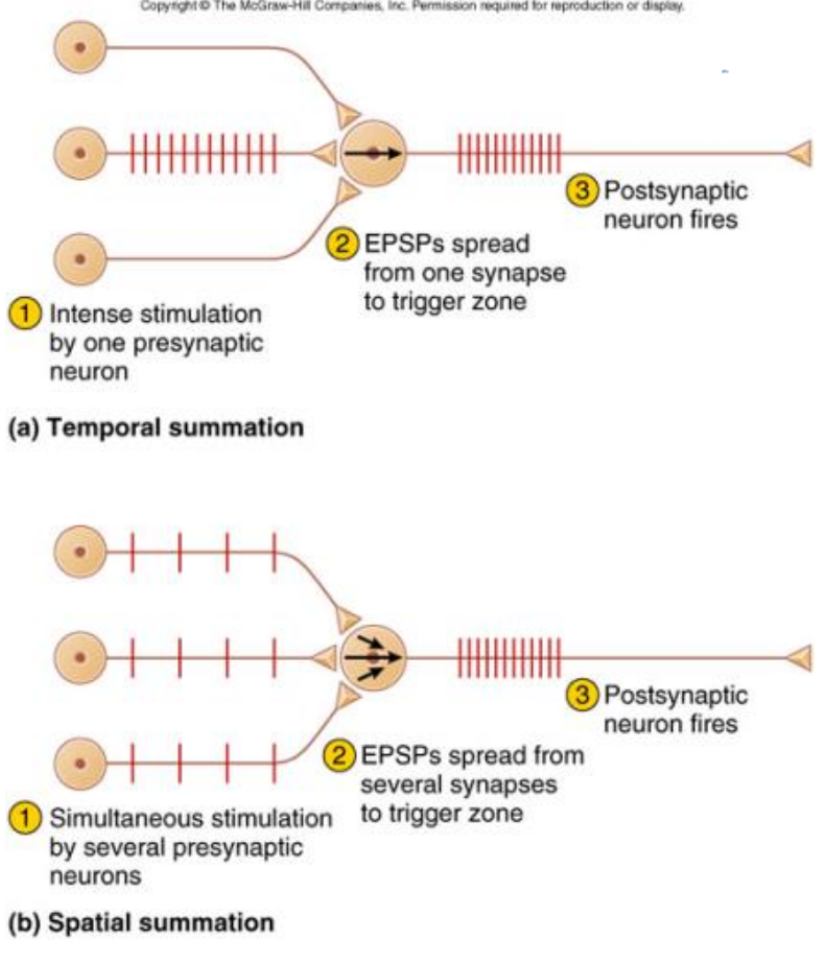
Summation - Postsynaptic Potentials
Net postsynaptic potentials in trigger zone
Firing depends on net input of other cells
Typical EPSP voltage = 0.5 mV and lasts 20 msec
30 EPSPs needed to reach threshold
Temporal Summation
Single synapse receives many EPSPs in short time
Spatial Summation
Single synapse receives many EPSPs from many cells
Pain Regulation
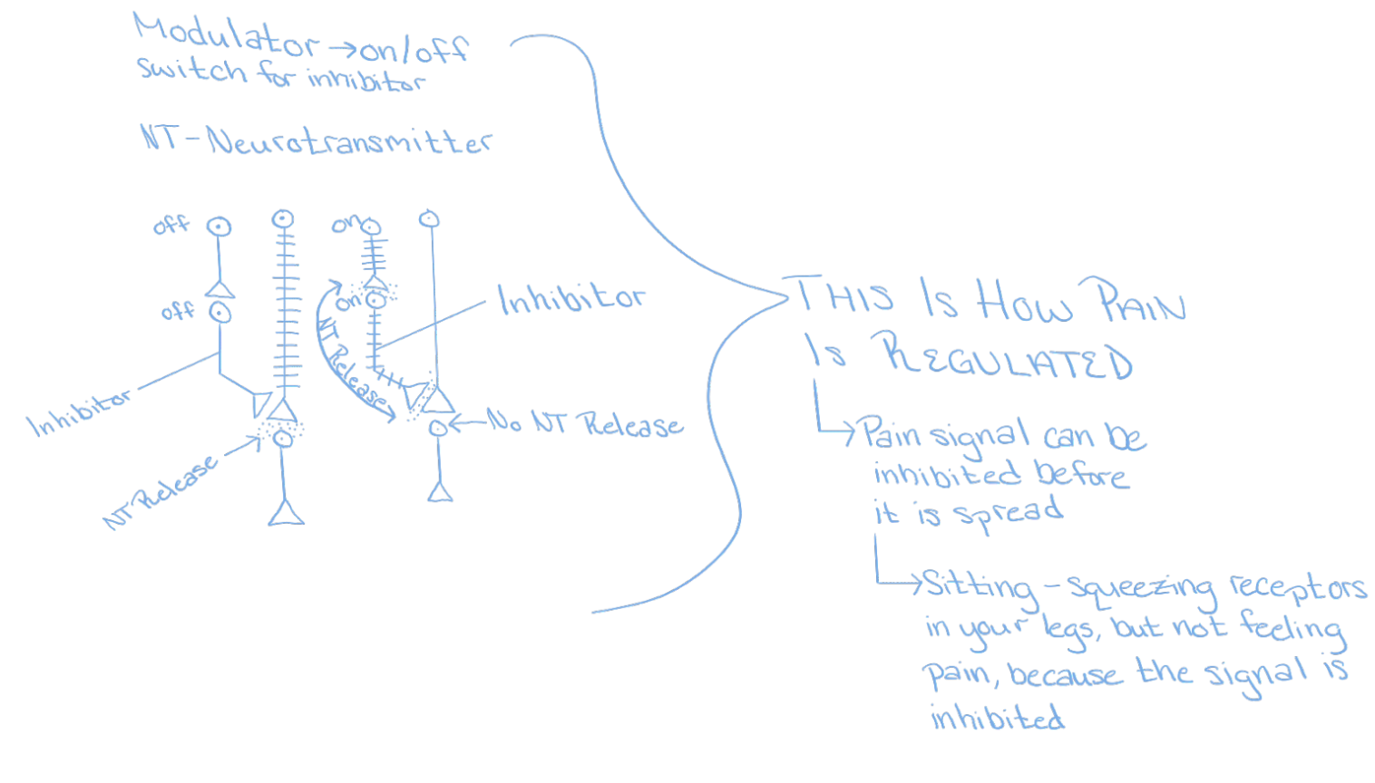
Presynaptic Inhibition
One presynaptic neuron suppresses another
Neuron I releases inhibitory GABA
Prevents voltage-gated calcium channels from opening -- it releases less or no neurotransmitter
Neural Coding and Integration
Qualitative information (taste or hearing) depends upon which neurons fire
Labeled Line Code: the brain knows what type of sensory information travels on each fiber
Quantitative information depends on:
Different neurons have different thresholds
Weak stimuli excite only specific neurons
Stronger stimuli cause a more rapid firing rate
CNS judges stimulus strength from the firing frequency of sensory neurons
Absolute refractory periods vary
Neural Pools and Circuits
Neural Pool: interneurons that share specific body function
Control rhythm of breathing
Facilitated vs. Discharge Zones
In Discharge Zone: a single cell can produce firing
In Facilitated Zone: single cell can only make it easier for the postsynaptic cell to fire
Neural Circuits
Diverging Circuit: one cell synapses on another, and each synapse on others
Converging Circuit: input from many fibers on one neuron (respiratory center)
Reverberating Circuits: neurons stimulate each other in a linear sequence, but one cell restimulates the first cell to start the process all over
Parallel After-Discharge Circuits: input neuron stimulates several pathways, which stimulate the output neuron to continue firing for a longer time after the input has truly stopped
Serial Processing
Input travels along one pathway to a specific destination
One neuron stimulates the next and so on to cause specific, anticipate response
Predictable all-or-nothing manner
Reflexes: rapid, automatic responses to stimuli, to produce a stereotyped and dependable response
Parallel Processing
Input travels along several different pathways to be integrated in different CNS regions
Inputs are segregated into many pathways
Different parts of the neural circuitry deal with the information delivered by each pathway simultaneously
Extremely important for higher-level mental functioning to put all parts together to understand the whole
Synaptic Plasticity
Synaptic Strength:
The amount or magnitude of the post-synaptic potential caused by activation of the pre-synaptic terminal
Plasticity: ability of brain to change synaptic strength as a result of experience
Plasticity: ability to learn and remember
Synaptic Plasticity Mechanisms
More Strength: Up Regulation:
Increase vesicles or transmitter
Increase Post-synaptic receptors
Less Strength: Down Regulation
Decrease vesicles or transmitter
Decrease post-synaptic receptors
Two Kinds of Synapses in the Brain
Compensatory:
Regulatory
Homeostatic
Underlies Addiction
Hebbian:
Intensifies with use!
Important for consolidating memory
Memory and Synaptic Plasticity
Physical basis of memory is a pathway
Called a memory trace or engram
New synapses or existing synapses modified to make transmission easier (synaptic plasticity)
Synaptic potentiation
Transmission mechanisms correlate with different forms of memory
Immediate, short and long-term memory
Immediate Memory
Ability to hold something in your thoughts for just a few seconds
Essential for reading ability
Feel for the flow of events (sense of the present)
Our memory of what just happened “echoes” in our minds for a few seconds
Reverberating circuits
Short-Term Memory
Lasts from a few seconds to several hours
Quickly forgotten if distracted
Search for keys, dial the phone
Reverberating circuits
Facilitation causes memory to last longer
Tetanic stimulation (rapid,repetitive signals) cause Ca2+ accumulation and cells more likely to fire
Post-tetanic potentiation (to jog a memory)
Ca2+ level in synaptic knob stays elevated
Little stimulation needed to recover memory
Long-Term Memory
Types of long-term memory
Declarative: retention of facts as text
Procedural: retention of motor skills
Physical remodeling of synapses
New branching of axons or dendrites
Molecular changes: long-term
Tetanic stimulation causes ionic changes
Neuron produces more neurotransmitter receptors
More protein synthesizes for synapse remodeling
Releases nitric oxide, then presynaptic neuron releases more neurotransmitter
Alzheimer’s Disease
100,000 deaths / year
11% of population over 65; 47% by age 85
Memory loss for recent events, moody, combative, lose ability to talk, walk, and eat
Diagnosis confirmed at autopsy
Atrophy of gyri (folds) in cerebral cortex
Neurofibrillary tangles and senile plaques
Degeneration of cholinergic neurons and deficiency of ACh and nerve growth factors
Genetic connection confirmed
Parkinson’s Disease
Progressive loss of motor function beginning in 50’s or 60’s -- no recovery
Degeneration of dopamine-releasing neurons
Prevents excessive activity in motor centers
Involuntary muscle contractions
Pill-rolling motion, facial rigidity, slurred speech, illegible handwriting, slow gait
Treatment: drugs and physical therapy
Dopamine precursor crosses brain barrier
MAO inhibitor slows neural degeneration
Surgical technique to relieve tremors