BSC2086L E.7 Respiratory System
0.0(0)
Card Sorting
1/58
Last updated 12:16 PM on 4/11/23
Name | Mastery | Learn | Test | Matching | Spaced | Call with Kai |
|---|
No analytics yet
Send a link to your students to track their progress
59 Terms
1
New cards
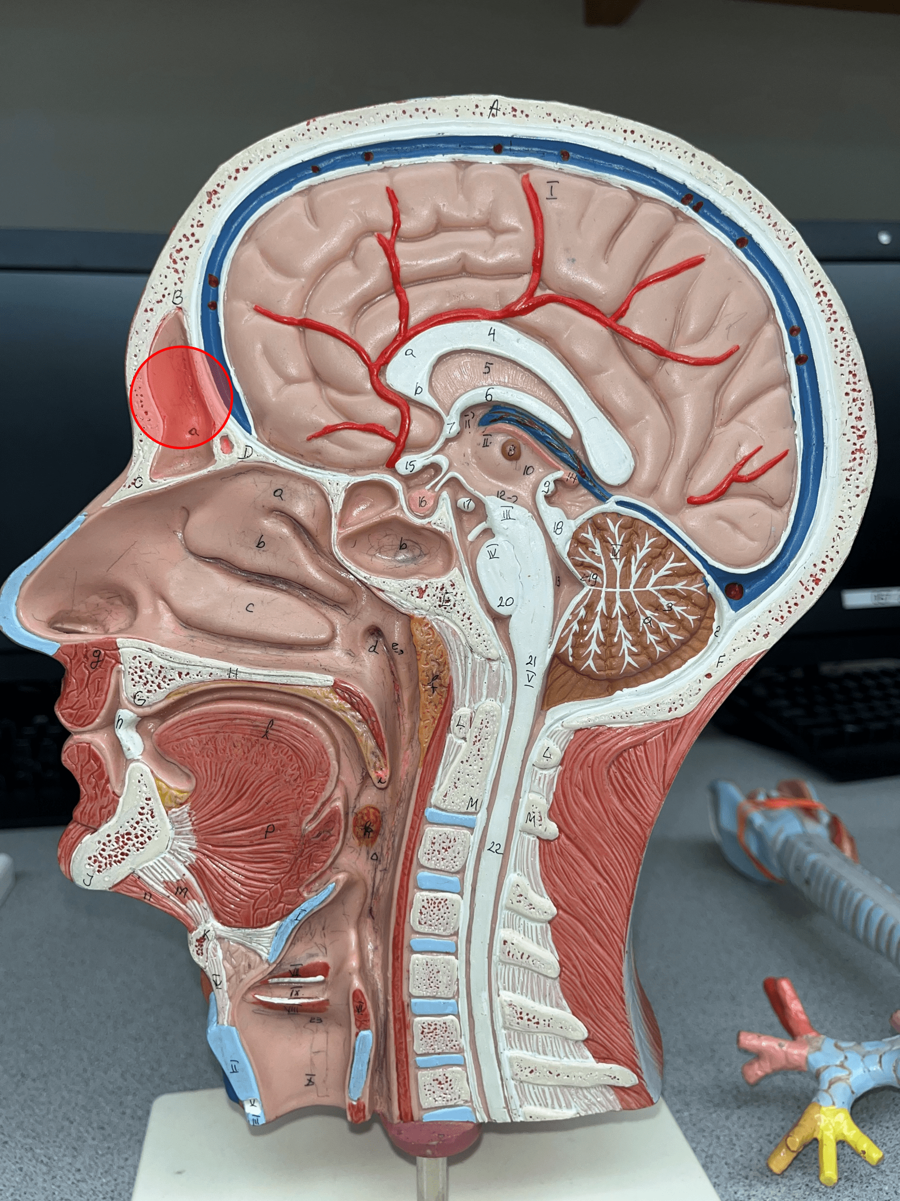
frontal sinus
• A sinus within the frontal bone.
• Located superior to the nose.
• Located superior to the nose.
2
New cards
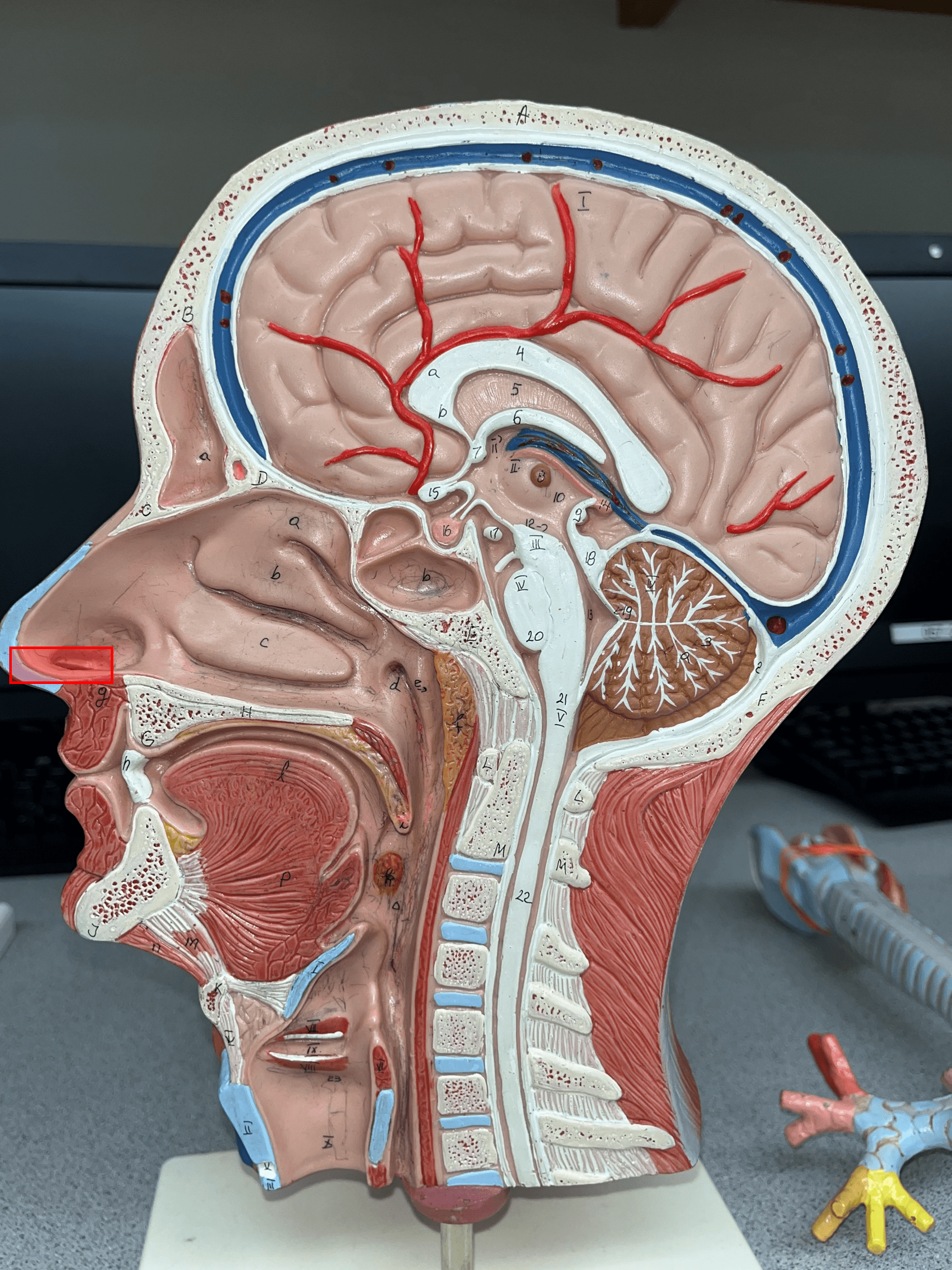
nose
• The chamber that initially receives air from the exterior.
• Composed of bone and hyaline cartilage.
• Composed of bone and hyaline cartilage.

3
New cards
external nare
• An opening that air passes through to enter the nose.
• Also known as a nostril.
• Just before the vestibule.
• Also known as a nostril.
• Just before the vestibule.
4
New cards
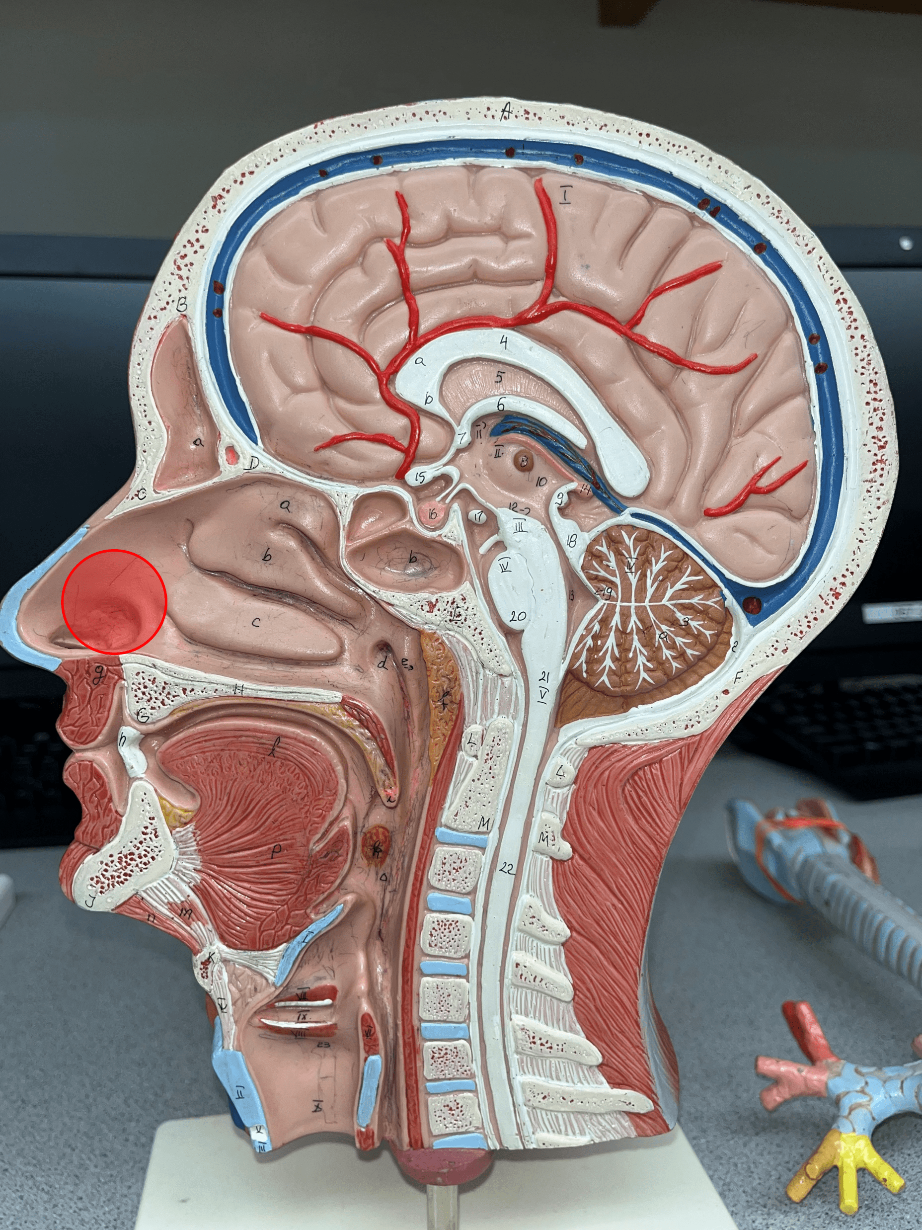
vestibule
• The first chamber that air passes through to enter the nasal cavity.
• Just after the external nare.
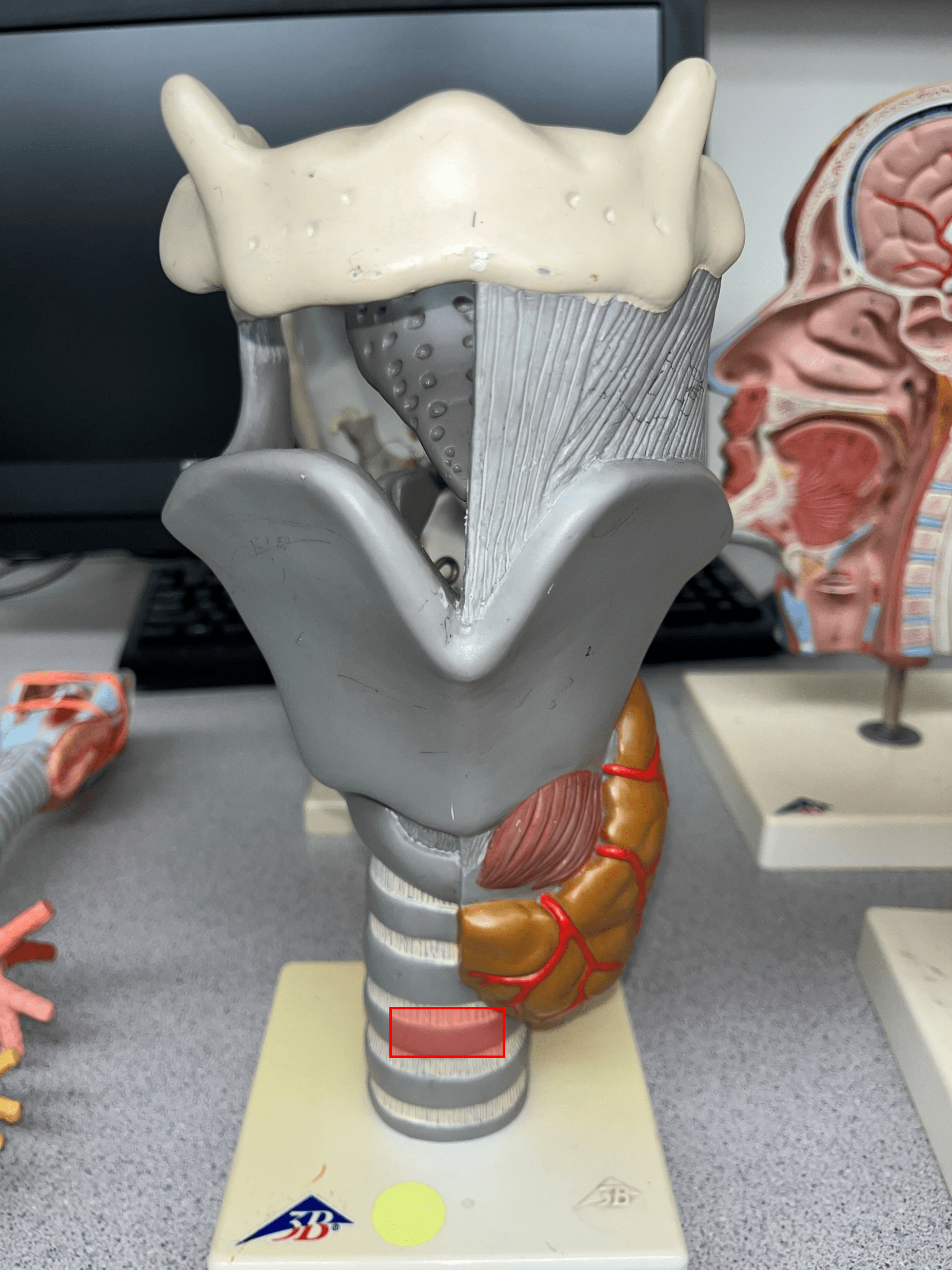
• Just before the nasal conchae.
• Just after the external nare.
• Just before the nasal conchae.
5
New cards
superior nasal concha
• The superior shelf-like structure in the nasal cavity.
• Just after the vestibule.
• Just before the internal nare.
• Just after the vestibule.
• Just before the internal nare.

6
New cards
middle nasal concha
• The middle shelf-like structure in the nasal cavity.
• Just after the vestibule.
• Just before the internal nare.
• Just after the vestibule.
• Just before the internal nare.
7
New cards
inferior nasal concha
• The inferior shelf-like structure in the nasal cavity.
• Just after the vestibule.
• Just before the internal nare.
• Just after the vestibule.
• Just before the internal nare.

8
New cards
superior meatus
The passage air flows through between the superior and middle conchae.

9
New cards
middle meatus
The passage air flows through between the middle and inferior conchae.

10
New cards
inferior meatus
The passage air flows through the inferior concha and the floor of the palate.

11
New cards
internal nare
• An opening that air passes through to enter the pharynx from the nasal cavity.
• Also known as a posterior nasal aperture.
• Just after the nasal conchae.
• Also known as a posterior nasal aperture.
• Just after the nasal conchae.

12
New cards
sphenoidal sinus
• A sinus within the sphenoid bone.
• Located superior to the internal nares.
• Located superior to the internal nares.
13
New cards
pharynx
• Located posterior to the nasal cavity.
• Divided into three segments.
• Also known as the throat.
• Divided into three segments.
• Also known as the throat.

14
New cards
nasopharynx
• The superior segment of the pharynx.
• Above the level of the soft palate.
• Receives air from the nose by the internal nares.
• Above the level of the soft palate.
• Receives air from the nose by the internal nares.
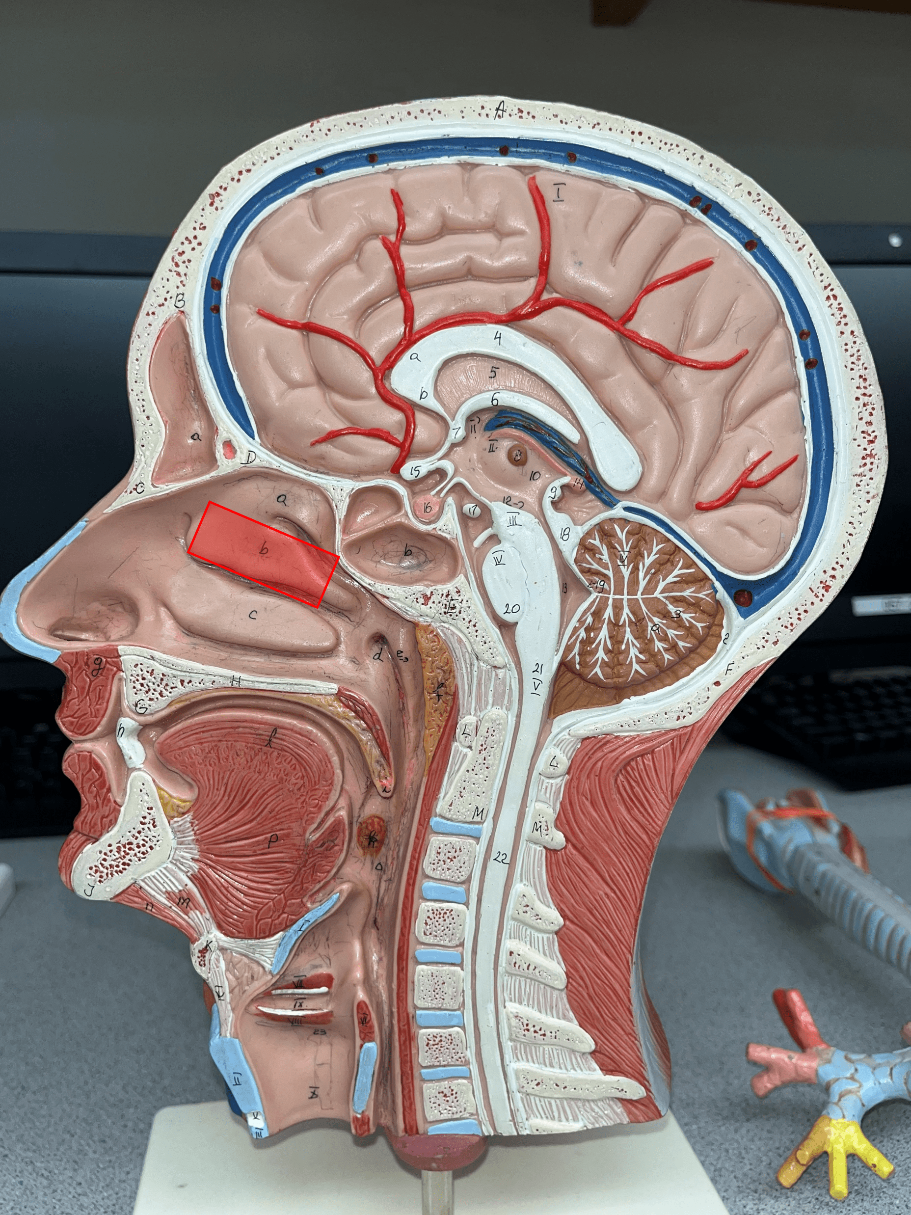
15
New cards
pharyngeal tonsil
• One of the three main tonsils.
• Located on the superoposterior corner of the wall of the nasopharynx.
• Located on the superoposterior corner of the wall of the nasopharynx.

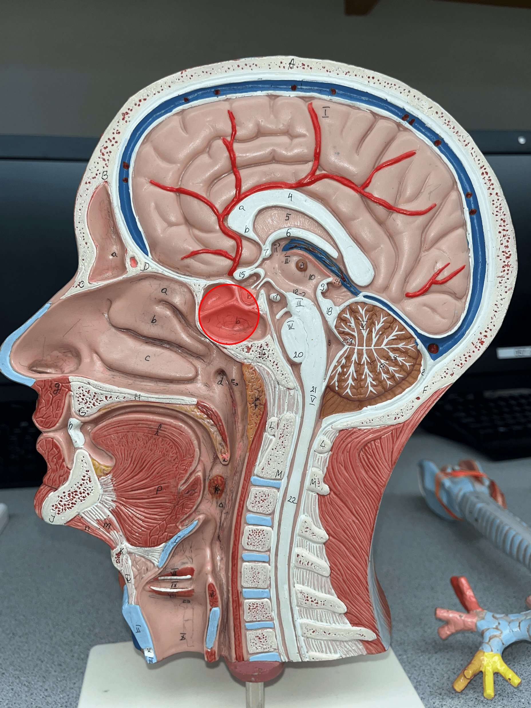
16
New cards
oropharynx
• The middle segment of the pharynx.
• Below the level of the soft palate and above the level of the hyoid bone.
• Receives food from the oral cavity.
• Below the level of the soft palate and above the level of the hyoid bone.
• Receives food from the oral cavity.

17
New cards
uvula
• A soft extension of tissue.
• Found at the distal end of the soft palate.
• Found at the distal end of the soft palate.

18
New cards
palatine tonsil
• One of the three main tonsils.
• Located between the oral cavity and the oropharynx and just inferior to the uvula.
• Located between the oral cavity and the oropharynx and just inferior to the uvula.

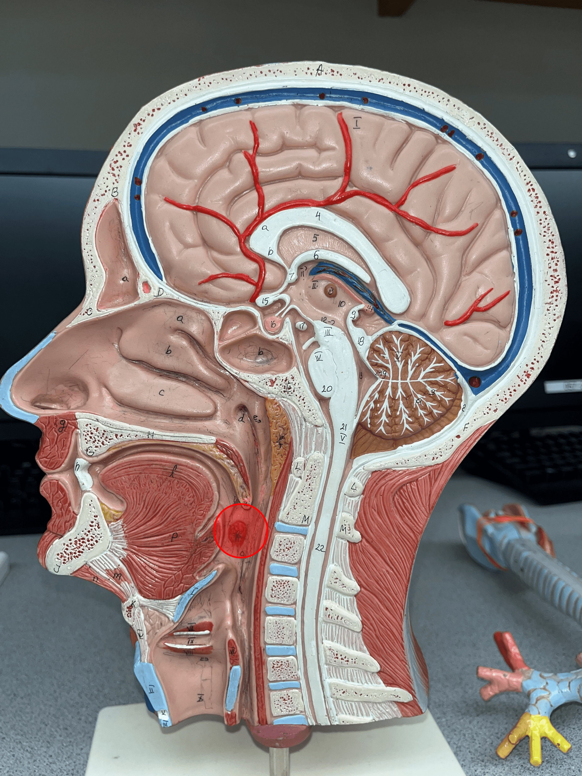
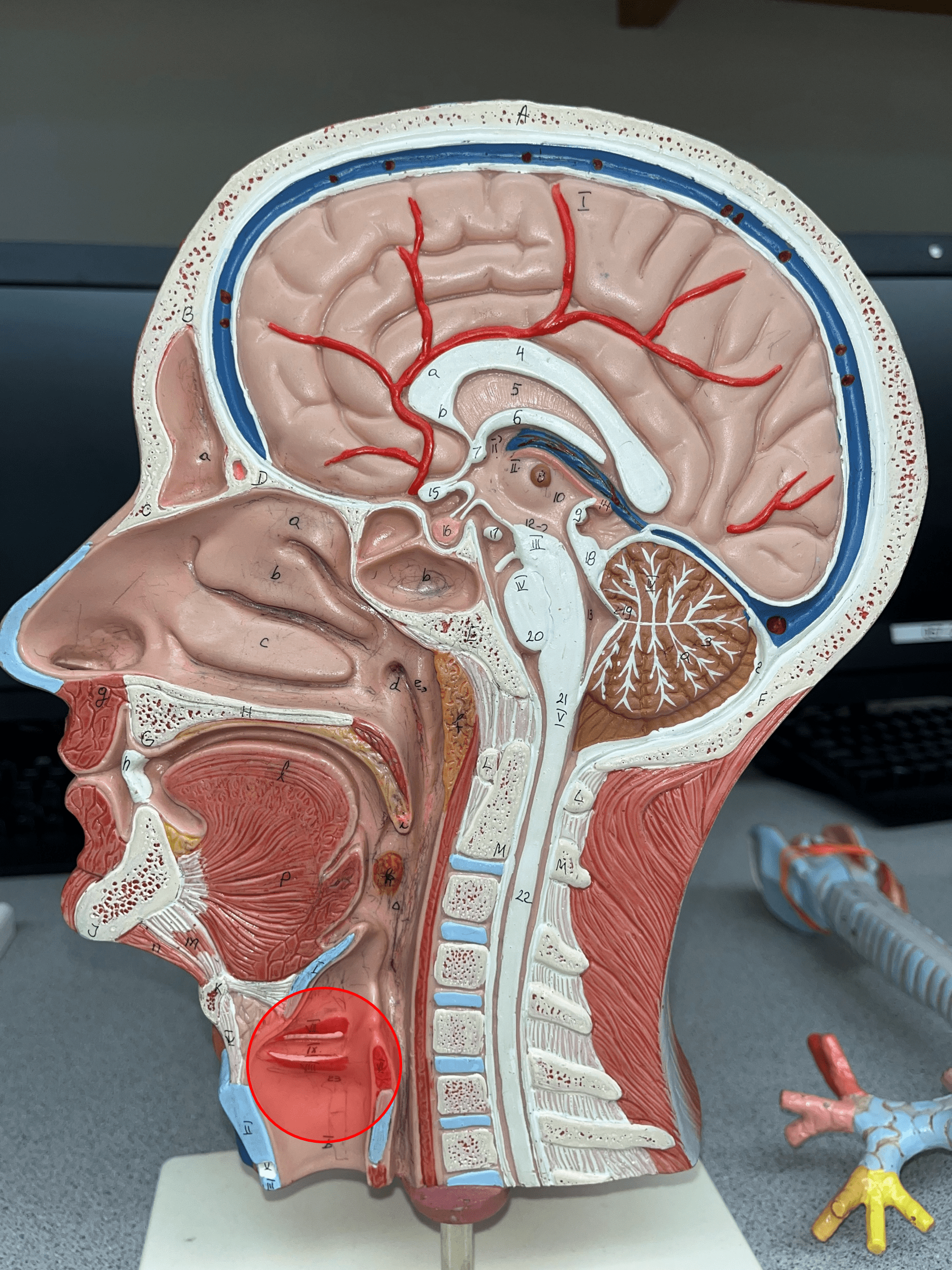
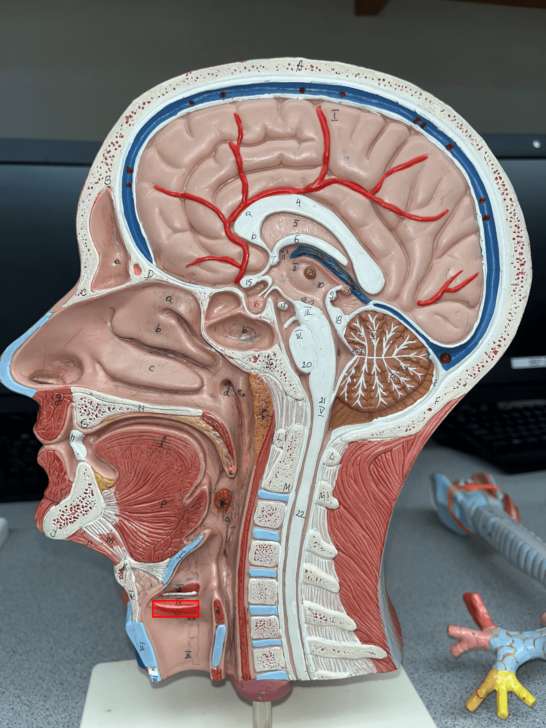
19
New cards
lingual tonsil
• One of the three main tonsils.
• Located at the base of the tongue.
• Located at the base of the tongue.
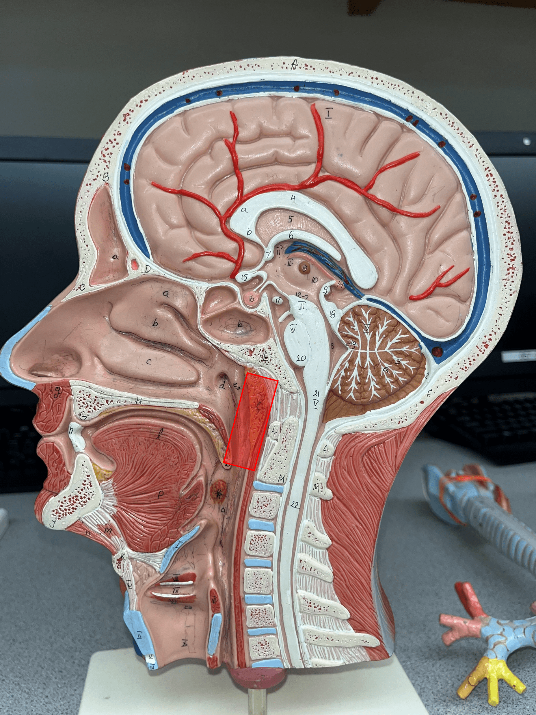
20
New cards
laryngopharynx
• The inferior segment of the pharynx.
• Below the level of the hyoid bone.
• Extends downward to meet the larynx.
• Below the level of the hyoid bone.
• Extends downward to meet the larynx.

21
New cards
larynx
• The box-like organ connecting the laryngopharynx and the trachea.
• Also known as the voicebox.
• Also known as the voicebox.
22
New cards
vestibular folds
• The superior pair of folds lateral to the glottis in the larynx.
• Also known as false vocal cords.
• Also known as false vocal cords.

23
New cards
vocal folds
• The inferior pair of folds lateral to the glottis in the larynx.
• Also known as true vocal cords.
• Also known as true vocal cords.
24
New cards
epiglottis
• A single cartilage of the larynx.
• Functions as a flap that closes over the glottis during swallowing.
• Functions as a flap that closes over the glottis during swallowing.
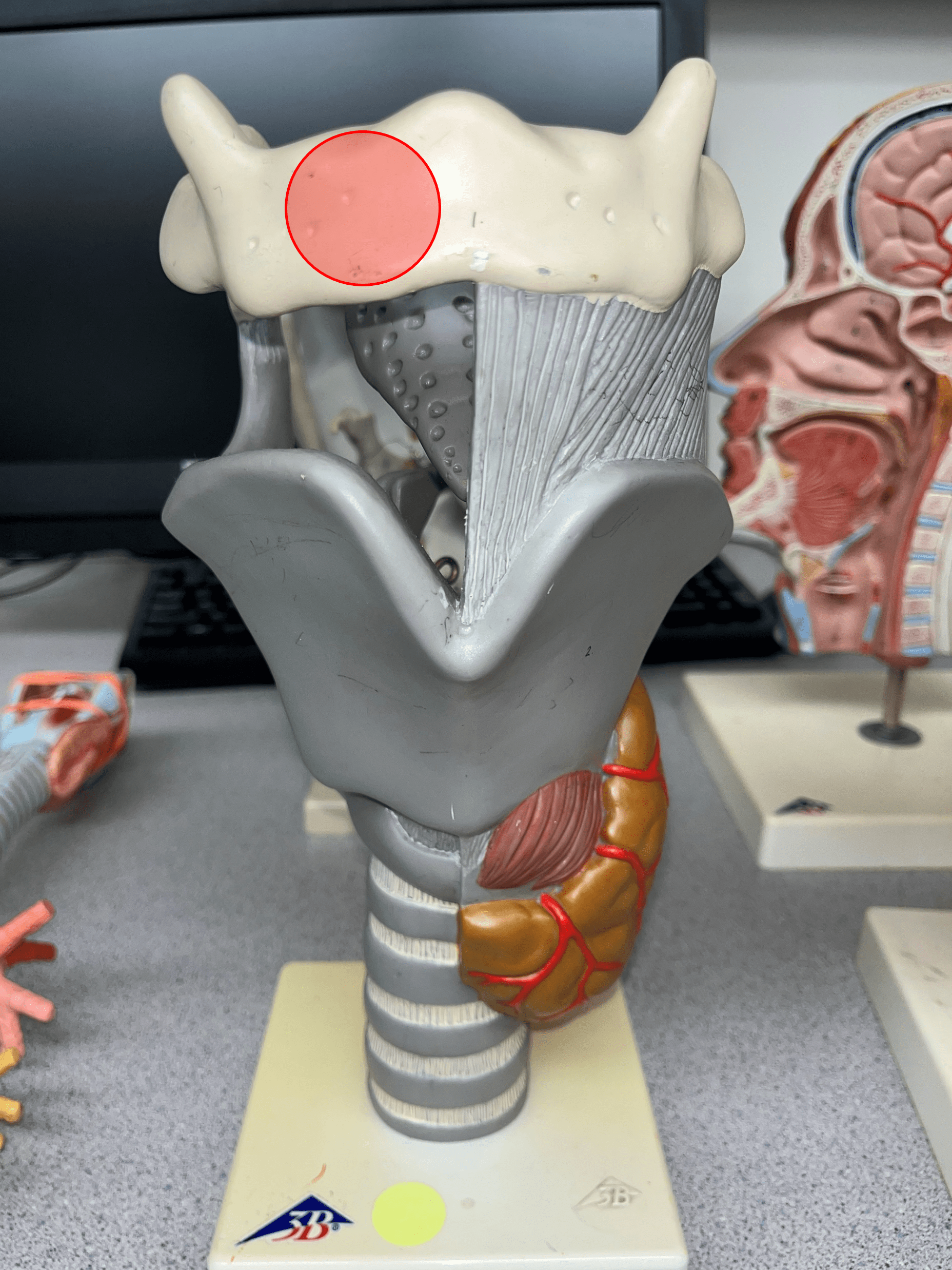
25
New cards
hyoid bone
• A bone located near the larynx.
• Anterior to the epiglottis.
• Anterior to the epiglottis.
26
New cards
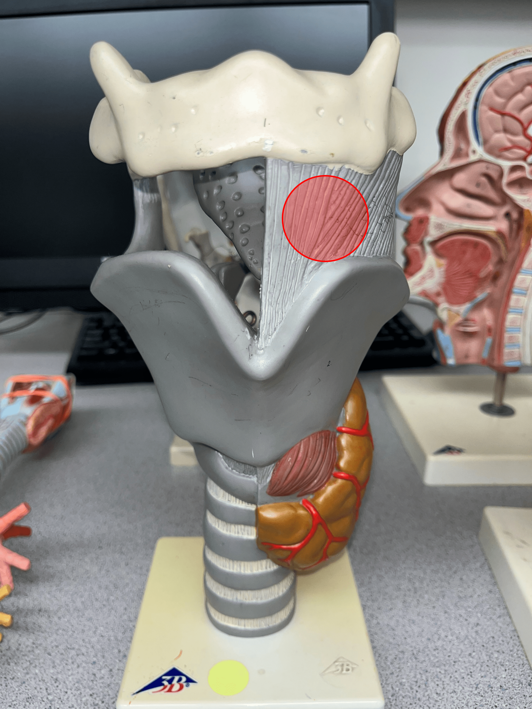
thyrohyoid membrane
The membrane that connects the thyroid cartilage to the hyoid bone.
27
New cards
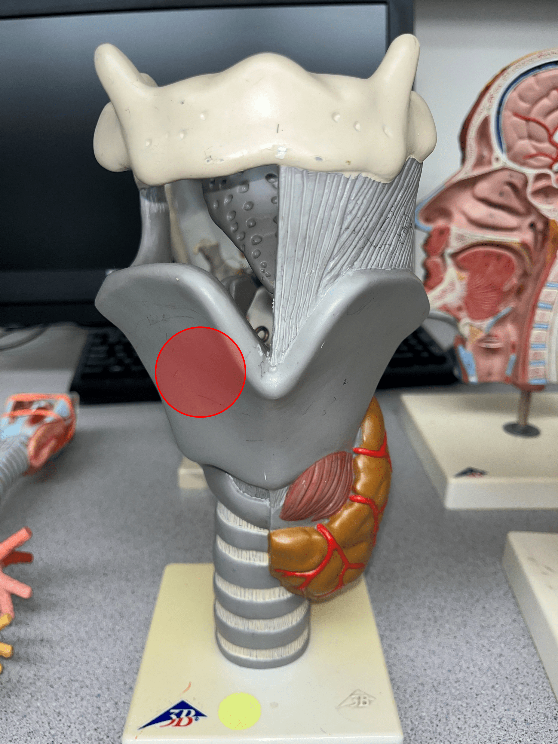
thyroid cartilage
• A single cartilage of the larynx.
• The largest cartilage of the larynx.
• Located on the anterior wall of the larynx.
• Inferior to the epiglottis and hyoid bon.
• The largest cartilage of the larynx.
• Located on the anterior wall of the larynx.
• Inferior to the epiglottis and hyoid bon.
28
New cards
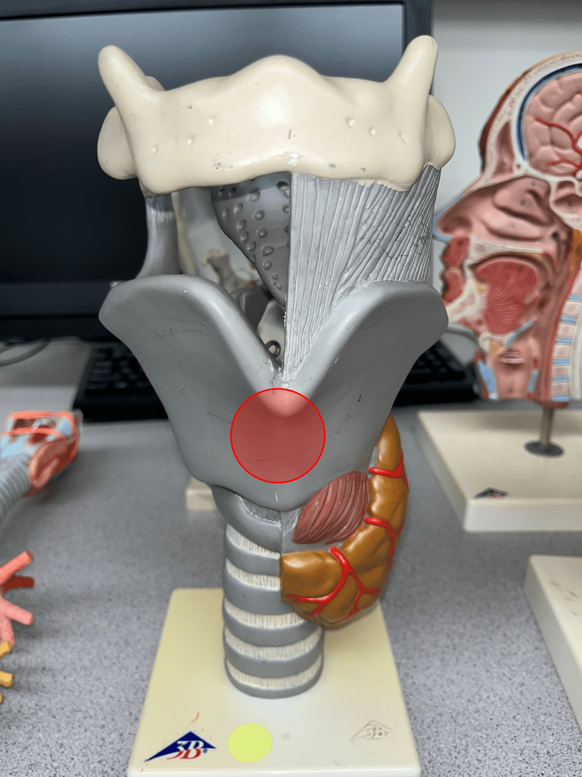
laryngeal prominence
• A prominence found on the thyroid cartilage of the larynx.
• Also known as the Adam’s apple.
• Also known as the Adam’s apple.
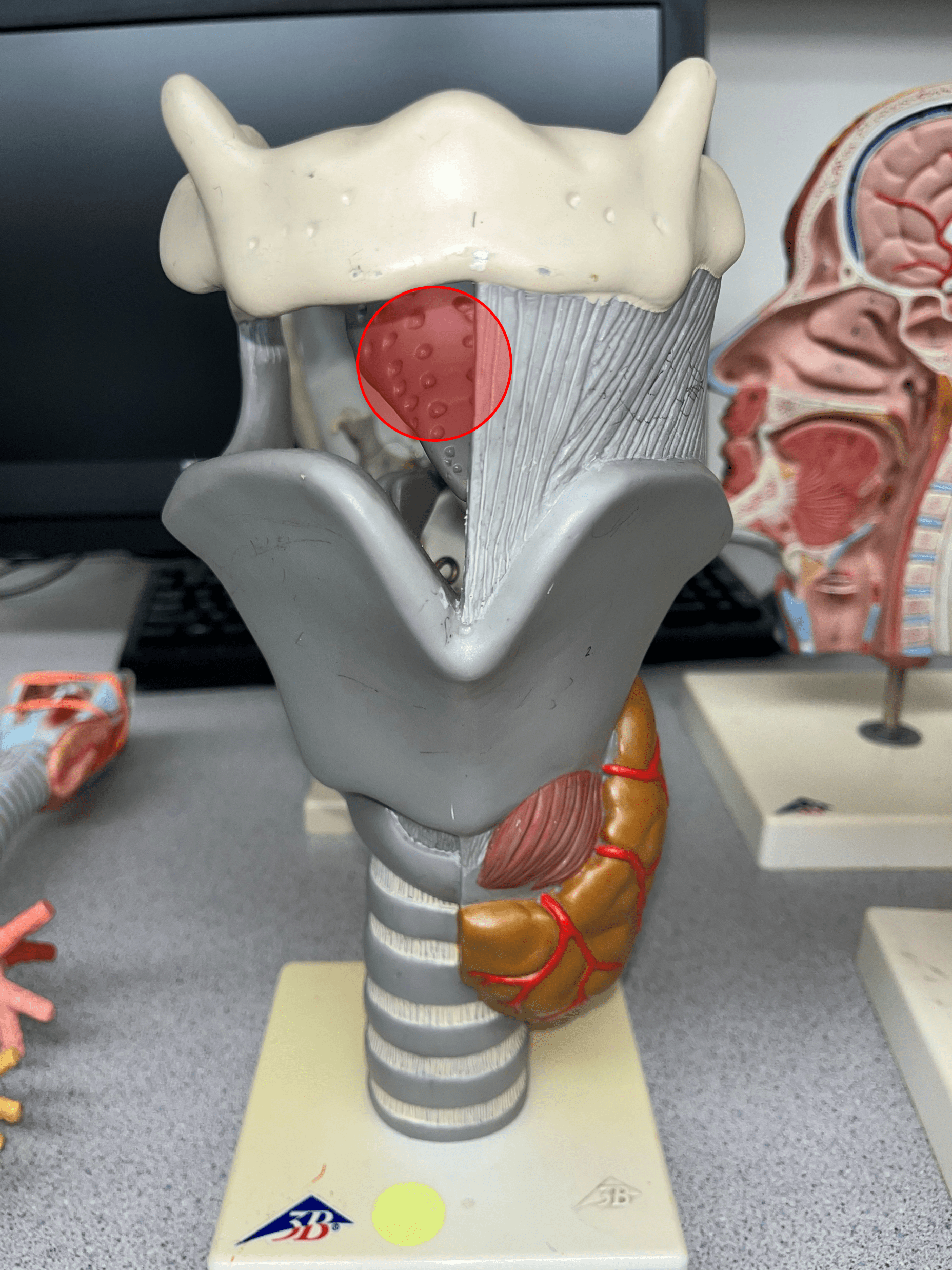
29
New cards
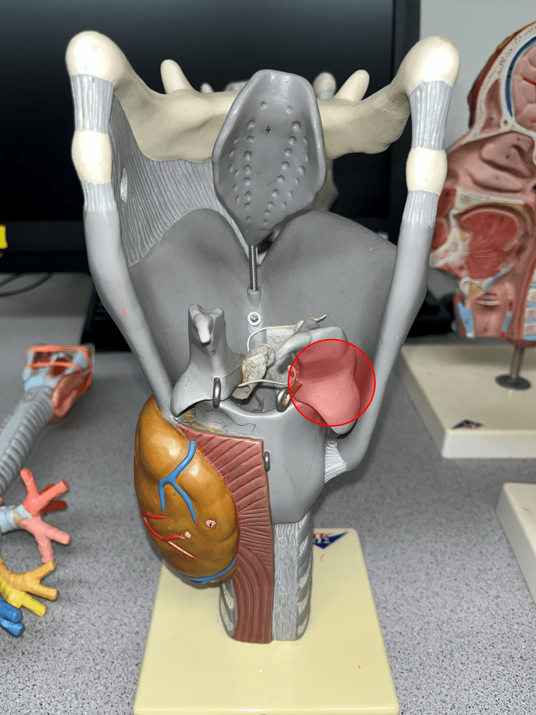
cuneiform cartilage
• A paired cartilage of the larynx.
• Located on the posterior wall of the larynx.
• The superior posterior cartilage.
• Located on the posterior wall of the larynx.
• The superior posterior cartilage.
30
New cards
corniculate cartilage
• A paired cartilage of the larynx.
• Located on the posterior wall of the larynx.
• The middle posterior cartilage.
• Not shown in image.
• Located on the posterior wall of the larynx.
• The middle posterior cartilage.
• Not shown in image.

31
New cards
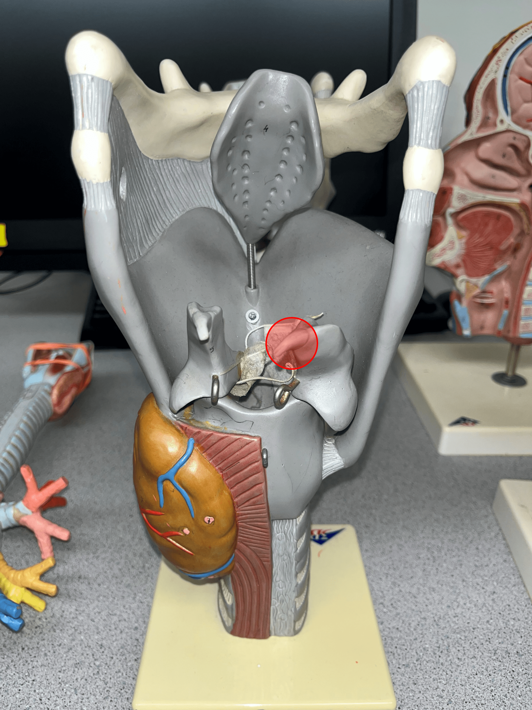
arytenoid cartilage
• A paired cartilage of the larynx.
• Located on the posterior wall of the larynx.
• The inferior posterior cartilage.
• Mostly surrounds on the arytenoid muscle on its anterior surface.
• Located on the posterior wall of the larynx.
• The inferior posterior cartilage.
• Mostly surrounds on the arytenoid muscle on its anterior surface.
32
New cards
arytenoid muscle
• A muscle encased by the arytenoid and cuneiform cartilages within the larynx.
• Not shown in image.
• Not shown in image.

33
New cards
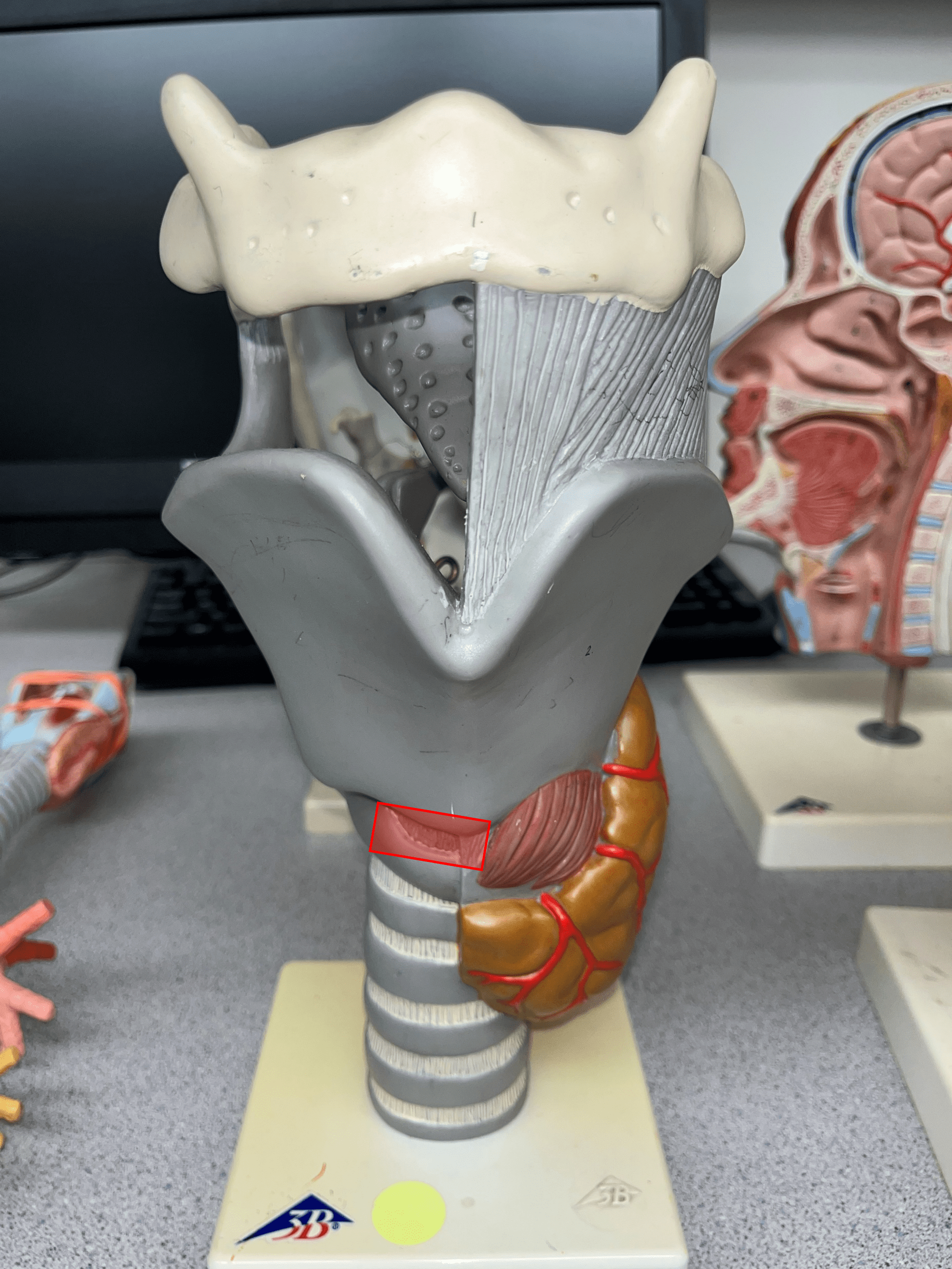
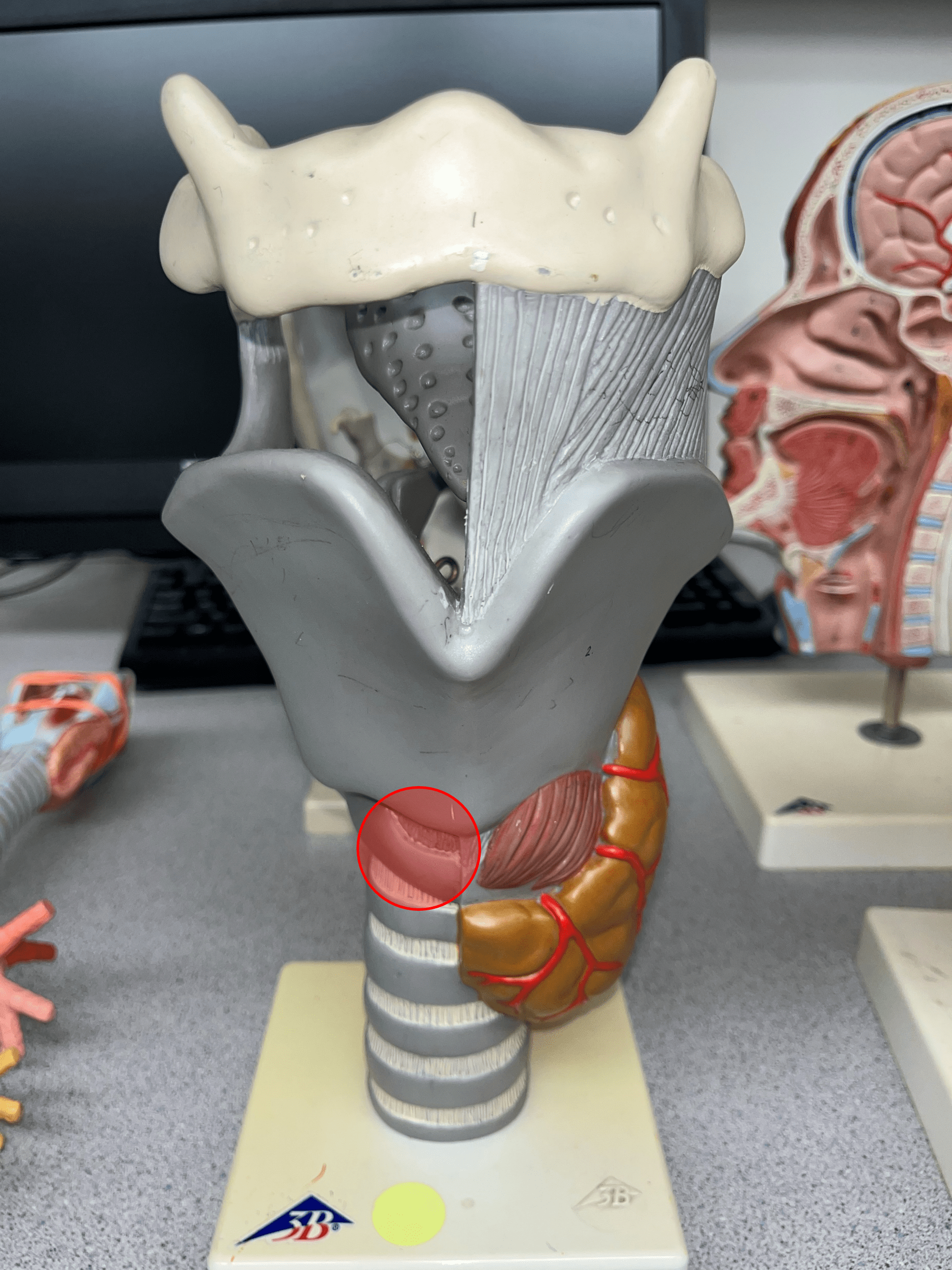
cricothyroid ligament
The ligament that connects the cricoid cartilage to the thyroid cartilage.
34
New cards
cricoid cartilage
• A single cartilage of the larynx.
• Inferior to the thyroid cartilage and superior to the tracheal cartilages.
• Inferior to the thyroid cartilage and superior to the tracheal cartilages.
35
New cards
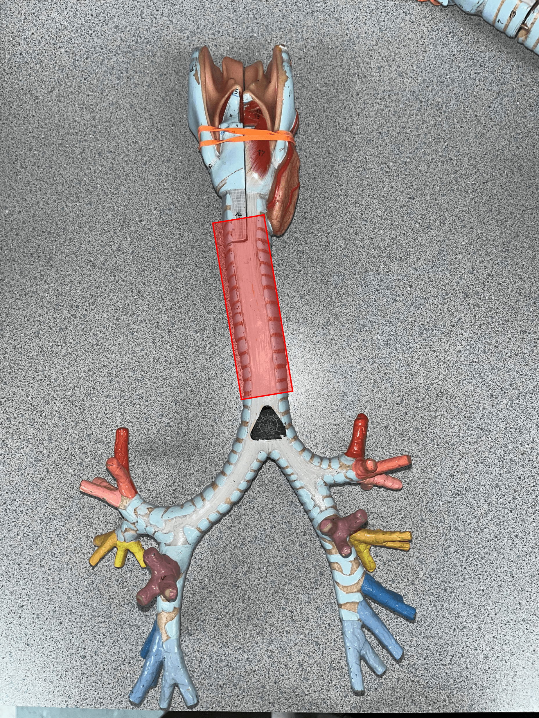
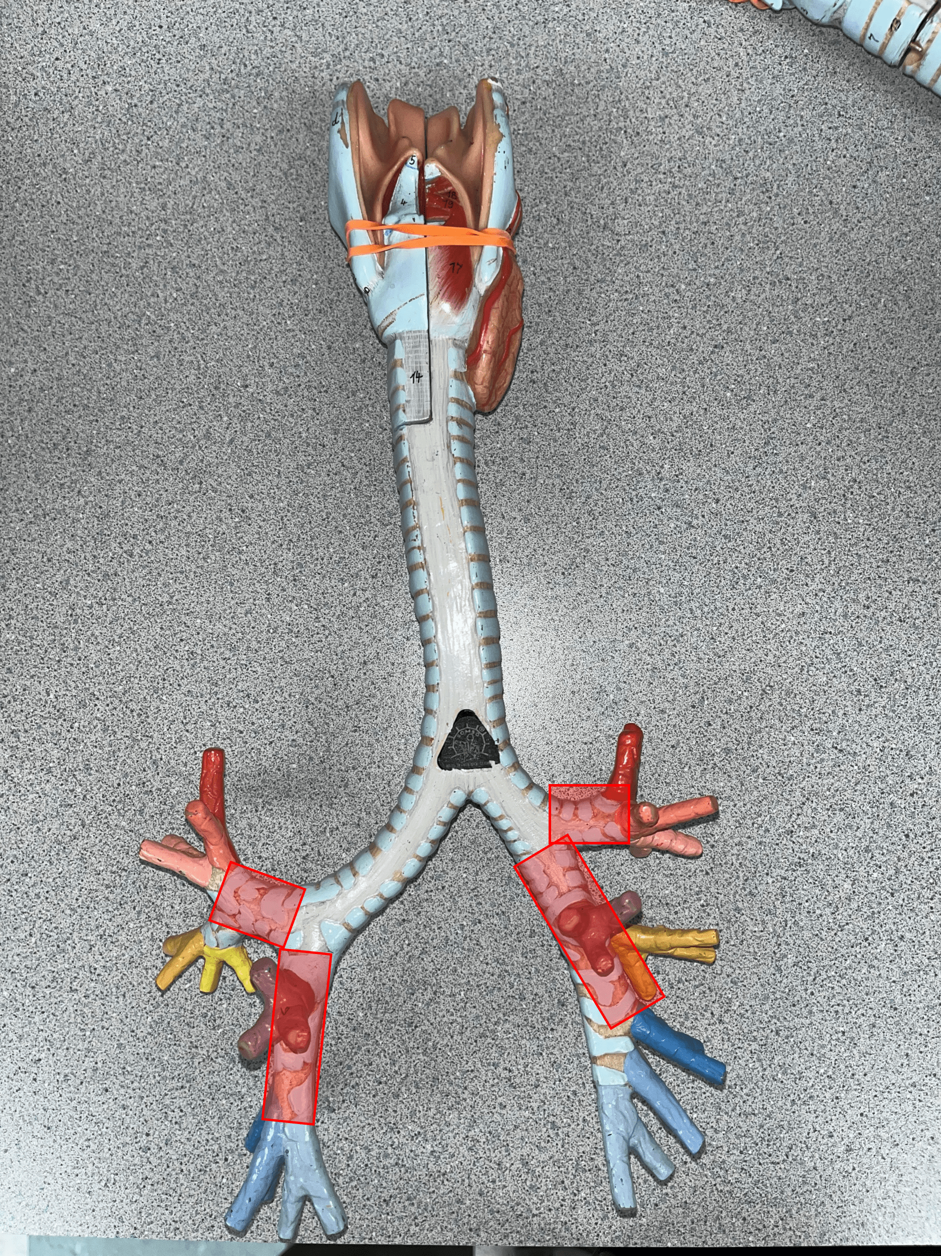
trachea
• The tube-shaped organ carrying air between the larynx and bronchial tree.
• Located inferior to the larynx.
• Also known as the windpipe.
• Located inferior to the larynx.
• Also known as the windpipe.
36
New cards
tracheal cartilage
• A C-shaped piece of a stack of cartilages running along the length of the trachea.
• The two ends of the cartilage are connected by the trachealis muscle.
• The two ends of the cartilage are connected by the trachealis muscle.
37
New cards
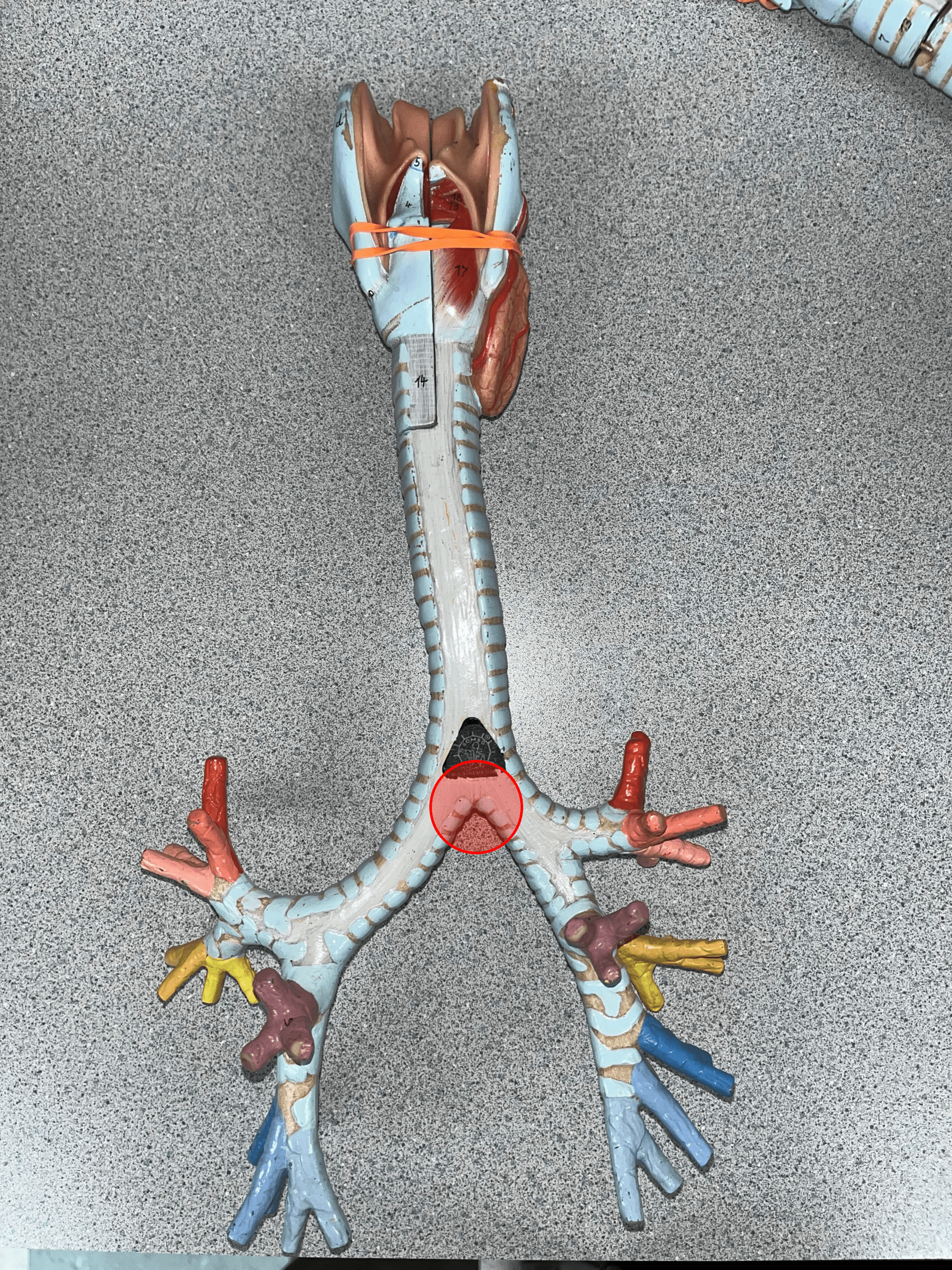
carina
• The termination of the trachea.
• The trachea splits into primary bronchi at this point.
• The trachea splits into primary bronchi at this point.
38
New cards
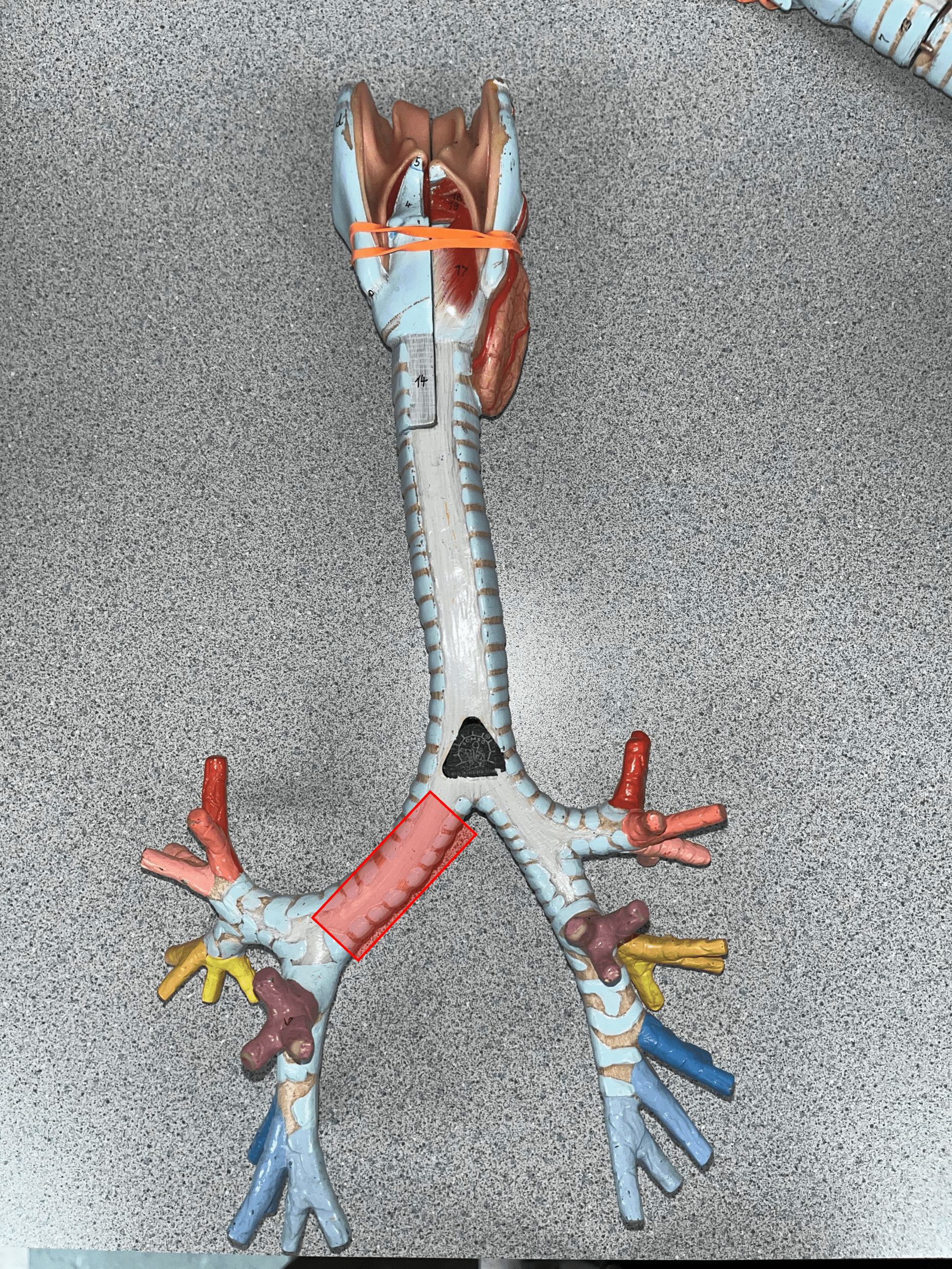
right primary bronchus
• The R. branch of the bronchial tree after the carina.
• Supplies the R. lung.
• Branches into three secondary bronchi.
• Also known as a main bronchus.
• Supplies the R. lung.
• Branches into three secondary bronchi.
• Also known as a main bronchus.
39
New cards
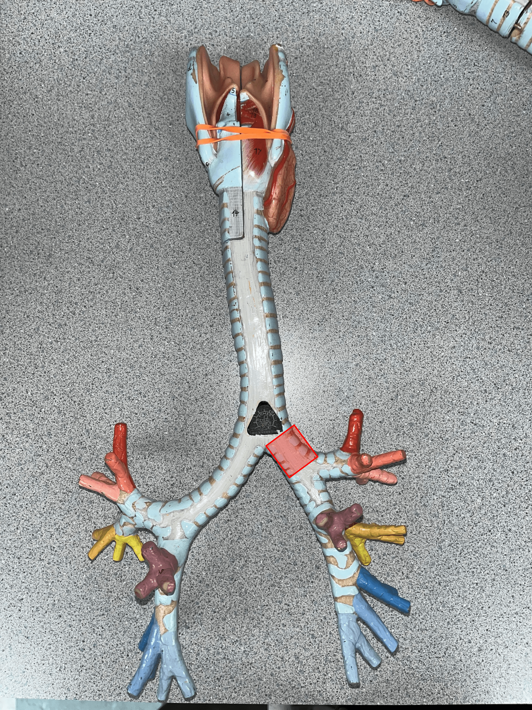
left primary bronchus
• The L. branch of the bronchial tree after the carina.
• Supplies the L. lung.
• Branches into two secondary bronchi.
• Also known as a main bronchus.
• Supplies the L. lung.
• Branches into two secondary bronchi.
• Also known as a main bronchus.
40
New cards
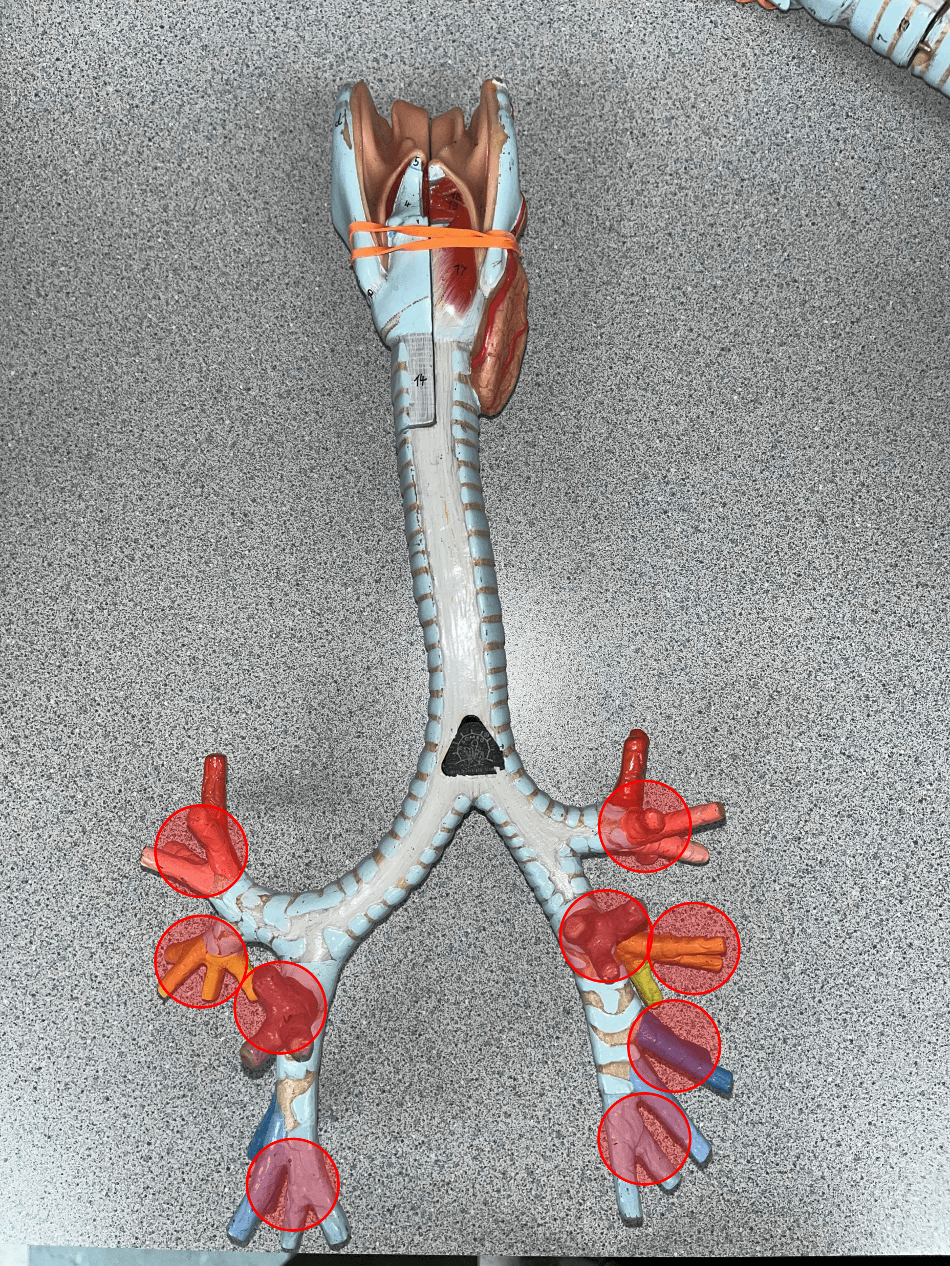
secondary bronchi
• Smaller branches of a primary bronchus.
• Supplies each lobe of a lung.
• Branches into tertiary bronchi.
• Also known as lobar bronchi.
• Supplies each lobe of a lung.
• Branches into tertiary bronchi.
• Also known as lobar bronchi.
41
New cards
tertiary bronchi
• Smaller branches of a secondary bronchus.
• Supplies bronchopulmonary segments of a lobe.
• Branches into bronchioles.
• Also known as segmental bronchi.
• Supplies bronchopulmonary segments of a lobe.
• Branches into bronchioles.
• Also known as segmental bronchi.
42
New cards
bronchioles
• Smaller branches of a tertiary bronchus.
• Supplies lobules of bronchopulmonary segments.
• Branches into terminal bronchioles.
• Not shown in image.
• Supplies lobules of bronchopulmonary segments.
• Branches into terminal bronchioles.
• Not shown in image.

43
New cards
terminal bronchioles
• Smaller branches of a bronchiole.
• Branches into respiratory bronchioles.
• Not shown in image.
• Branches into respiratory bronchioles.
• Not shown in image.

44
New cards
respiratory bronchioles
• Smaller branches of a terminal bronchiole.
• Branches into alveolar ducts.
• Not shown in image.
• Branches into alveolar ducts.
• Not shown in image.
45
New cards
alveolar ducts
• Smaller branches of a respiratory bronchiole.
• Leads into alveolar sacs.
• Not shown in image.
• Leads into alveolar sacs.
• Not shown in image.

46
New cards
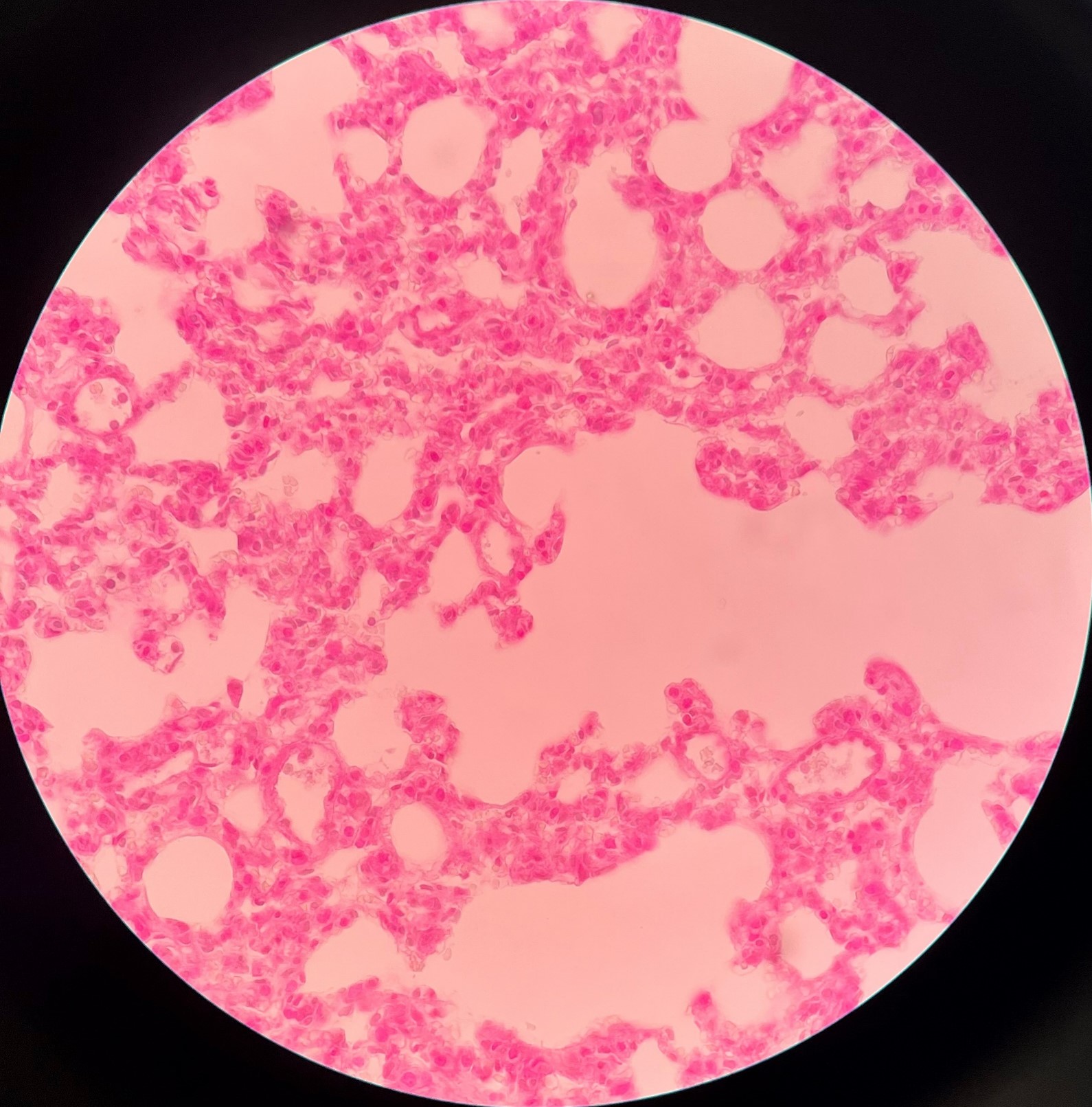
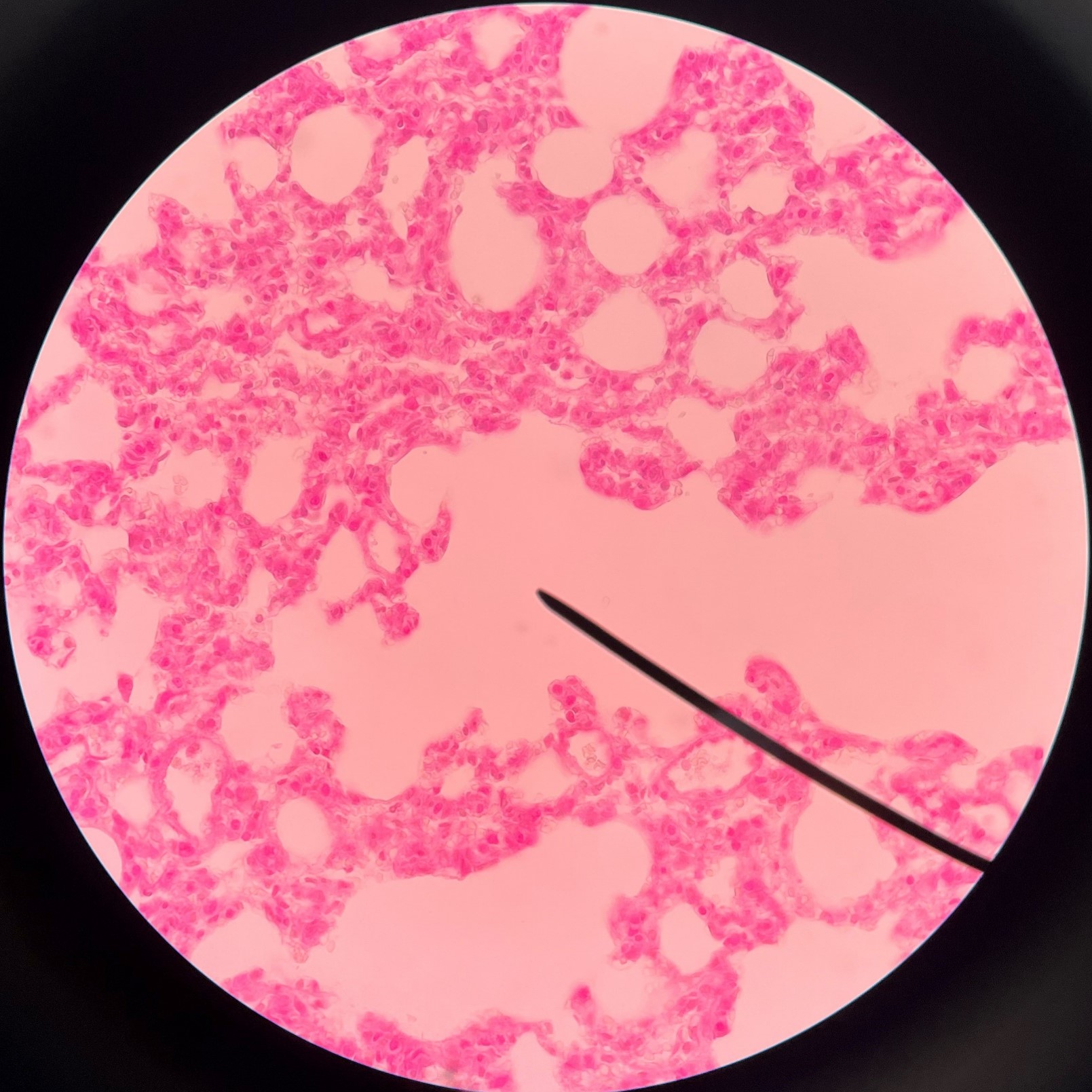
alveolar sac
• Clusters of alveoli.
• Led into by the alveolar ducts.
• Led into by the alveolar ducts.
47
New cards
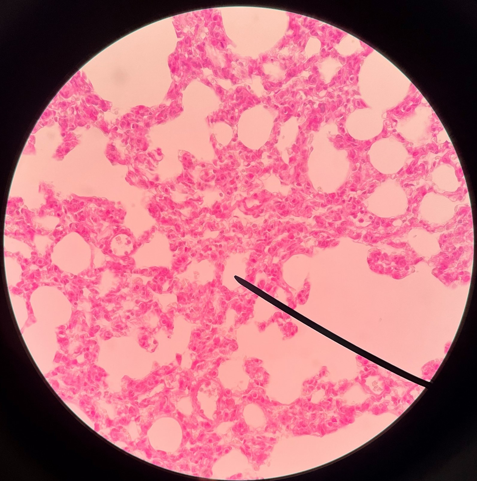
alveoli
• Where respiration occurs in the lungs.
• Budded on alveolar sacs.
• Budded on alveolar sacs.
48
New cards
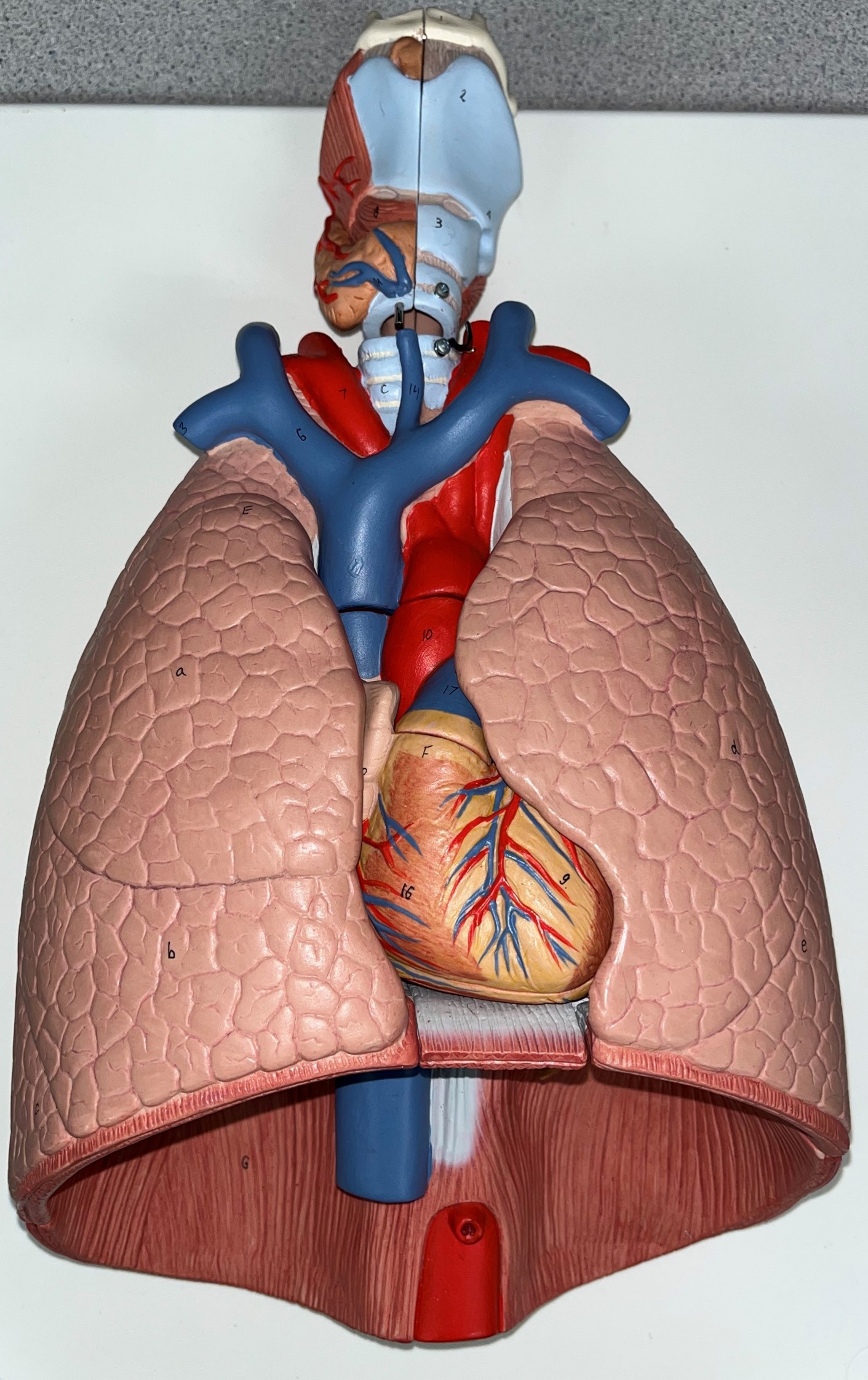
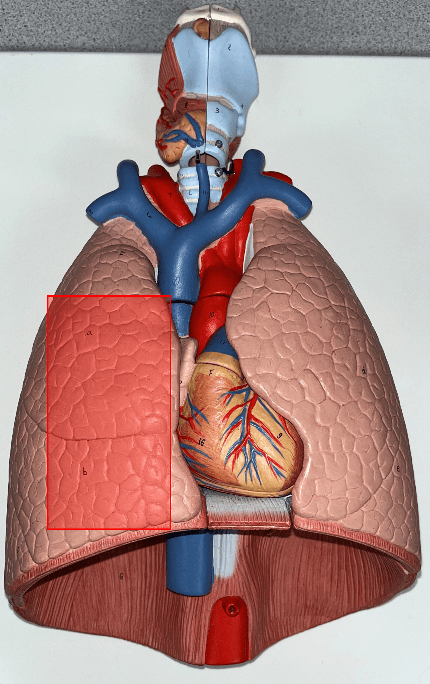
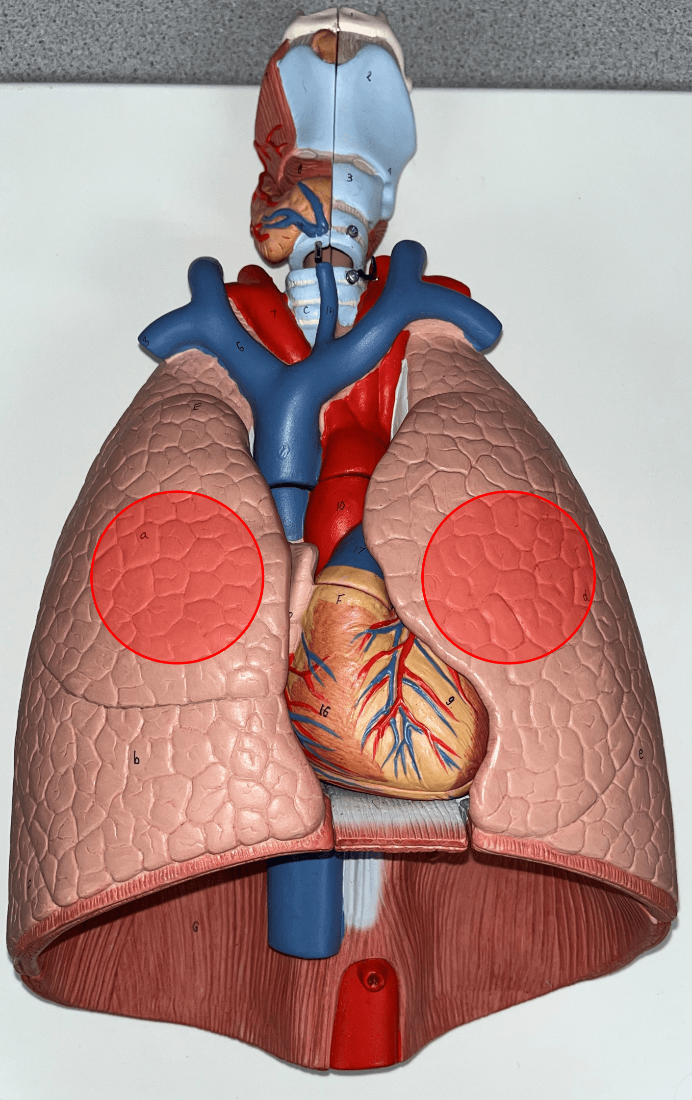
right lung
• The R. principal respiratory organ of the thoracic cavity.
• Divided into three lobes.
• Divided into three lobes.
49
New cards
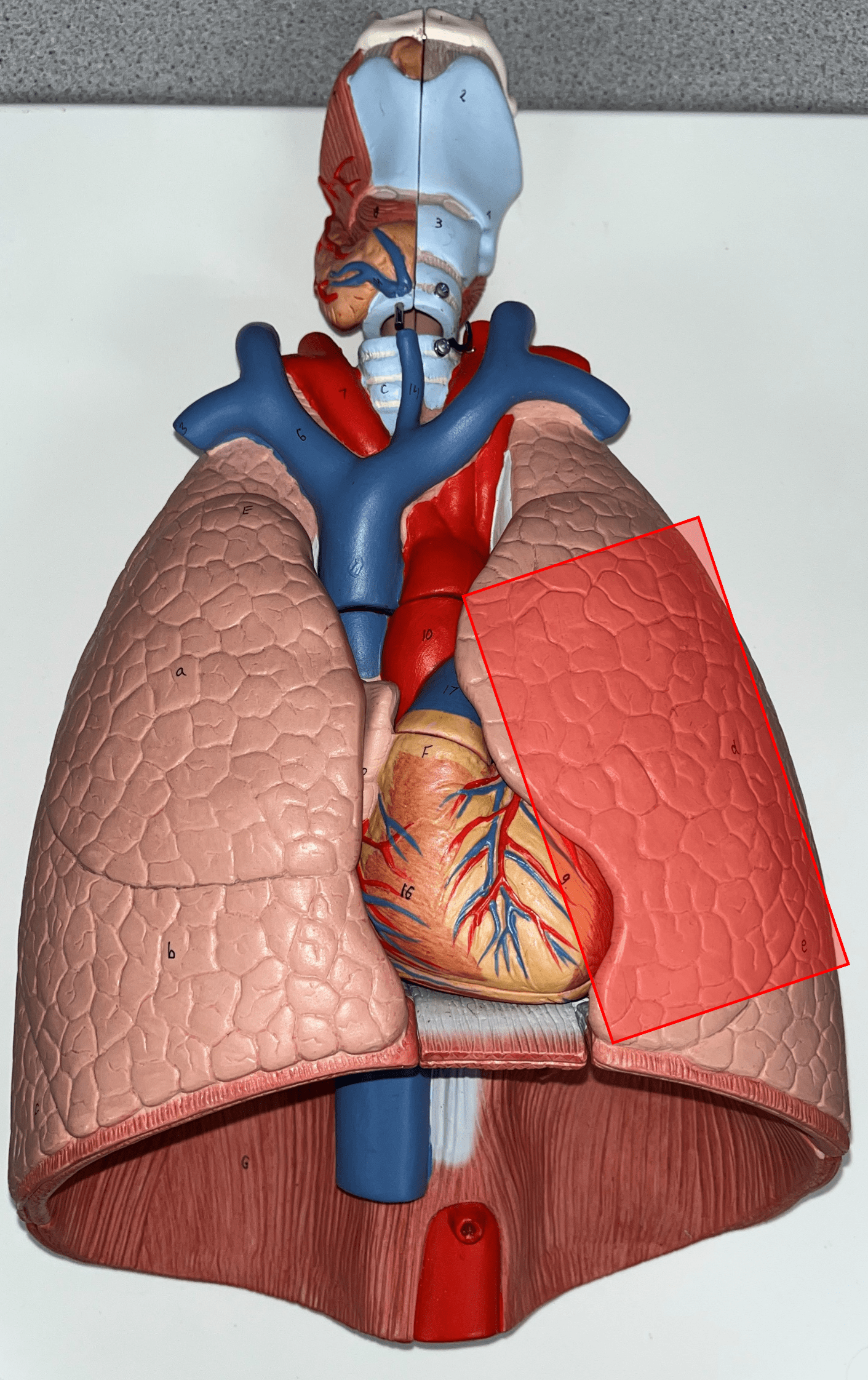
left lung
• The L. principal respiratory organ of the thoracic cavity.
• Divided into two lobes.
• Divided into two lobes.
50
New cards
superior lobe
The uppermost lobe of the R. and L. lungs.
51
New cards
middle lobe
The middlemost lobe of the R. lung.
52
New cards
inferior lobe
The bottommost lobe of the R. and L. lungs.
53
New cards
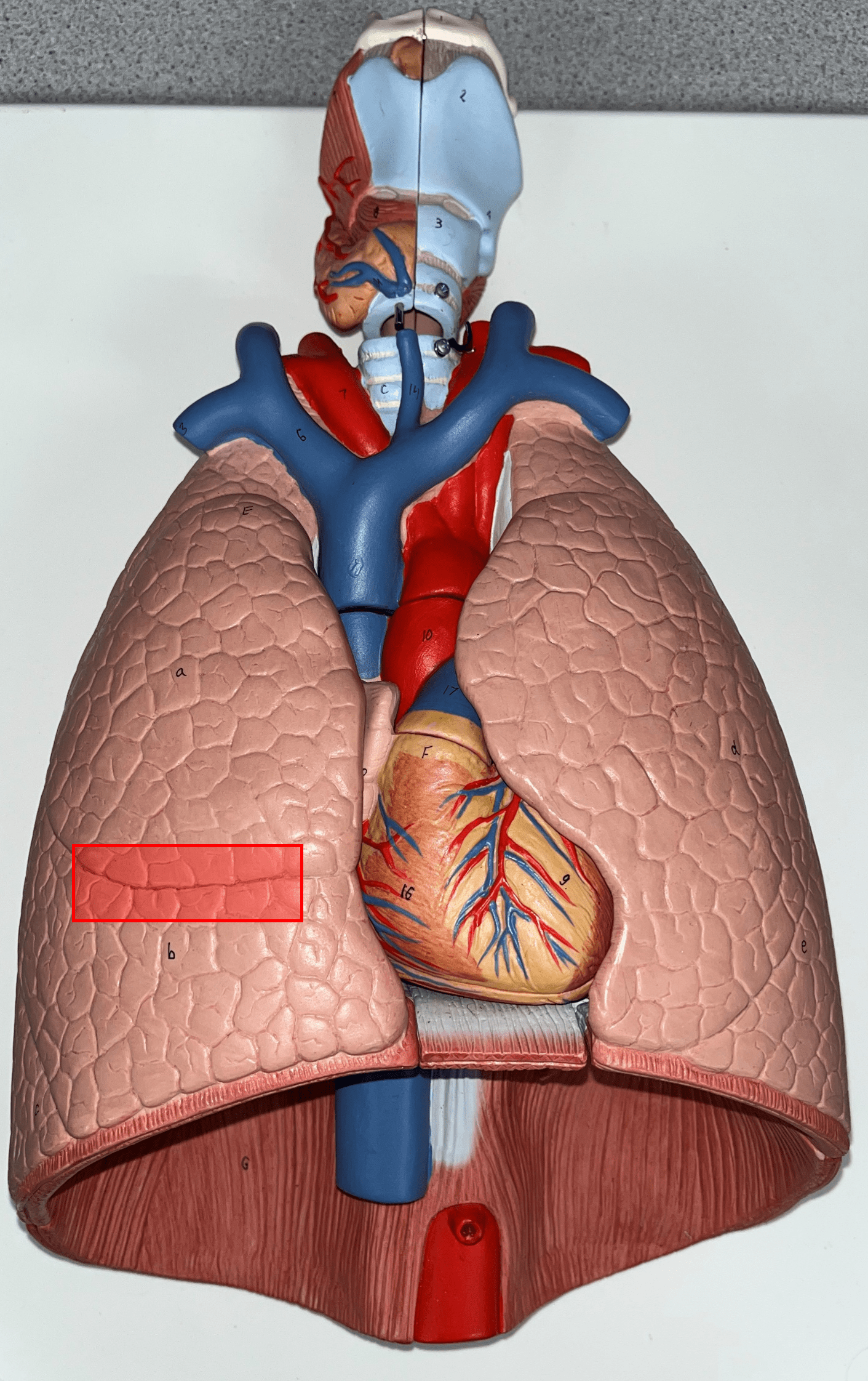
horizontal fissure
Separates the superior and middle lobes of the R. lung.
54
New cards
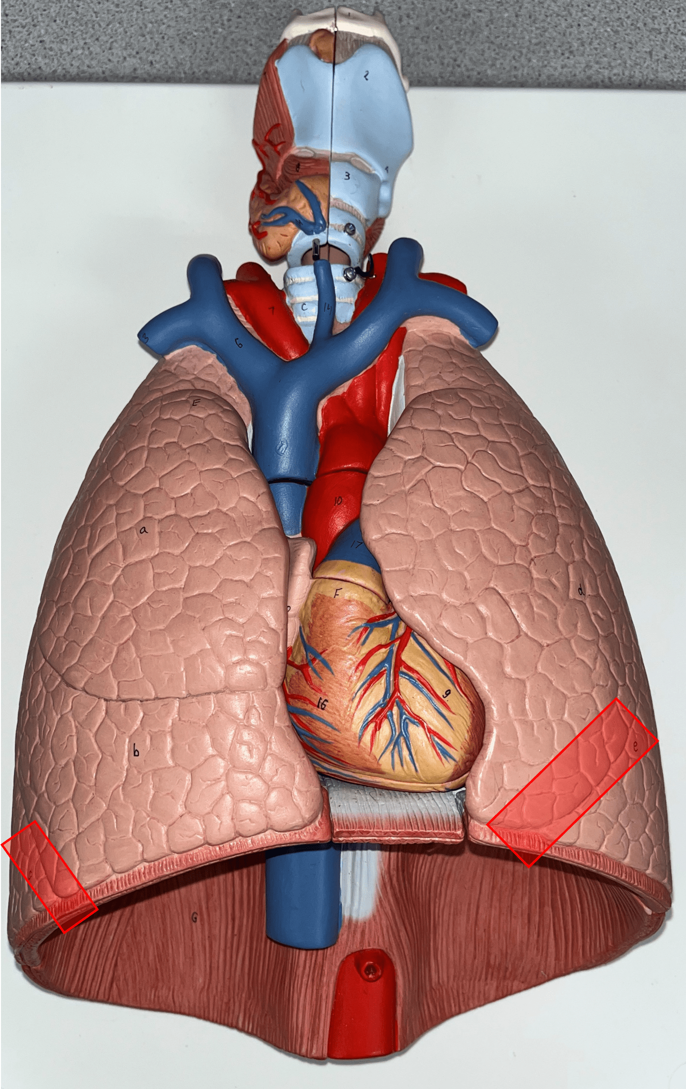
oblique fissure
• Separates the middle and inferior lobes of the R. lung.
• Separates the superior and inferior lobes of the L. lung.
• Separates the superior and inferior lobes of the L. lung.
55
New cards
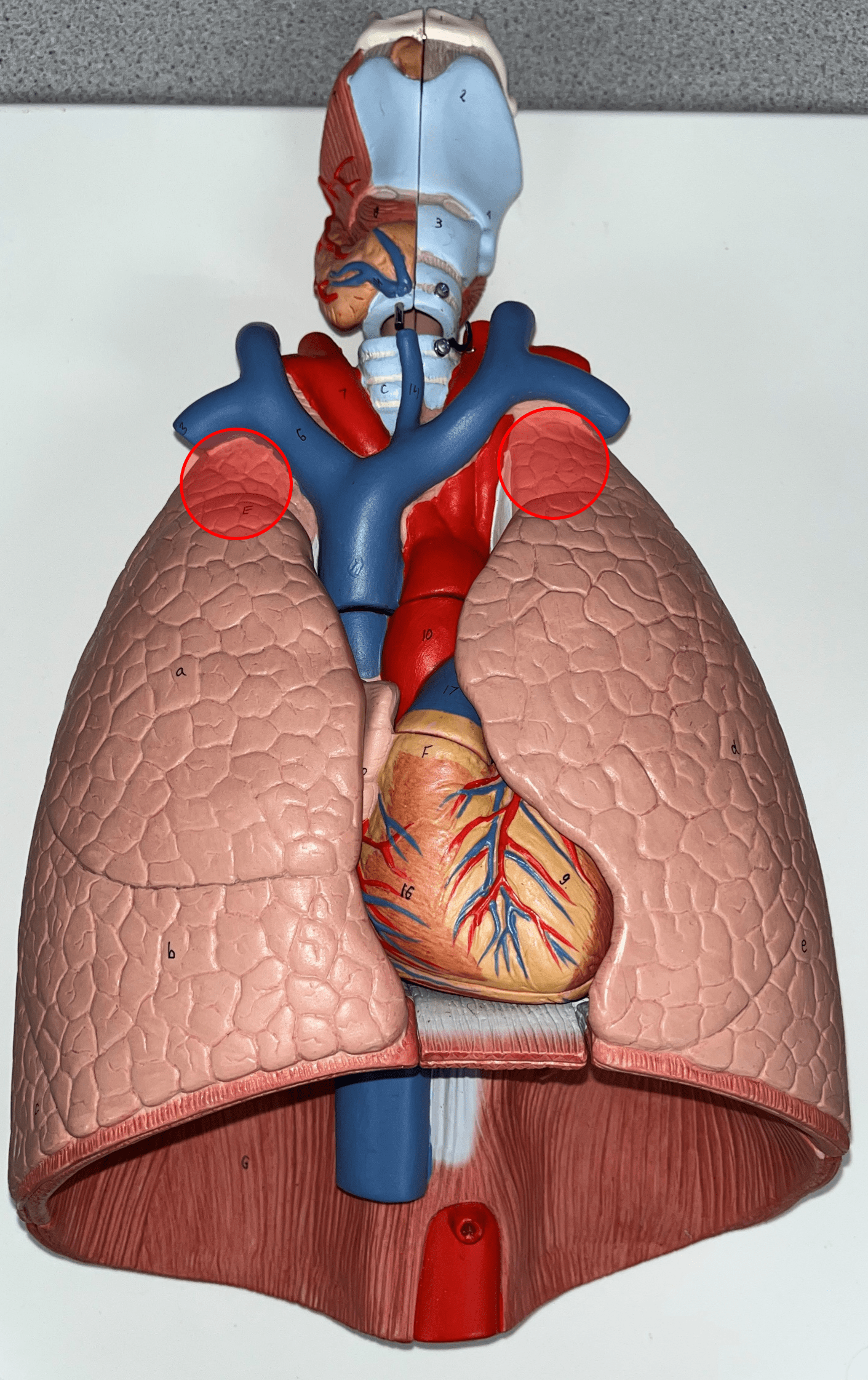
apex
The rounded superior portion of the R. and L. lungs.
56
New cards
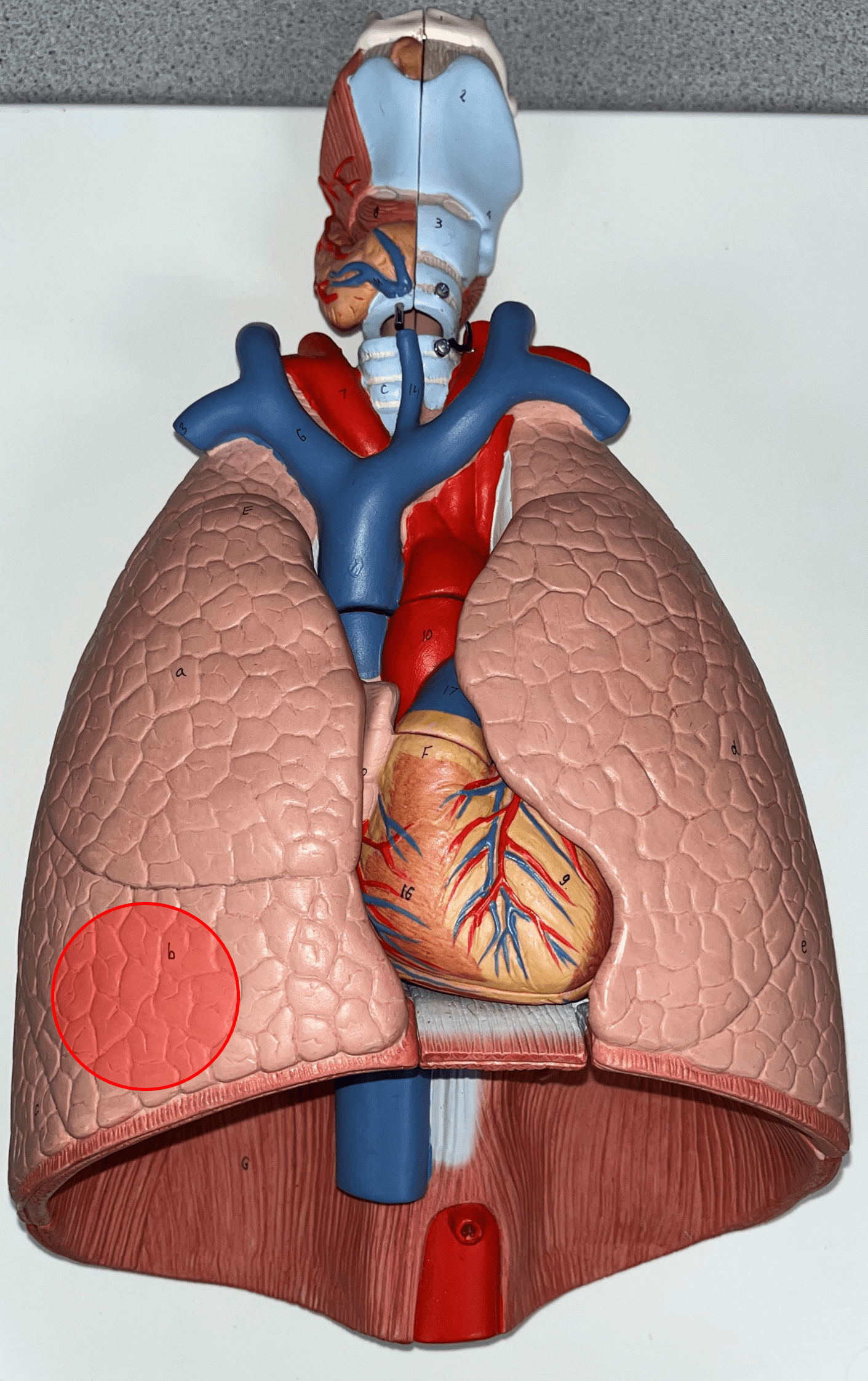
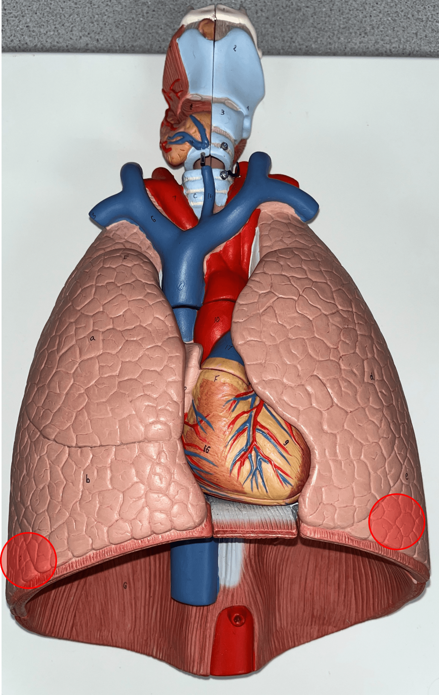
base
The broad inferior portion of the R. and L. lungs.

57
New cards
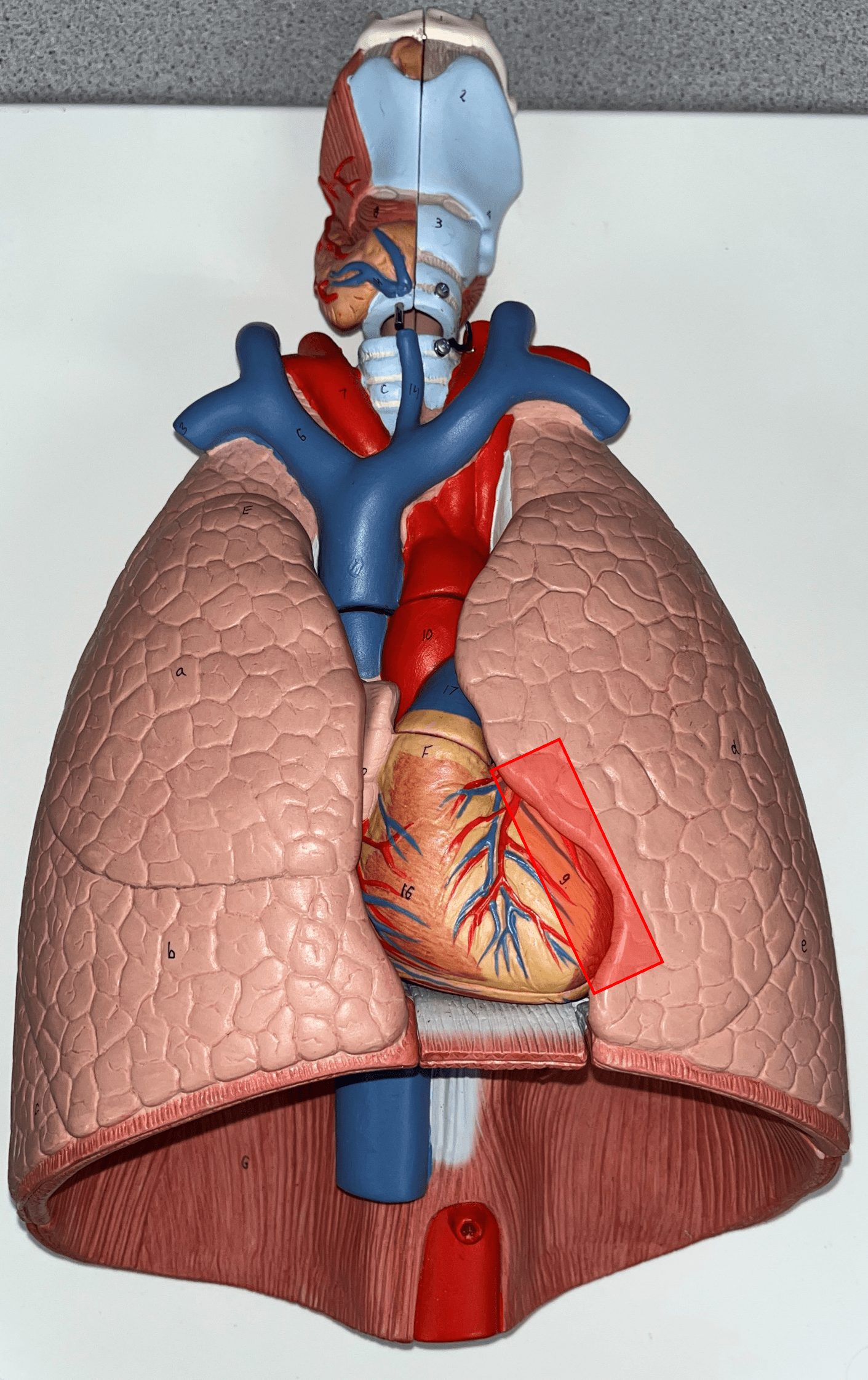
cardiac impression
The concave surface of the L. lung that accommodates the apex of the heart.
58
New cards
root
• The common passage of bronchi, bronchial veins and arteries, and lymphatic vessels on the medial side of the R. and L. lungs.
• Not shown in image.
• Not shown in image.

59
New cards
hilum
• The area of the R. and L. lungs surrounding their roots.
• Not shown in image.
• Not shown in image.