Topic 6 - Human Physiology
0.0(0)
Card Sorting
1/119
Earn XP
Description and Tags
Last updated 1:19 AM on 11/30/22
Name | Mastery | Learn | Test | Matching | Spaced | Call with Kai |
|---|
No analytics yet
Send a link to your students to track their progress
120 Terms
1
New cards
Epiglottis
Covers opening of trachea when eating
2
New cards
Peristalsis
- Contraction of circular and longitudinal muscle layers
- Begins in esophagus, continues into the intestines
- Occurs in 1 direction
- Churns food, mixes with enzymes for digestion
- Begins in esophagus, continues into the intestines
- Occurs in 1 direction
- Churns food, mixes with enzymes for digestion
3
New cards
Anti-peristalsis
Vomiting, stomach contracts to remove food
4
New cards
Stomach acid
- Kills bacteria and other pathogens
- Mucus protects the lining of the stomach from acid
- Neutralized by bicarbonate in the small intestine
- Mucus protects the lining of the stomach from acid
- Neutralized by bicarbonate in the small intestine
5
New cards
Villi
- Small, finger-like projections
- Aid in nutrient absorption
- Aid in nutrient absorption
6
New cards
Pancreas
Produces bicarbonate and other enzymes
7
New cards
Liver
Produces bile and other enzymes
8
New cards
Lipid digestion
Lipids are emulsification by bile salts to form tiny drops of triglycerides.
Lipase breaks it down into fatty acids and glycerol.
Lipase breaks it down into fatty acids and glycerol.
9
New cards
Emulsification
A process in which large lipid molecules are broken down into several small lipid molecules.
Churning action of stomach and use of bile salts in begining of small intestine
Churning action of stomach and use of bile salts in begining of small intestine
10
New cards
Lipase
Enzyme used to break down triglycerides
11
New cards
Protein digestion
Endopeptidases break peptide linkages in the protein interior to form shorter polypeptide chains.
Exopeptidases act on the ends of the chains to form dipeptides.
Dipeptidases break down dipeptides into amino acids.
Exopeptidases act on the ends of the chains to form dipeptides.
Dipeptidases break down dipeptides into amino acids.
12
New cards
Exopeptidases examples
Aminopeptidases act on the NH2 end and carboxypeptidases act on the COOH end.
13
New cards
Dipeptides
2 amino acids
14
New cards
Enzymes used for digestion
Amylase, Lipase, Protease
15
New cards
Cellulose digestion
Humans do not have the enzyme needed to digest cellulose.
Is excreted as waste, but provides diet with fibre, keeping digestive track healthy.
Is excreted as waste, but provides diet with fibre, keeping digestive track healthy.
16
New cards
Starch digestion
Starch cannot pass through the villi, too large.
Begins in the mouth with salivary enzyme amylase, however it stops working in the stomach due to the acidity.
Pancreatic amylase continues digestion in small intestine by breaking 1-4 bonds into glucose fragments; maltose and maltriose.
Amylopectin 1-6 bonds cannot be broken down by amylase, and are called dextrins.
Enzymes from small intestine membrane turn glucose fragments into glucose.
17
New cards
Enzymes used to form glucose
Maltase (for maltose), Glucosidase (for maltotriose), Dextrinase (for dextrins)
18
New cards
Glucose absorption
Glucose is absorbed into villus by cotransport, enters into villus capillaries, moves into venules in sub-mucosa, blood is carrier to the liver, excess glucose becomes glycogen for energy storage.
19
New cards
Surface area increase in digestive tracts
The inner intestine wall is folded
Villi are projections on the inner wall
Epithelial cells on the villi fold into microvilli
Villi are projections on the inner wall
Epithelial cells on the villi fold into microvilli
20
New cards
Methods of absorption
Co-transport (for glucose and amino acids), Facilitated diffusion (for monosaccharides), Osmosis (for water), Simple diffusion (for triglycerides)
21
New cards
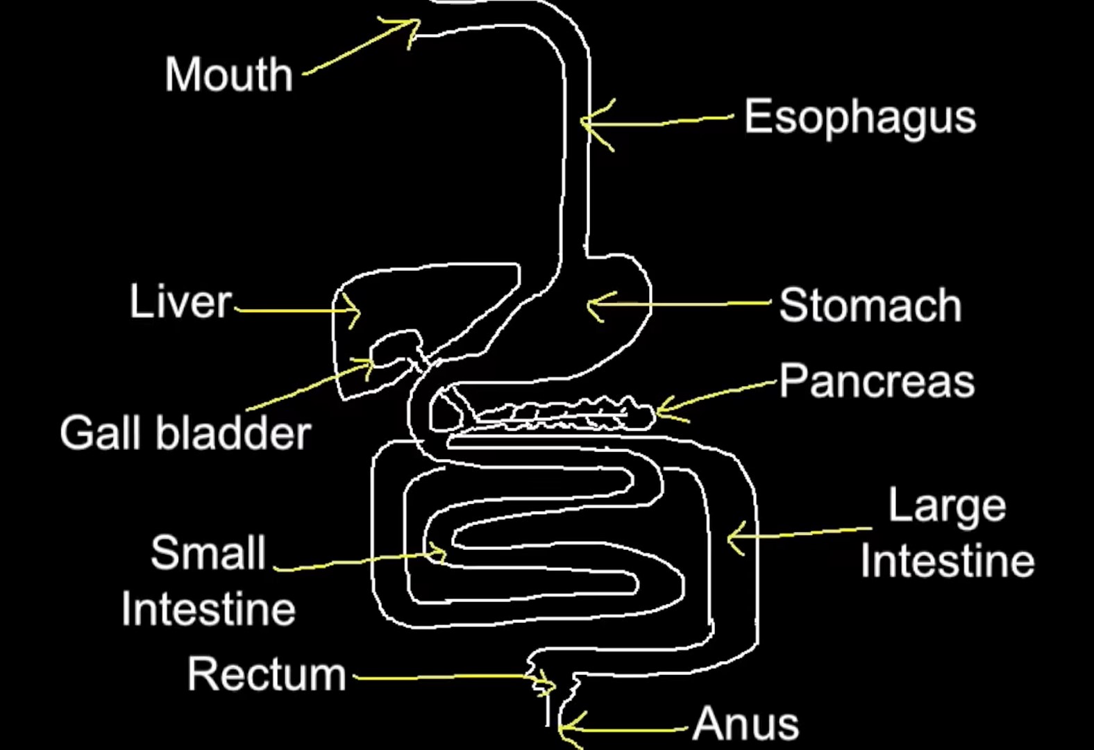
Digestive System
22
New cards
Gaseous exchange
the exchange of gases between an organism and it's surroundings, the uptake of oxygen and release of carbon dioxide
23
New cards
Factors that speed up diffusion
Surface area: greater surface area results in greater diffusion
Difference in concentration: greater concentration gradient results in greater diffusion
Length of diffusion path: shorter path results in greater diffusion
Difference in concentration: greater concentration gradient results in greater diffusion
Length of diffusion path: shorter path results in greater diffusion
24
New cards
Components of Respiration
Breathing, External respiration, Internal respiration, Cellular respiration
25
New cards
4. Cellular respiration
Breakdown of sugar molecules into energy molecules
- Requires presence of O2, Results in production of CO2
- Requires presence of O2, Results in production of CO2
26
New cards
3. Internal respiration
Exchange of gases between the blood in capillaries and individual cells
- O2 diffuses into cell, CO2 diffuses out of cell
- O2 diffuses into cell, CO2 diffuses out of cell
27
New cards
1. Breathing
Inhalation - taking in oxygen from external environment
Exhalation - expelling carbon dioxide
Exhalation - expelling carbon dioxide
28
New cards
2. External respiration
Exchange of gases between alveoli and the blood
- O2 diffuses into blood, CO2 diffuses out of lungs
- O2 diffuses into blood, CO2 diffuses out of lungs
29
New cards
Nasal and Oral Cavities
Warm, moisten and filter the air entering through the nose. Mucus traps small particles, Nose hairs sweep out larger particles, Cilia moves trapped particles to nose opening or down esophagus
30
New cards
Pharynx
Common path for food and air. Branches into esophagus (food) and trachea (air).
31
New cards
Larynx
Known as the voice box. Sound is produced through vibrations of the vocal cord and in the air.
32
New cards
Trachea
Connects pharynx to the lungs. Lined with mucus and Cilia. Branches into left and right bronchus.
33
New cards
Bronchi and Bronchioles
Bronchi branch into smalller tubes called bronchioles. Lined with mucus and cilia for more filtering.
34
New cards
Alveoli
Hollow air sac clusters at the end of the smallest bronchioles where gas exchange occurs.
35
New cards
Advantages of alveoli shape
1 cell thick, decreases the diffusion path
Large combined surface area due to spherical shape
Large combined surface area due to spherical shape
36
New cards
Maintanence of concentration gradients
Maintained by blood flow on one side and air flow on the other.
37
New cards
Pneumocytes
Cells in alveoli, 2 types
38
New cards
Type 1 Pneumocytes
Most of the flattened epethelial cells in alveoli walls are type 1 cells
39
New cards
Type 2 Pneumocytes
Occupy small portion of alveoli surface.
Secrete fluids which coat the inner surface of alveoli to reduce surface tension.
Fluid allows oxygen to dissolve and diffuse into blood.
Secrete fluids which coat the inner surface of alveoli to reduce surface tension.
Fluid allows oxygen to dissolve and diffuse into blood.
40
New cards
Causes of lung cancer
smoking, second hand smoke, air pollution, radon gas, asbestos, silica
41
New cards
Consequences of lung cancer
Very few patients survive more than 5 years.
DIfficulty breathing, coughing, chest pain, loss of appetite, weight.
Tumors can form in many parts of the body.
DIfficulty breathing, coughing, chest pain, loss of appetite, weight.
Tumors can form in many parts of the body.
42
New cards
Emphysema
Gradual deconstruction of alveoli - thin inner walls rupture to create larger air sacs with thick walls.
The surface are is reduced - the lungs become less elastic.
Damage is irreversible.
The surface are is reduced - the lungs become less elastic.
Damage is irreversible.
43
New cards
Types of Blood Vessels
Artery, Capillary, Vein
44
New cards
Arteries
Thick wall, narrow lumen, has 3 layers, can stretch, is thick and muscular, carries blood away from heart
45
New cards
Capilaries
Thin wall, narrow lumen, wall is 1 cell thick, no muscle/elastic fibres in wall, site of gas exchange
46
New cards
Veins
Thinner wall than arteries, wide lumen, elastic fibres in wall, has valves in lumen, carries blood towards heart
47
New cards
Endothelial
Cells which line the inside of blood vessels
48
New cards
Arterioles
Smaller arteries
49
New cards
Layers of the heart
Endocardium, Myocardium, Pericardium
50
New cards
Endocardium
Smooth, inner linning of heart
51
New cards
Myocardium
Muscle, middle layer of heart
52
New cards
Pericardium
Fluid filled sac, outer layer of heart
53
New cards
Pericardial fluid
Fluid withing the pericardium, reduces friction created by the heart when beating
54
New cards
Coronary arteries
Carry blood supply to the heart (heart needs blood to function)
55
New cards
Heart Chambers
Left ventricle, right ventricle, Left atrium, right atrium
56
New cards
Heart valves
Tricuspid valve, Bicuspid valve, Pulmonary valve, Aortic valve, prevents blood backflow
57
New cards
Tricuspid valve
Separates right atrium and ventricle
58
New cards
Bicuspid valve
Separates left atrium and ventricle
59
New cards
Pulmonary valve
Separates pulmoary artery and right ventricle
60
New cards
Aortic valve
Separates aorta and left ventricle
61
New cards
Semilunar valves
Valve at the base of arteries (aortic and pulmonary valves)
62
New cards
Atrioventricular valves
Valves between atria and ventricles (trsicuspid and bicuspid valves)
63
New cards
Blood flow
Deoxygenated blood enters right atrium through the Vena Cava. Enters right ventricle and leaves through the pulmonary arteries. Goes to lungs, where gas exchange occurs. Oxygenated blood enters left atrium through pulmonary veins. Enters left ventricle and leaves through the aorta. Goes to the rest of the body. Repeat.
64
New cards
Sinoatrial Node (SV)
Pacemaker, sets a rythm (approx 72 beats per minute), found near right atrium
65
New cards
Atrioventricular Node (AV)
Passes nerve impulses to the ventricles , found on middle edge of right atrium
66
New cards
Cardiac Cycle
A heartbeat. 3 stages; Atrial systole, Ventricular systole, and Diastole.
67
New cards
Atrial stystole
SA node sends nerve impulse, causing blood in the right and left atria to be forced into the right and left ventricles.
68
New cards
Ventricular systole
Blood pressure in the ventricles is high, causes the atrioventricular valves to shut. Nerve impulse reaches the AV node, causing blood in the right and left ventricles to be forced into the pulmonary artery and aorta.
69
New cards
Diastole
Semilunar valves close to by back pressure of blood in the arteries. Atria and Ventricles relax.
Blood flows into the atria, the atrioventricular valves open and blood enters the ventricles passively.
Blood flows into the atria, the atrioventricular valves open and blood enters the ventricles passively.
70
New cards
Heart Rate
Frequency of the cardiac cycle
71
New cards
Blood Pressure
Force of blood on the walls of the arteries. A ratio of systolic and diastolic pressure of the blood filling and being expelled from the heart (avg is 122mm Hg / 80mm Hg).
72
New cards
Factors that affect Heart Rate
pH of blood, O2 concentrations, bloos pressure
73
New cards
pH of blood
Reflected in CO2 concentrations (combines with water to form carbonic acid, which lowers the pH). If there is too much CO2 it means the blood is too acidic and heart rate will increase.
74
New cards
Cardiovascular center
Located in the medullar oblongata. Receives input from recpetors in the body which monitor the factors that affect heart rate. Sends nerve impulses to the SA node which tell it whether to increase or decrease frequency of heart beats.
75
New cards
Atherosclerosis
Development of fatty tissue (atheroma) in lumen of arteries.
76
New cards
Causes of Arterioscleriosis
- High concentration of LDL (low density lipoprotein)
- High blood glucose levels (due to obesity/over eating)
- High blood pressure
- Consumption of trans fats
- High blood glucose levels (due to obesity/over eating)
- High blood pressure
- Consumption of trans fats
77
New cards
Consequences of Arterioscleriosis
- Affects the coronary arteries, can lead to a heart attack
- Affects the brain arteries, can lead to a stroke
- Affects the renal arteries, can lead to kidney disease
- Affects the aorta, can lead to aortic aneurysm
- Affects the brain arteries, can lead to a stroke
- Affects the renal arteries, can lead to kidney disease
- Affects the aorta, can lead to aortic aneurysm
78
New cards
Epinephrene
- Hormone produced by the adrenal gland (also known as adrenaline).
- Level rises when physical activity is needed to increase heart rate
- Part of the fight or flight response
- Level rises when physical activity is needed to increase heart rate
- Part of the fight or flight response
79
New cards
Galen
Believed the circulatory system consisted of two separate one-way systems of distribution, blood was consumed by all organs of the body, then regenerated in either the liver or the heart.
80
New cards
Harvey
Tied a ligature around the upper part of a man's arm. He observed that when tied tightly, blood flow into the arm's lower portion was cut off. When loosened slightly, blood flowed into the lower part of the arm but was unable to escape back to the upper part. As the veins swelled, small bumps in the veins became more noticeable and were distinguished as valves.
Showed that blood flow was unidirectional, with arteries carrying blood away from the heart and veins returning to the heart.
Showed that blood flow was unidirectional, with arteries carrying blood away from the heart and veins returning to the heart.
81
New cards
Immune System
Protects the body against foreign invaders using physical and chemical mechanisms.
82
New cards
Physical Mechanisms
Cilia, Mucous, Skin
83
New cards
Chemical Mechanisms
Phagocytosis, Antibodies/Antigens, Inflammatory response
84
New cards
Innate immune response
Nonspecific defence, 1st and 2nd line
85
New cards
Acquired immune response
Specific defence, 3rd line
86
New cards
First line of defence
External.
- Skin is an almost impenetrable barrier
- Enzymes in mucus can trap and destroy bacteria cells
- Cilia sweep debris out of the throat and nose (cough/sneeze)
- Stomach acid kills bacteria in stomach
- Enzymes in tears destroy bacteria
- Eyelashes sweep over the eye to keep bacteria out
- Skin is an almost impenetrable barrier
- Enzymes in mucus can trap and destroy bacteria cells
- Cilia sweep debris out of the throat and nose (cough/sneeze)
- Stomach acid kills bacteria in stomach
- Enzymes in tears destroy bacteria
- Eyelashes sweep over the eye to keep bacteria out
87
New cards
Second line of defence
Internal.
- Lymphatic system
- Phagocytosis of a cell
- Natural Killer cells
- Antimicrobial proteins
- Fever
- Lymphatic system
- Phagocytosis of a cell
- Natural Killer cells
- Antimicrobial proteins
- Fever
88
New cards
Lymphatic system organs
Spleen, Appendix, Thymus, Tonsils
89
New cards
Lymph Nodes
Specialized cells in the body which produce lymphocytes, located throughout the body (under chin, armpits, intestinal tract)
90
New cards
Lymph
Clear fluid which circulates in the lymphatic vessels. Contains lymphocytes (white blood cells).
91
New cards
Lymphatic vessels
Transports lymph from organs to the circulatory system.
92
New cards
Lymphocytes
Type of white blood cells; Made in bone marrow (spongy tissue in bones)
93
New cards
Macrophages
Type of white blood cells; Engulf pathogens, destroy pathogens with digestive enzymes.
94
New cards
Phagocytosis
1. Macrophages detect the presence of a bacterium
2. Bacterium attaches to the receptor on the membrane
3. Bacterium is englufed into a food vacuole
4. Lysosome in the cytoplasm fuses with the vacuole
5. Bacterium is broken up into its essential components which are disposed into the cytoplasm
2. Bacterium attaches to the receptor on the membrane
3. Bacterium is englufed into a food vacuole
4. Lysosome in the cytoplasm fuses with the vacuole
5. Bacterium is broken up into its essential components which are disposed into the cytoplasm
95
New cards
Third line of defence
Creation of antibodies through lymphocytes (specialized white blood cells)
96
New cards
Antigens
On the surface of every foreign material which enters the body.
97
New cards
Examples of Antigens
- Potentially damaging microbes and their toxins
- Pollen, Flea, Dust mite feces
- Blood cell surface proteins
- Surface proteins of transplanted tissues/organs
- Pollen, Flea, Dust mite feces
- Blood cell surface proteins
- Surface proteins of transplanted tissues/organs
98
New cards
Antigen Receptors
Located on both B cells and T cells, used to recognize antigens
99
New cards
Antibodies
Proteins that attach to specific antigens
100
New cards
Antigen/Antibody specificity
Tip of each antibody becomes specialized for a specific antigen. i.e. Only 1 antigen can bond to the tip of a specific antibody