lab practical 5: eye and ear anatomy
1/20
There's no tags or description
Looks like no tags are added yet.
Name | Mastery | Learn | Test | Matching | Spaced | Call with Kai |
|---|
No analytics yet
Send a link to your students to track their progress
21 Terms
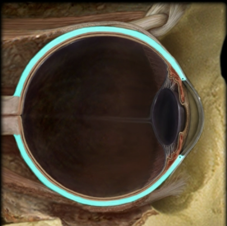
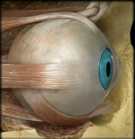
name the part and layer of the eye
sclera, fibrous layer
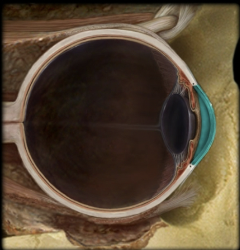
name the part and layer of the eye
cornea, fibrous layer
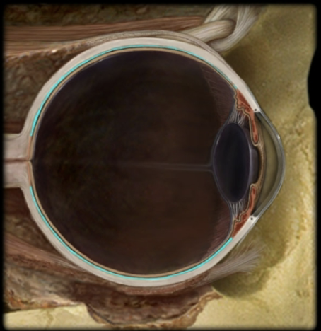
name the part and layer of the eye
choroid, vascular layer
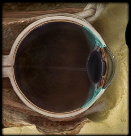
name the part and layer of the eye
ciliary body, vascular layer
name the part and layer of the eye
ciliary processes, vascular layer
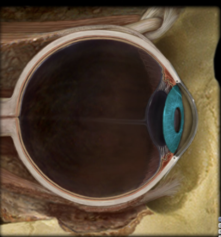
name the part and layer of the eye
iris, vascular layer
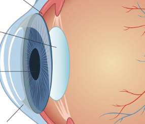
name the three important anatomical parts of the eye
iris, pupil, lens
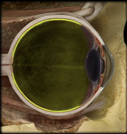
name the part and layer of the eye
retina, inner layer
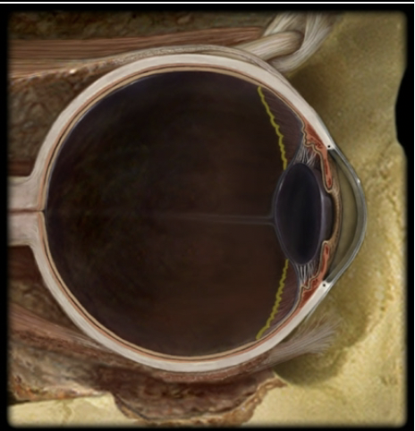
name the part and layer of the eye
ora serrata, inner layer
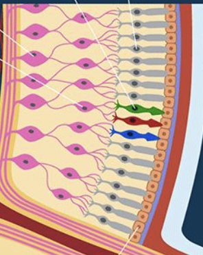
name the retinal layers and the cells found in each layer
neural layer: bipolar cells, ganglion cells
pigmented layer: rods, cones
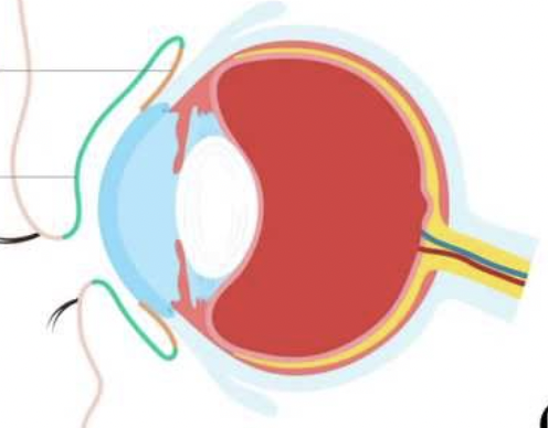
name the types of conjunctiva
orange: bulbar conjunctiva
green: palpebral conjunctiva
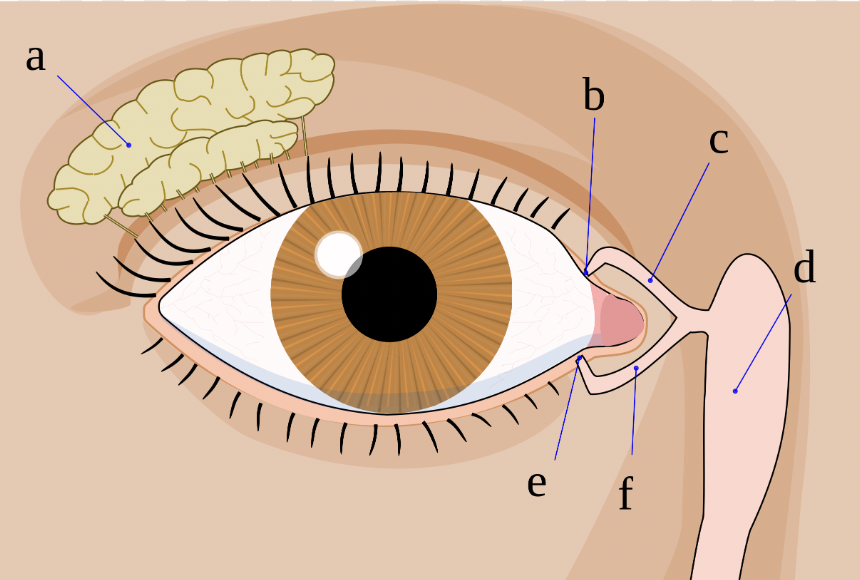
label structures a, c, f, d and g
a: lacrimal gland
c and f: lacrimal canaliculi
d: lacrimal sac
g: nasal duct
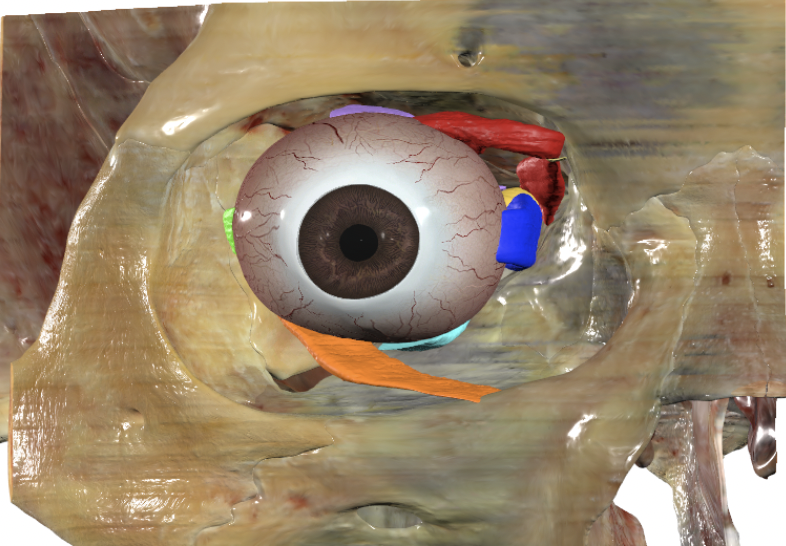
identify all colored structures from the anterior view
red: superior oblique
orange: inferior oblique
green: lateral rectus
light blue: inferior rectus
navy blue: medial rectus
purple: superior rectus
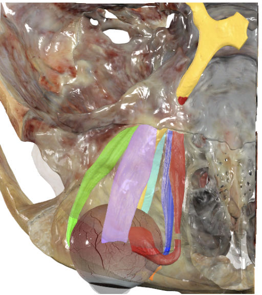
identify all colored structures from the superior view
red: superior oblique
orange: inferior oblique
yellow: optic nerve
green: lateral rectus
light blue: inferior rectus
navy blue: medial rectus
purple: superior rectus
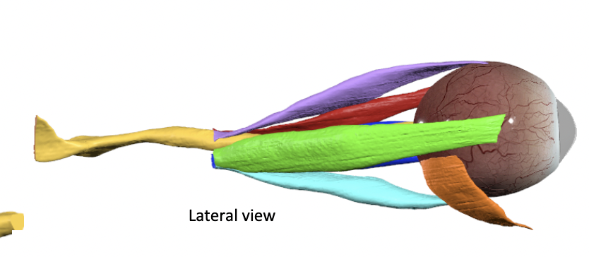
identify all colored structures from the lateral view
red: superior oblique
orange: inferior oblique
yellow: optic nerve
green: lateral rectus
light blue: inferior rectus
navy blue: medial rectus
purple: superior rectus
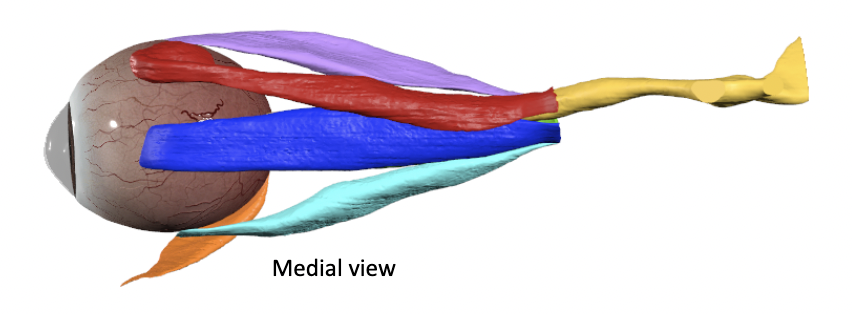
identify all colored structures from the medial view
red: superior oblique
orange: inferior oblique
yellow: optic nerve
light blue: inferior rectus
navy blue: medial rectus
purple: superior rectus
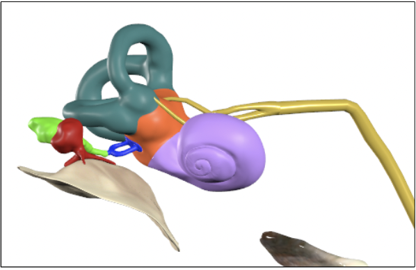
identify all colored structures in the anterior view
red: malleus
orange: vestibule
yellow: vestibulocochlear nerve
green: incus
teal: semicircular canals
navy blue: stapes
purple: cochlea
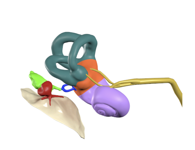
identify all colored structures from the superior view
red: malleus
orange: vestibule
yellow: vestibulocochlear nerve
green: incus
teal: semicircular canals
navy blue: stapes
purple: cochlea
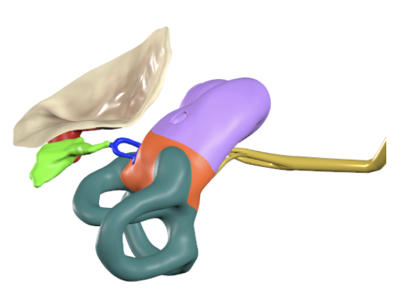
identify all colored structures in the inferior view
red: malleus
orange: vestibule
yellow: vestibulocochlear nerve
green: incus
teal: semicircular canals
navy blue: stapes
purple: cochlea
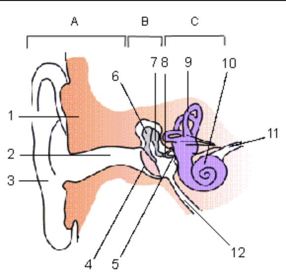
label all numbered and lettered structures
a: external ear
b: middle ear
c: inner ear
1: temporal bone
2: external acoustic meatus
3: auricle
4: tympanic membrane
5: oval window
6: malleus
7: incus
8: stapes
9: semicircular canals
10: cochlea
11: vestibulocochlear nerve
12: pharyngotympanic (auditory) tube
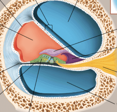
label all colored structures of the cochlear anatomy
orange: scala media (cochlear duct)
yellow: vestibulocochlear nerve
green: inner ear hair cells
blue: scala vestibule and tympani
light purple: tectorial membrane
black: basilar membrane