OB Final Exam - Anomalies, Syndromes, & Trisomy
1/97
There's no tags or description
Looks like no tags are added yet.
Name | Mastery | Learn | Test | Matching | Spaced | Call with Kai |
|---|
No analytics yet
Send a link to your students to track their progress
98 Terms
Type 1 Thanatophoric Dysplasia
Most common form of Thanatophoric Dysplasia
Short bowed femurs
Flat vertebral bodies
Frontal bossing
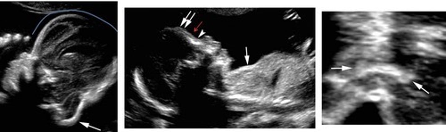
Type 2 Thanatophoric Dysplasia
Short straight femurs
Clover skull
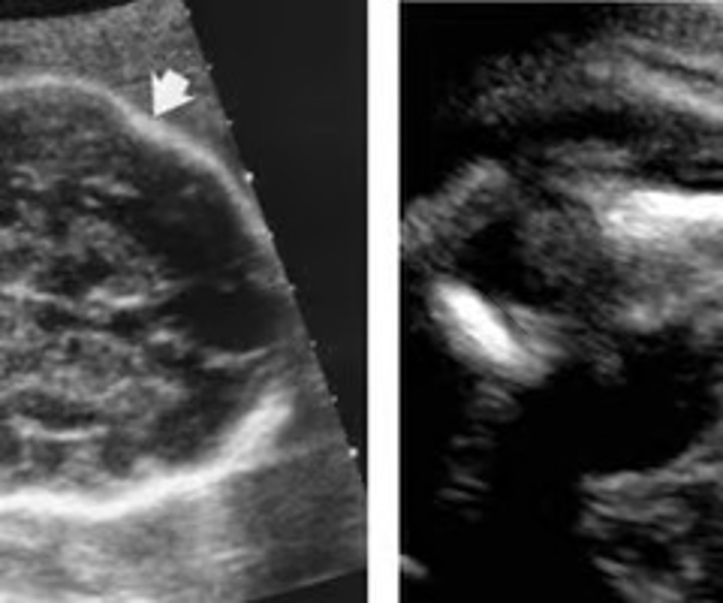
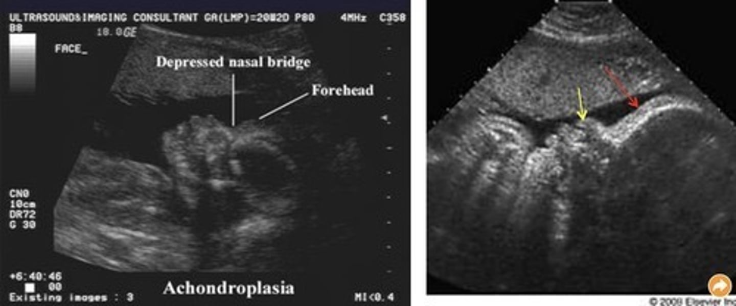
Achondroplasia
Most common non-lethal skeletal dysplasia
Fetus looks normal until 24 weeks
Rhizomelia with bowing femurs
Frontal bossing
Trident hands
Achondrogenesis
Bone hypomineralization - deceased/absent ossification of spine/skull
Lethal
Narrow thorax and ribs
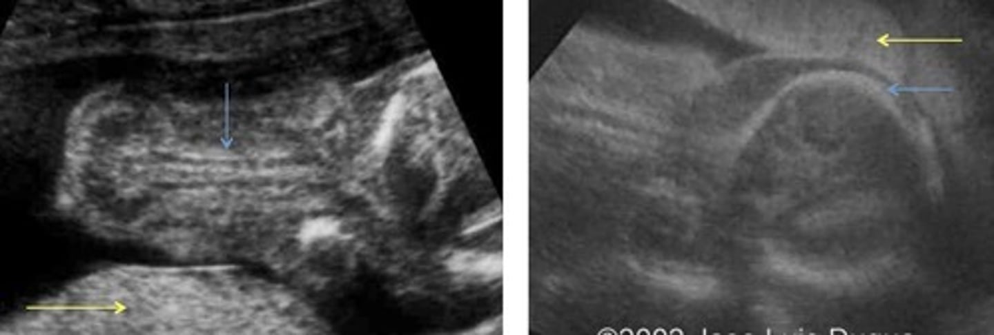
Osteogenesis Imperfecta
Connective tissue disorder
Decreased mineralization of bones - brittle bones
Increased visualization of near field brain
Possibly abnormally shaped head and/or skull fractures
Least to most severe: 4, 1, 3, 2
Caudal Regression
Incomplete development of lower half of body
Sacral agenesis
Talipes - cubed feet
Short lower extremities
GU and GI anomalies
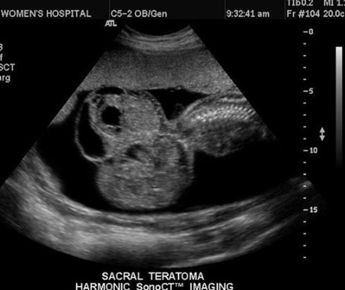
Sacrococcygeal Teratoma
Large complex mass
Arises from sacral spine
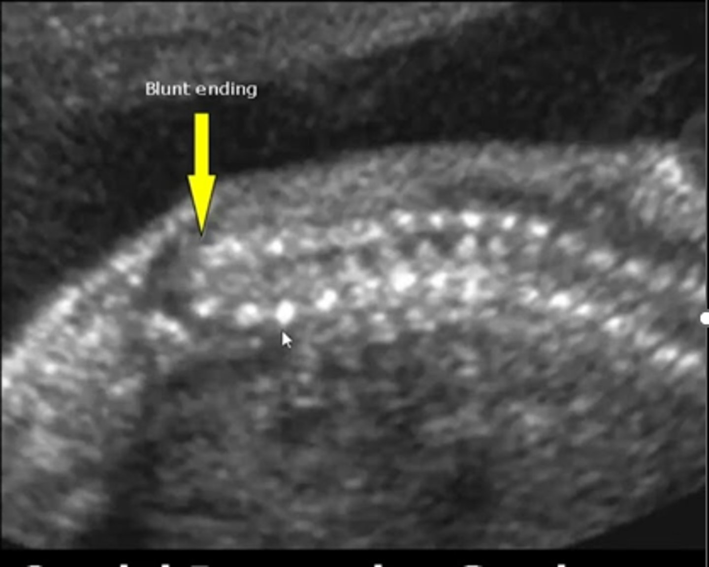
Spina Bifida
Spinal cord and/or vertebral column do not close properly
Associated with... Lemon-shaped head & Banana-shaped cerebellum
Spina Bifida Occulta
Cleft covered only by skin
Meningomyelocele
Meninges and neural elements herniate
Encephalocele
Brain herniates
Frontal Bossing
Protruding forehead
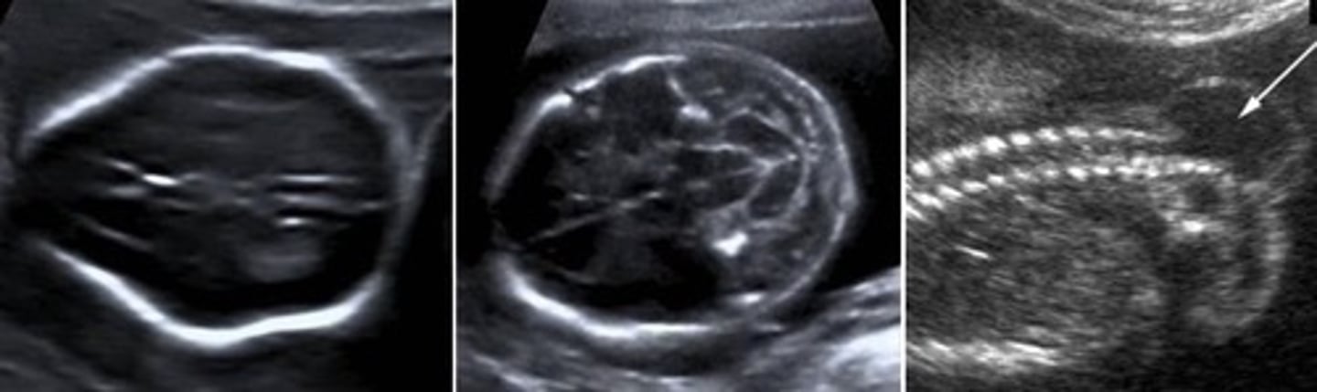
Lemon Sign
Sunken frontal bones
Strawberry Sign
Sunken frontal bones
Flattened occipital bone
Cloverleaf Sign
Sign of craniosynostosis
Spaulding Sign
Overlapping of cranial structures
Skull collapsing on itself
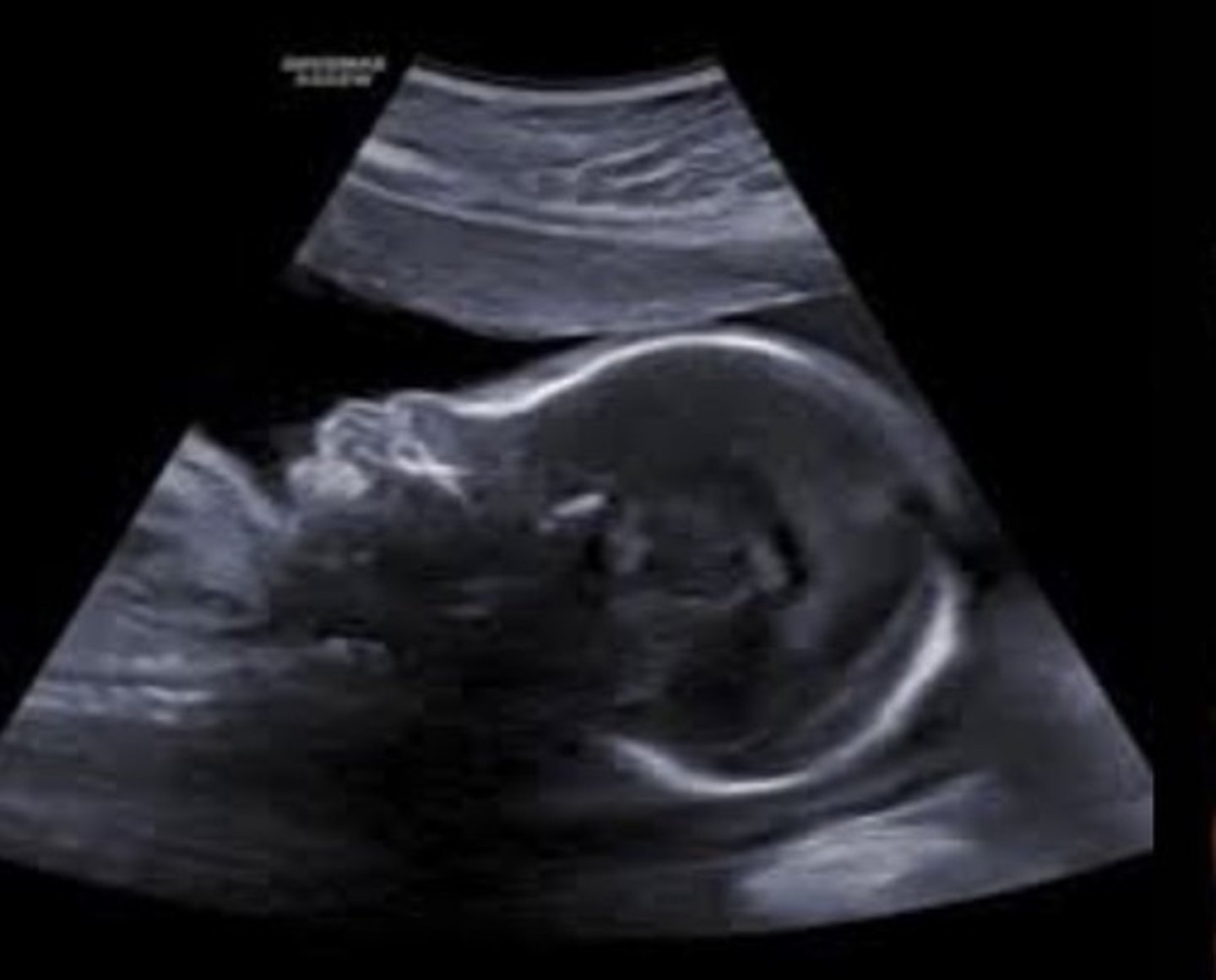
Anencephaly
Absent cranial vault
Absent/incomplete forebrain
Presence of brain stem, midbrain, skull base, face - has stromal covering
Bulging eyes
Lethal
Acrania
Partial or complete absence of cranium
May degenerate to anencephaly
Lethal
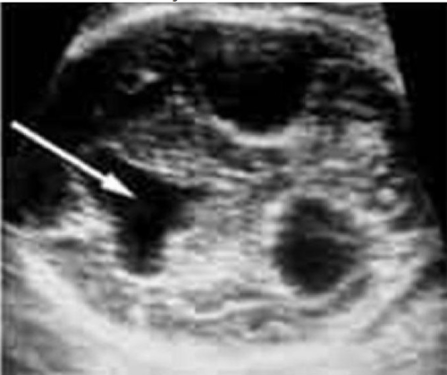
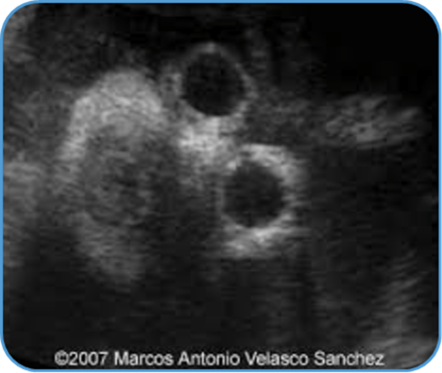
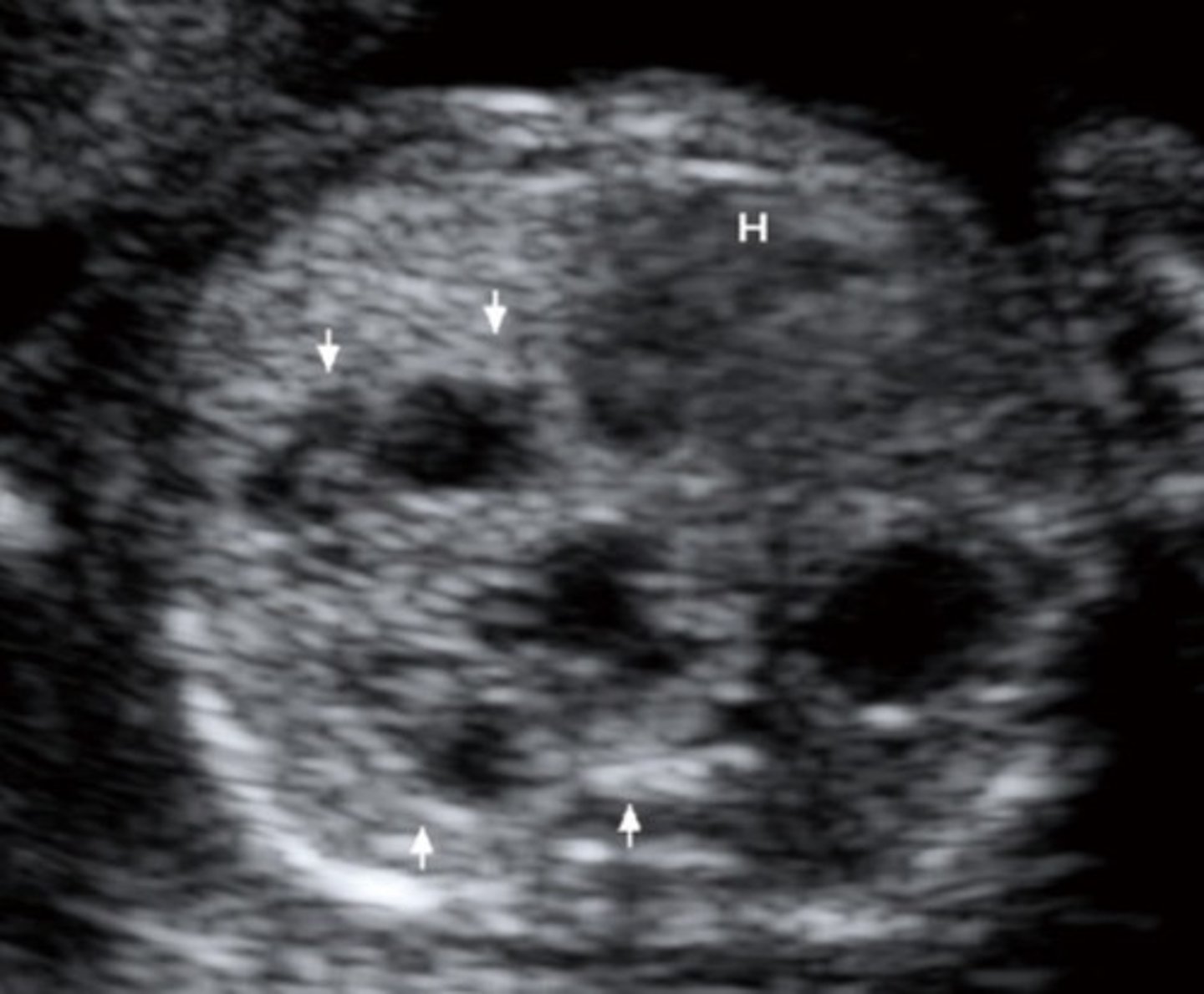
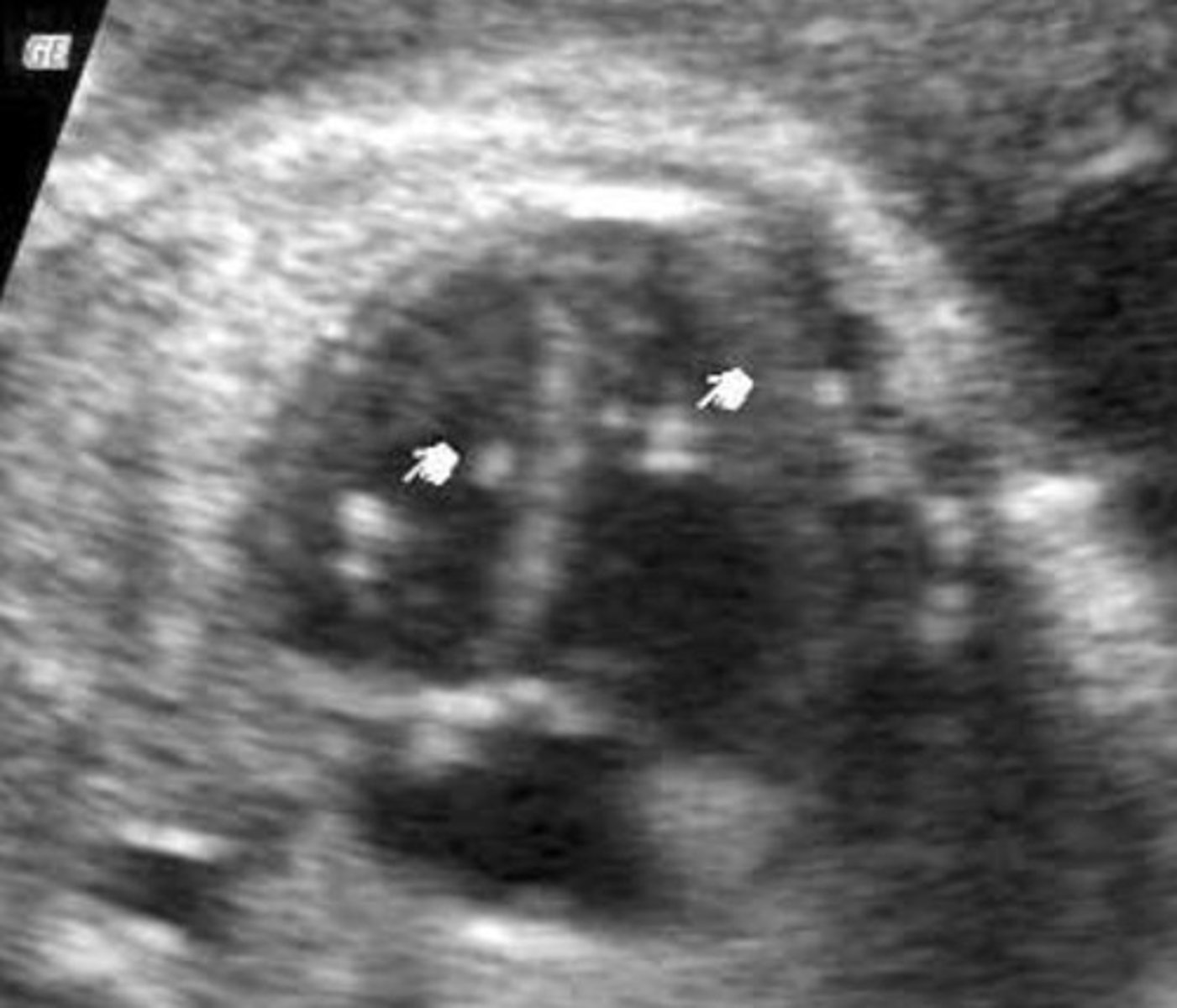
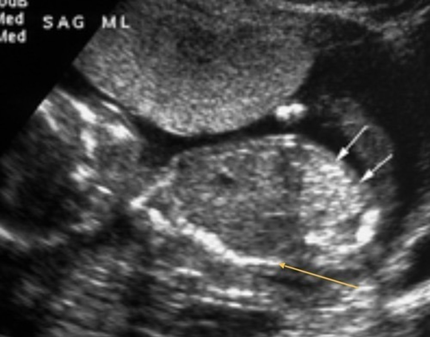
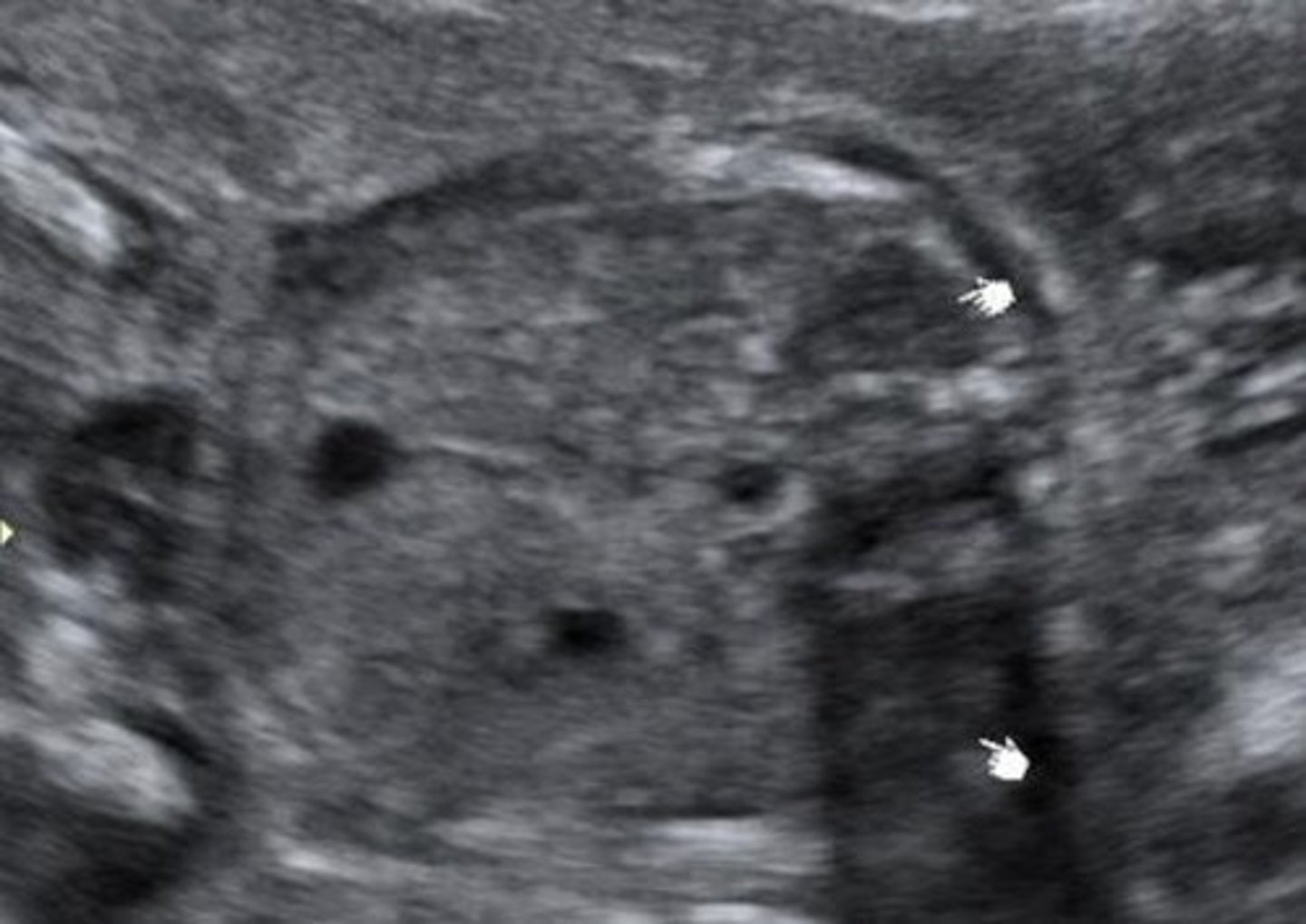
Choroid Plexus Cysts
Must be at least 2mm
Associated with Trisomy 18
Dandy-Walker Malformation
Defect in vermis of cerebellum that dilates the 4th ventricle
Splaying of cerebral hemispheres - enlarged cisterna magna
Alobar Holoprosencephaly
Single ventricle
Fusion of thalamus
Semilobar Holoprosencephaly
Single ventricle
Fusion of the thalamus
Partial formation of occipital horns
Remaining falx
Lobar Holoprosencephaly
Complete division of ventricles
Corpus callosum is present
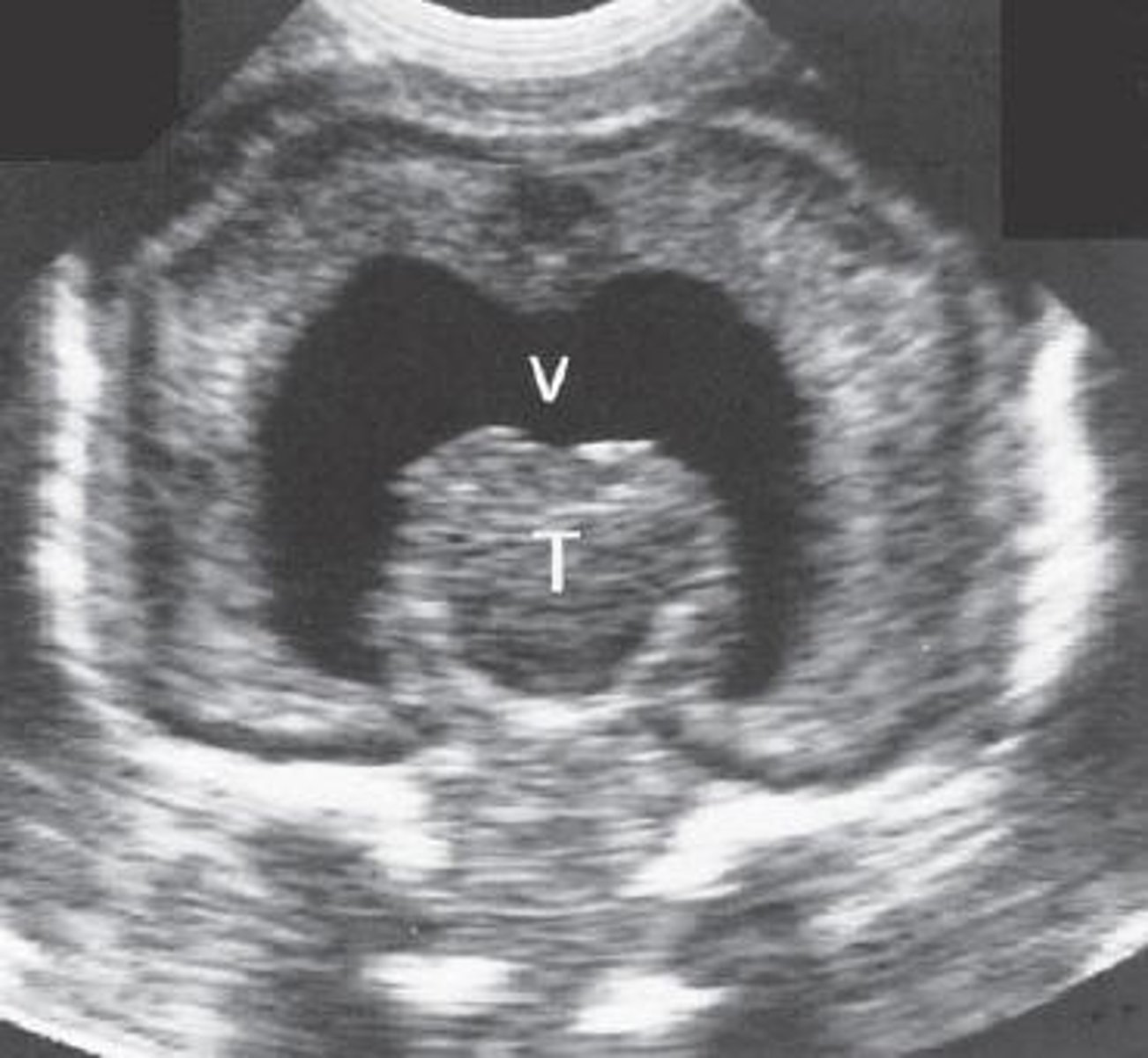
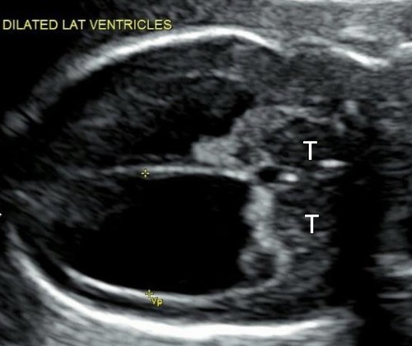
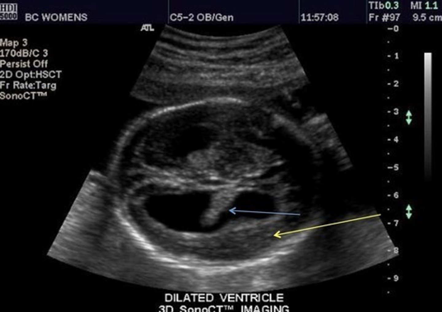
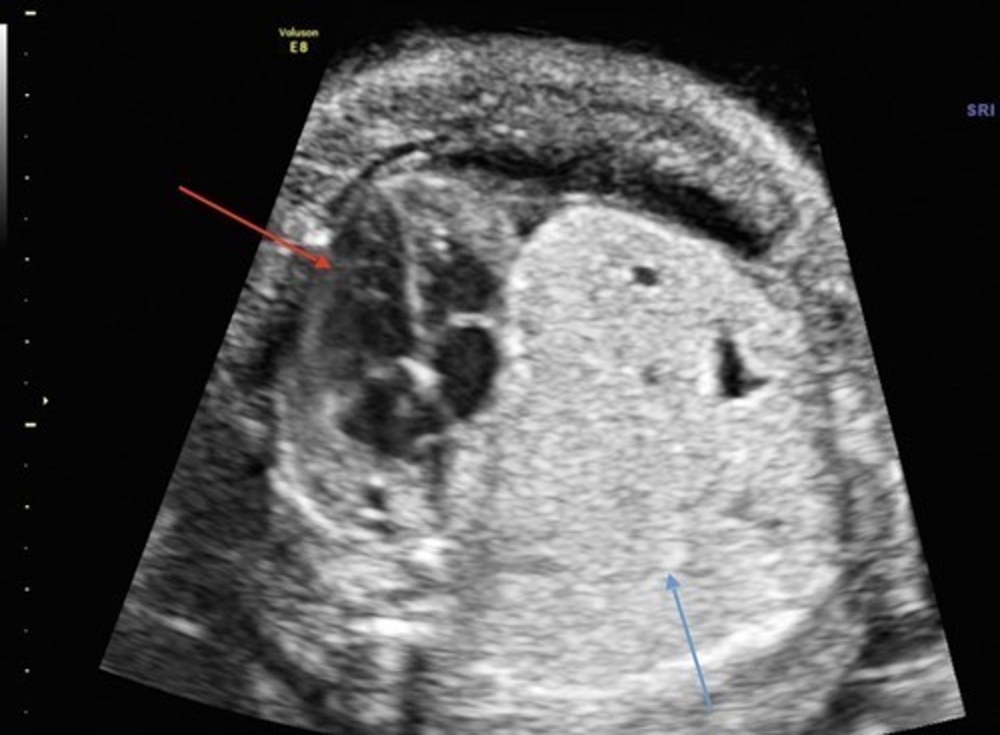
Hydrocephalus
Dilated ventricles
Dangling choroid plexus
Enlarged head
Arnold Chiari
Lemon-shaped head
Banana-shaped cerebellum
No cisterna magna
Mass protruding from spine
Midface Dysplasia
Jaw and cheekbones do not grow well
Depressed or absent nasal bridge
Eyes and forehead appearing to bulge
Hypertelorism
Wide spread eyes
Common with frontal cephalocele
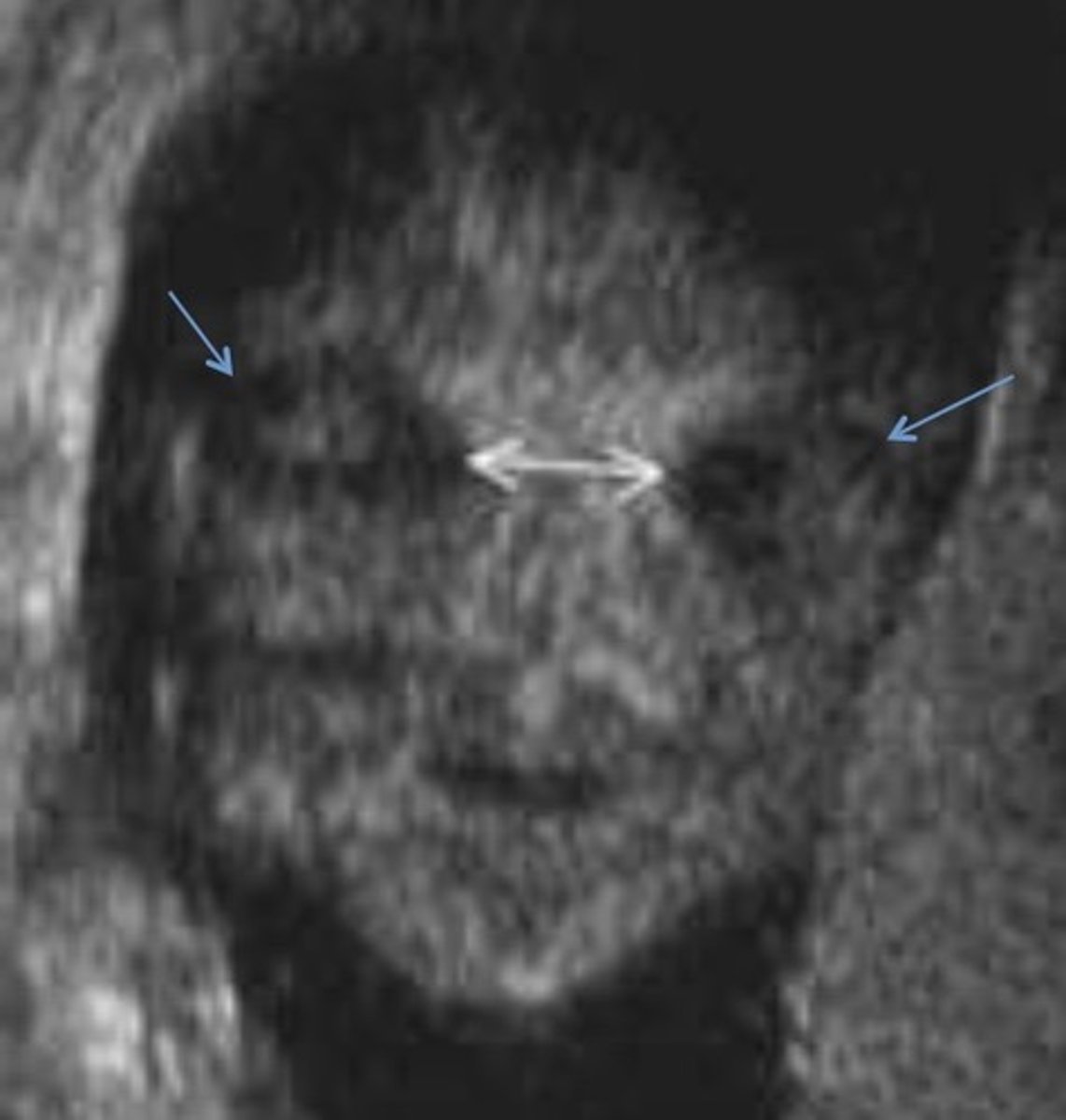
Hypotelorism
Decreased spaced between eyes
Common with skull/brain anomalies & syndromes
Proboscis
Flap of tissue superior to eyes - nose is usually absent
Arhinia
Absent nasal bone
Common with Trisomy 21
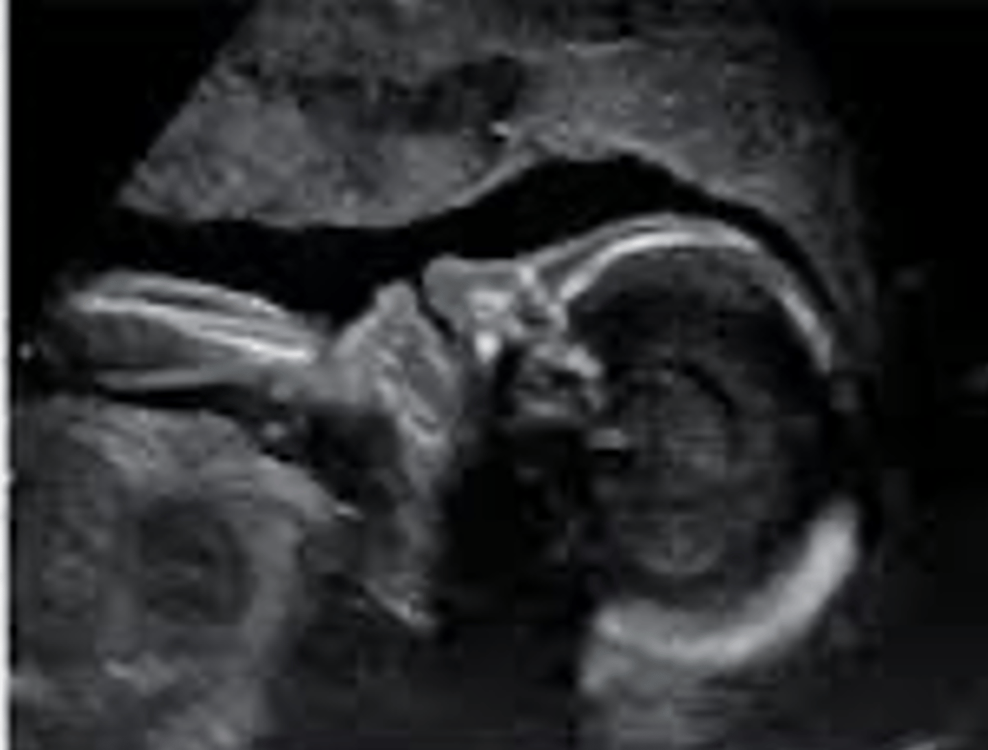
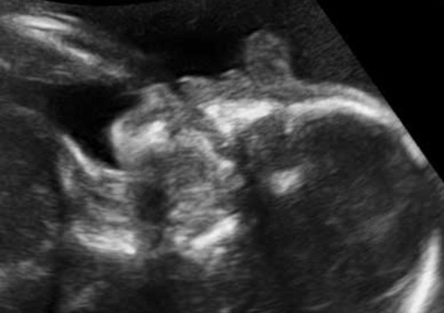
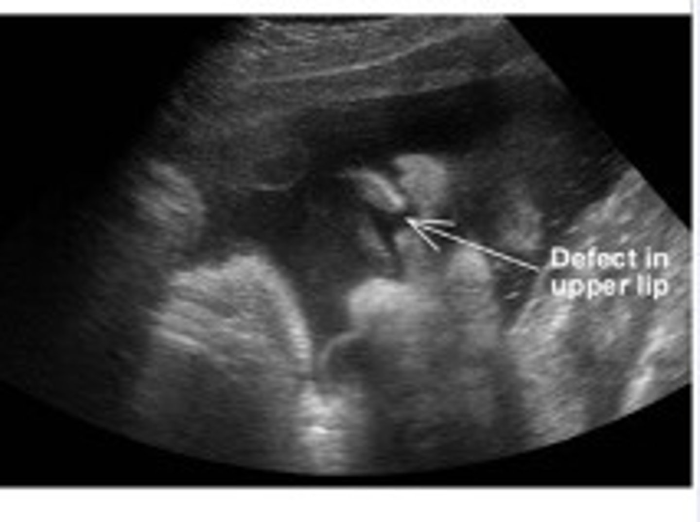
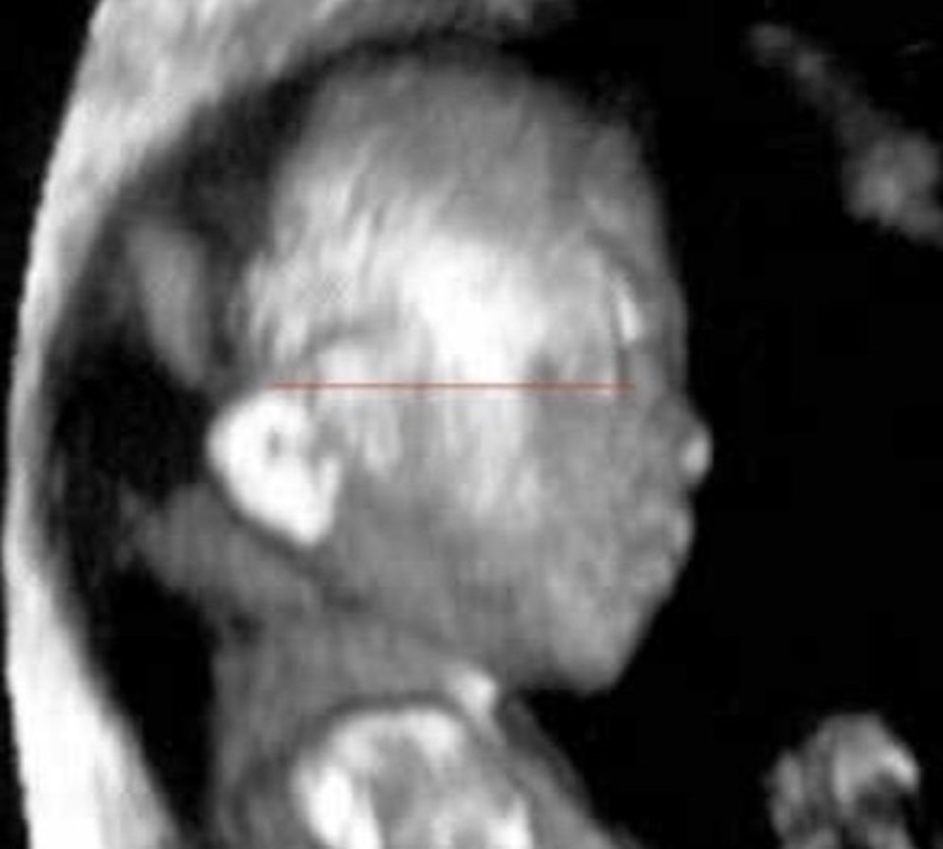
Cleft Lip
Most common facial anomaly
Palate doesn't fuse
Bilateral is slightly more common than unilateral
Low-Set Ears
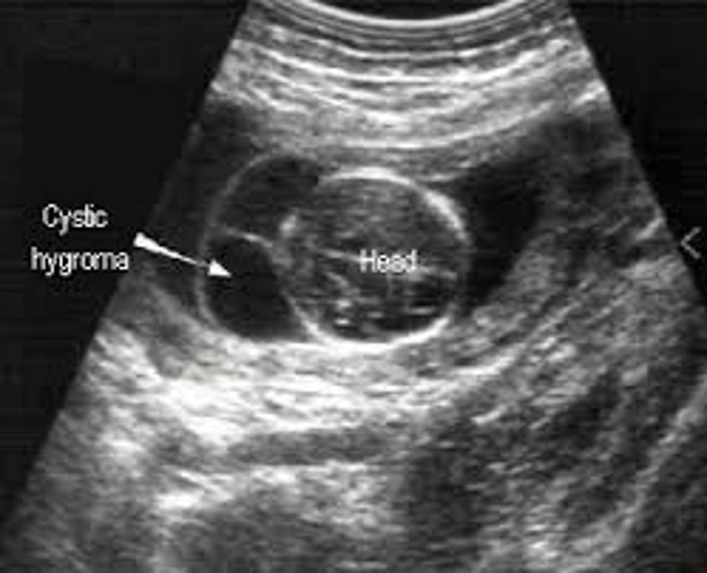
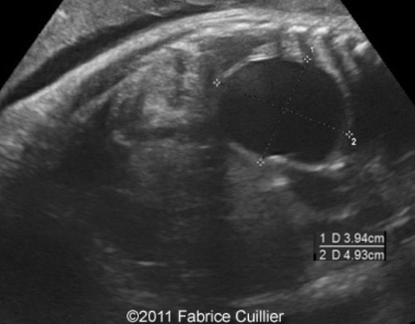
Cystic Hygroma
Most common neck mass
Abnormal connection of lymphatic vessels
Associated with Turner's syndrome & aneuploidies
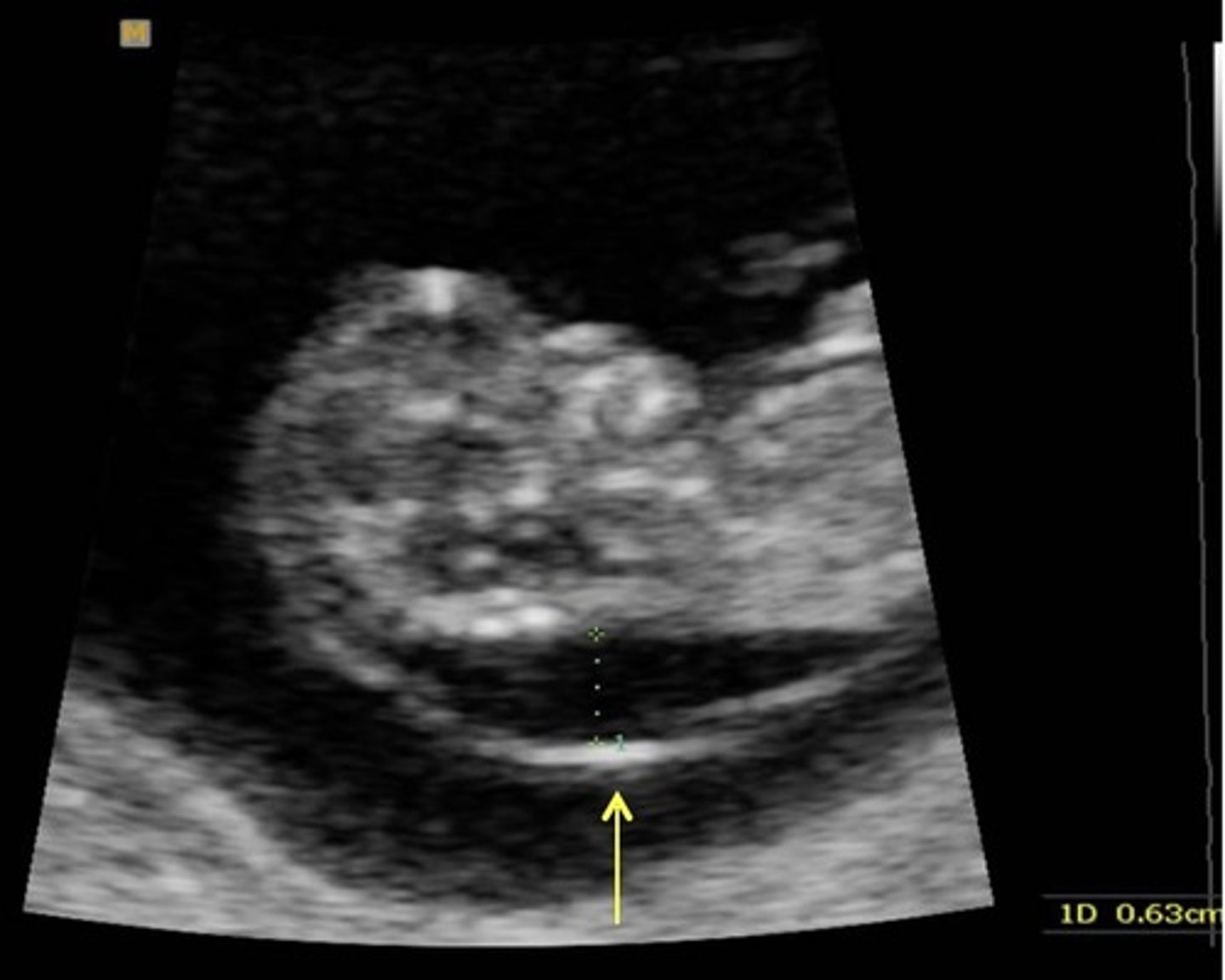
Thickened Nuchal Translucency
Anechoic fluid area in posterior neck region
Associated with Trisomy 21
> 3.4 mm
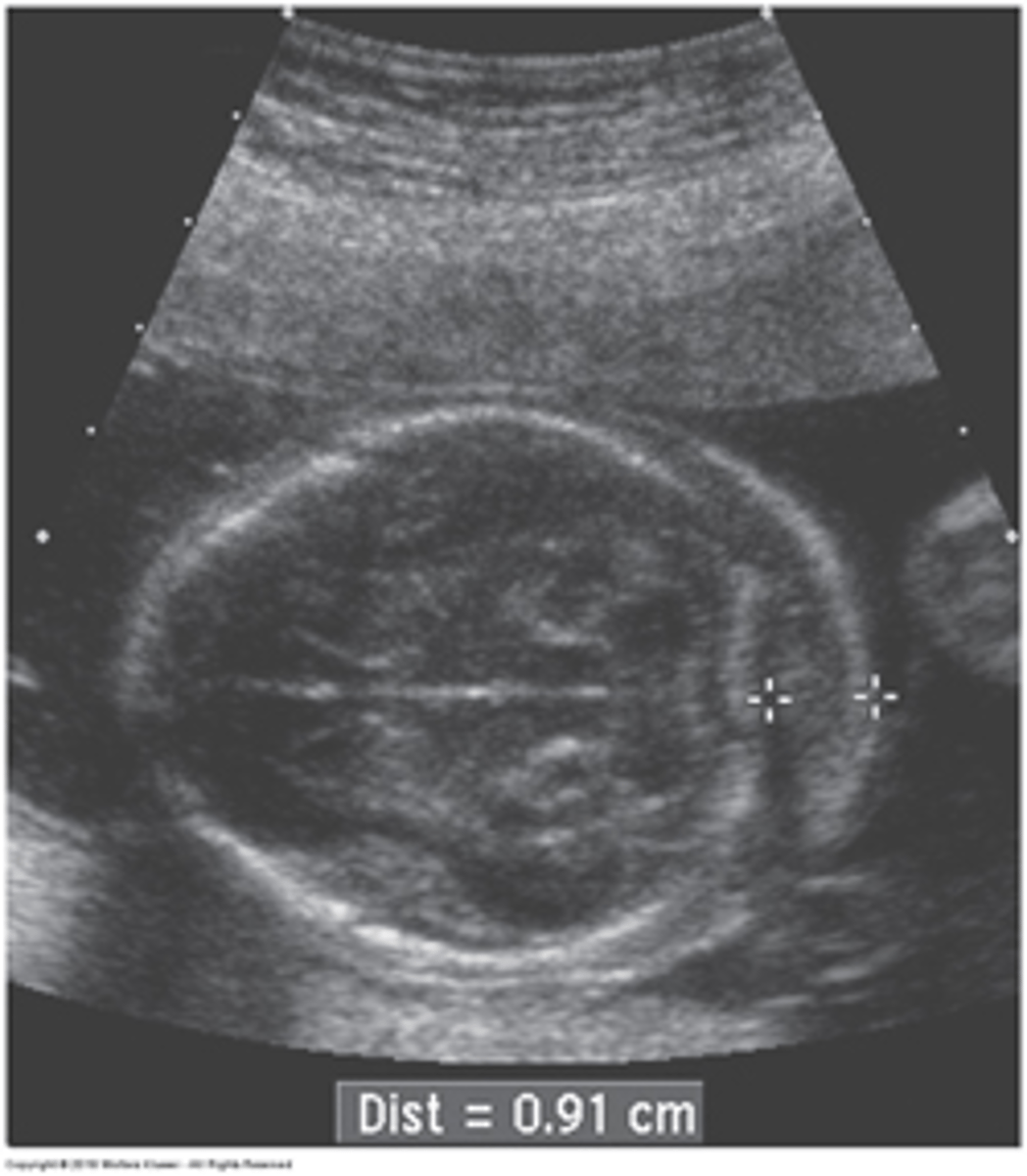
Thickened Nuchal Fold
> 6 mm
Pulmonary Hypoplasia
Lungs do not form properly
Small lung size
Bell-shaped chest
Oligohydramnios
Skeletal Dysplasia
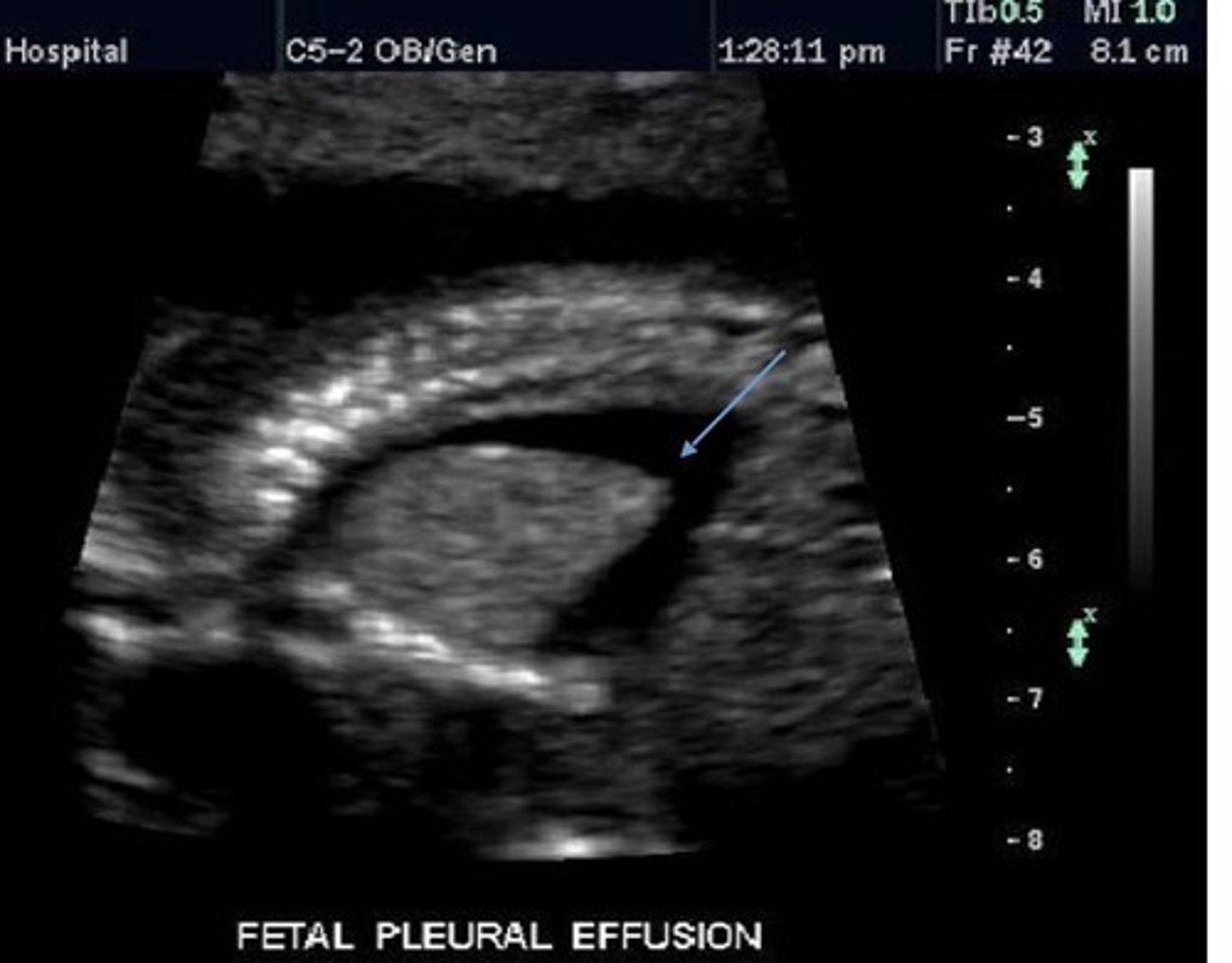
Pleural Effusion or Hydrothorax
Congenital Bronchial Atresia
Pulmonary Sequestration
Bronchogenic Cyst
Cyst within lung tissue
Pleural Effusion
Fluid in pleural cavity
Pulmonary Sequestration
Extra lung lobe
Not connected to bronchial tree - nonfunctional
Has its own blood supply
Type 1 CPAM
Most common CPAM type
Large Cysts
Good prognosis
Type 2 CPAM
Multiple smaller cysts within echogenic lung
Poorer prognosis
Type 3 CPAM
Very bright lung parenchyma with few/no cysts
Lungs often have mass effect
Poorest prognosis - poor lung function
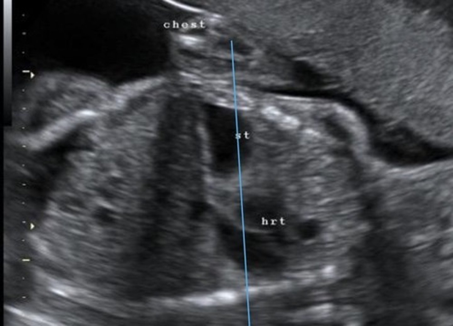
Diaphragmatic Hernia
Herniation of abdominal organs into thoracic cavity through defect in diaphragm
Echogenic Foci
Small echogenic area in heart
Usually in LV on papillary muscles or chordae
Cardiomegaly
Enlarged heart
Ectopia Cordis
Heart is partially/completely outside chest wall
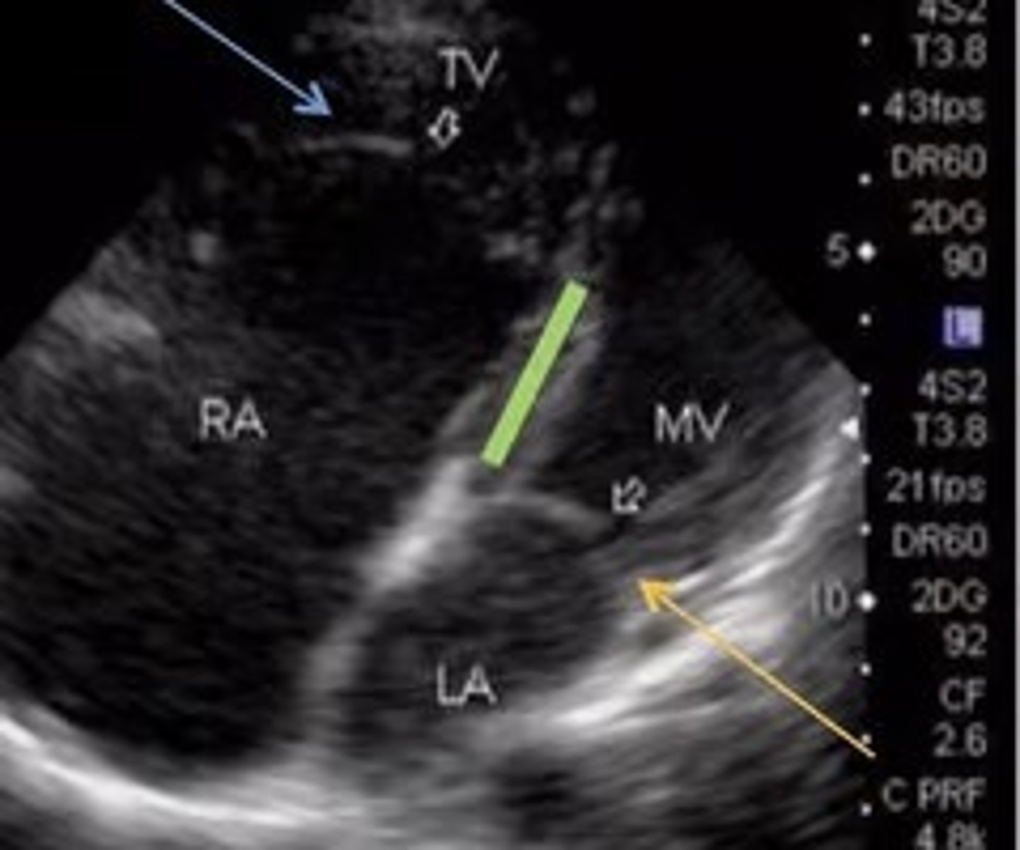
Ebstein's Anomaly
Best seen in 4 Chamber
Apical displacement of tricuspid valve -> TV is lower than it should be
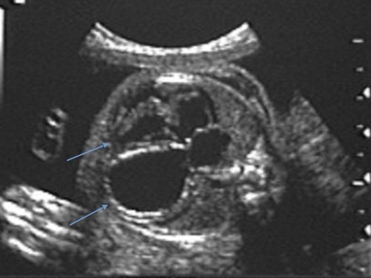
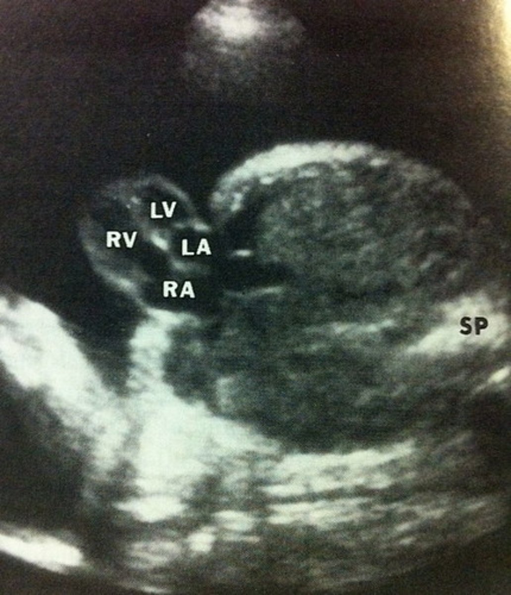
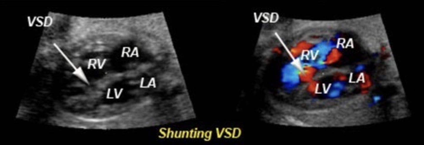
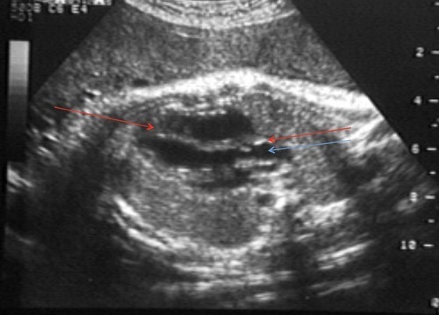
VSD
Most common cardiac defect
Best seen in LVOT view or 4ch. if septum is perpendicular to sound beam
Hole in ventricular septum
Commonly in membranous septum
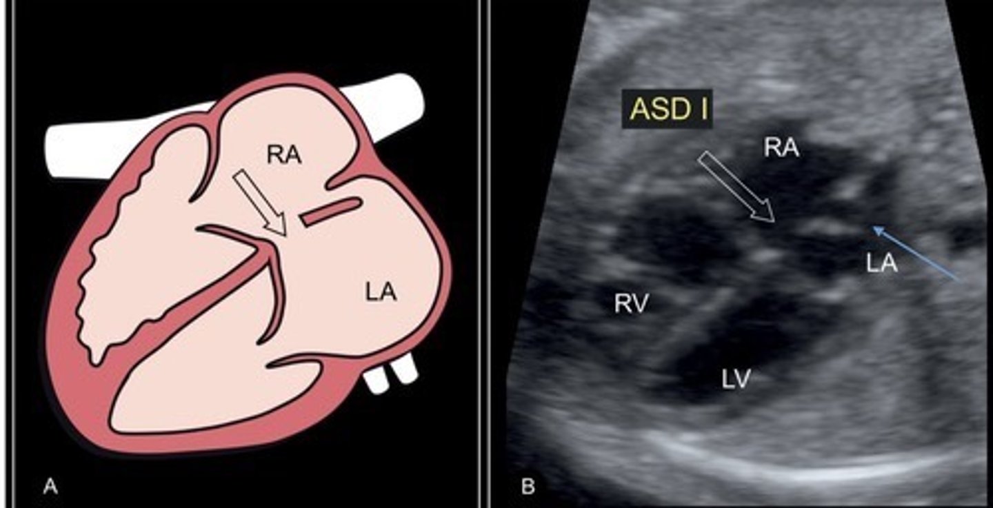
ASD
Best seen when IAS is perpendicular to sound beam
Hole in atrial septum
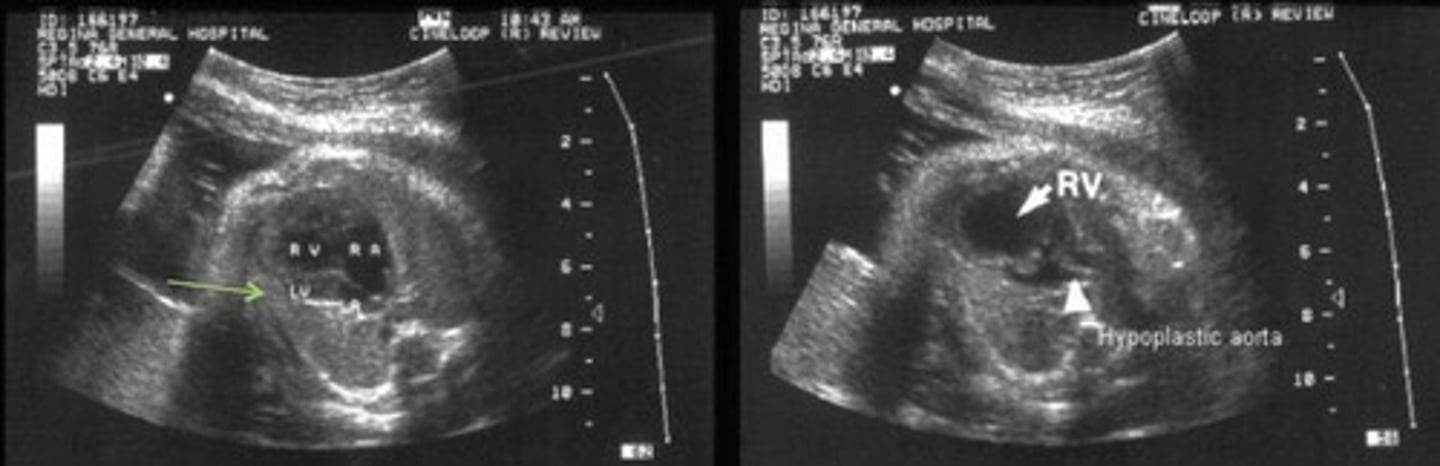
HLHS - Hypoplastic Left Heart Syndrome
Most common cause of death from heart disease during the neonatal period
Failure for LV to develop - left side of heart is small or absent
Small LV Little color flow through MV
Small AO
Reversed flow through foramen ovale and ductus arteriosus
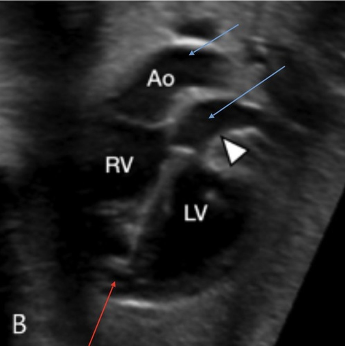
Tetralogy of Fallot
Best seen in LVOT view
VSD
Overidding/large AO
Pulmonary stenosis
RVH
DORV - Double RV Outlet
Best seen in PSAX or LVOT view
Pulmonary A and AO originate from RV
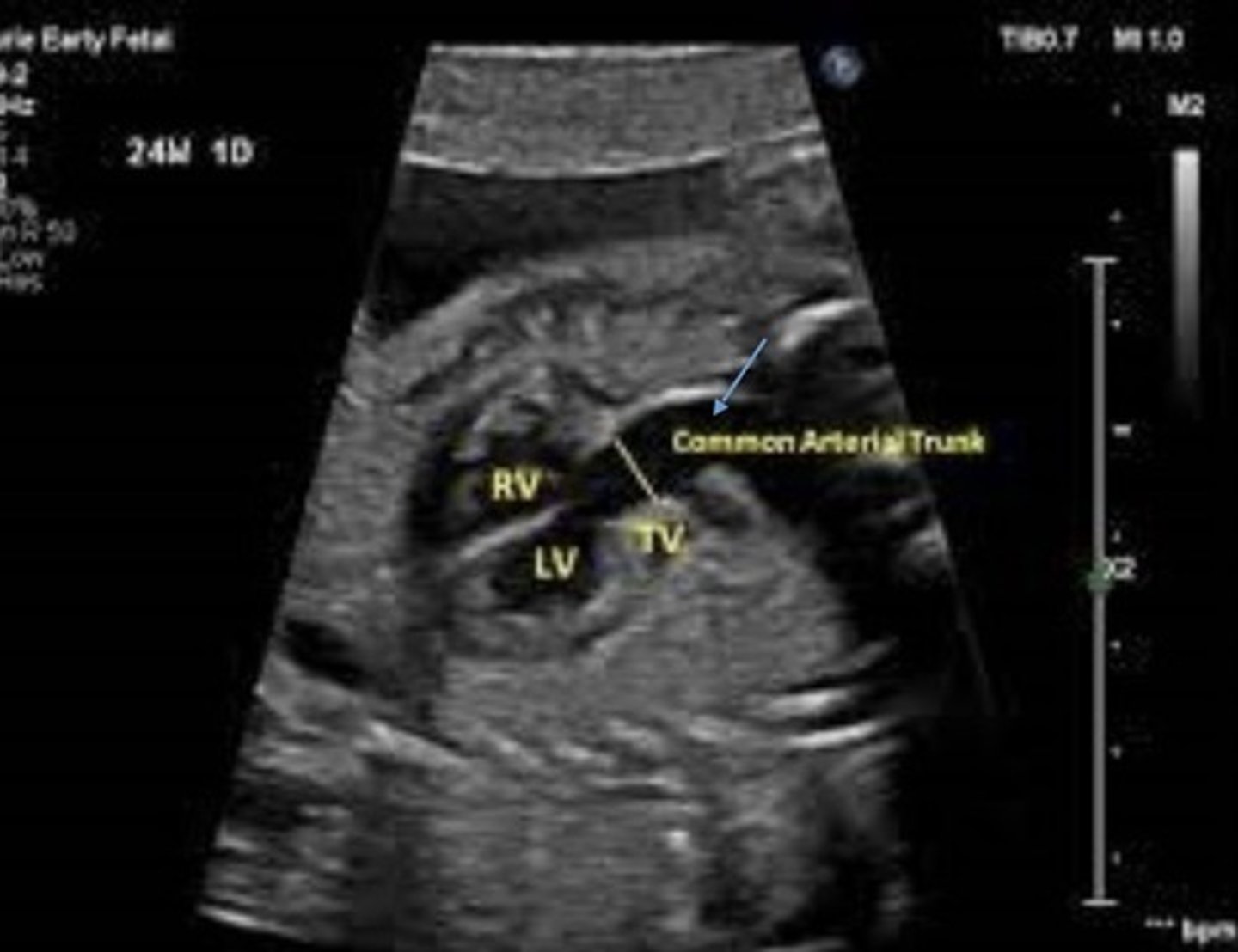
Truncus Arteriosus
Best seen in LVOT view
Truncoconal ridge fails to fuse
Appears as single great vessel with VSD
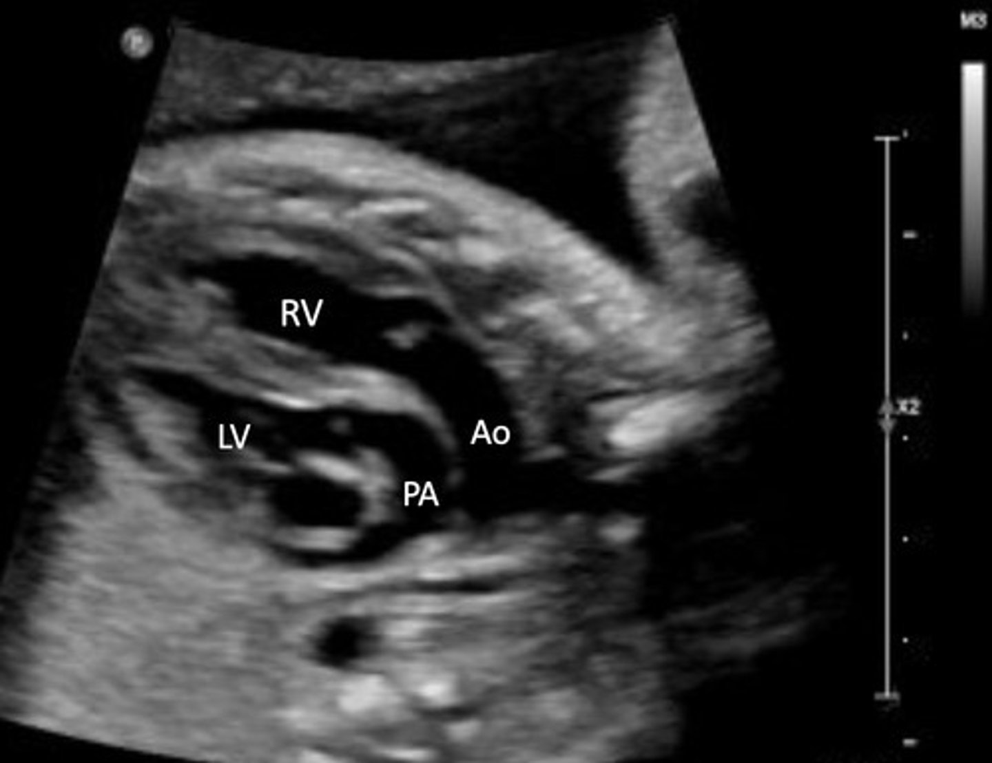
Transposition of Great Vessels
Best seen in PSAX view
Pulmonary A originates from LV
AO originates from RV
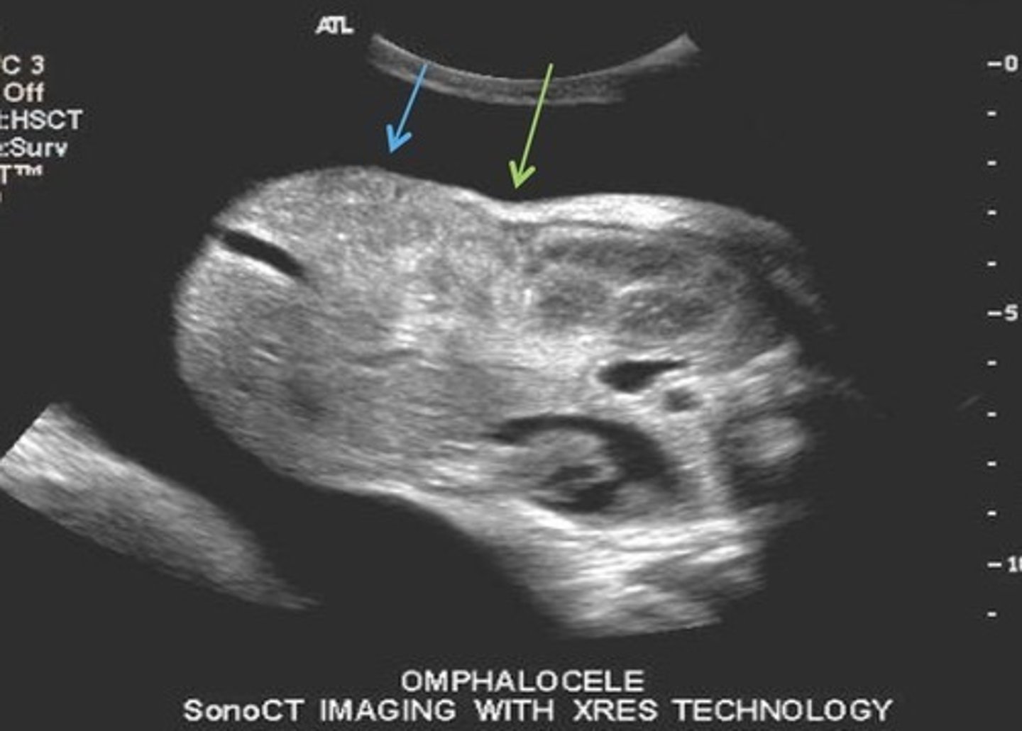
Omphalocele
Herniation of bowel into umbilical cord after 13 weeks
Type 1 Omphalocele
Contains only bowel
80% association with aneuploidy's
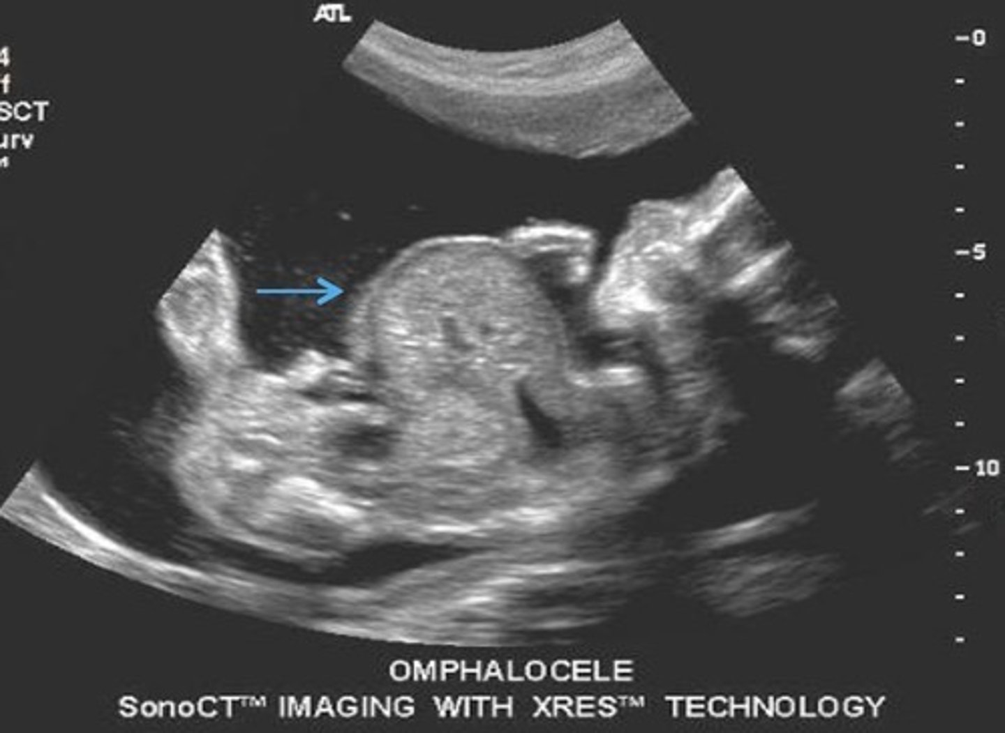
Type 2 Omphalocele
Contains bowel and liver
20% association with aneuploidy's
Gastroschisis
Open defect to the side of umbilical cord insertion
Bowel is outside of body
Bladder Exstrophy
Urinary bladder protrudes through lower abdominal wall defect
No bladder seen
Lower anterior abdominal wall defect
Pentalogy of Cantrell
Consists of 5 Defects:
Cleft sternum
Diaphragmatic hernia
Upper anterior wall defect
Cardiac defects -> ectopic cordsis (heart outside body) Pericardial defect -> creates pericardial/pleural effusion
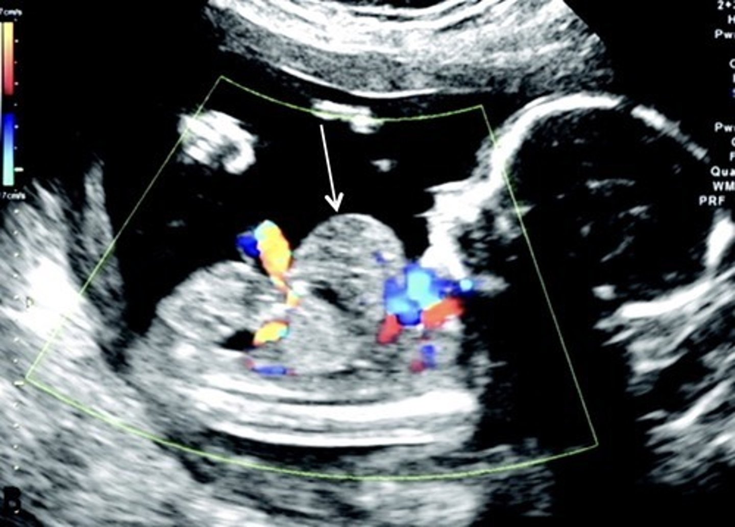
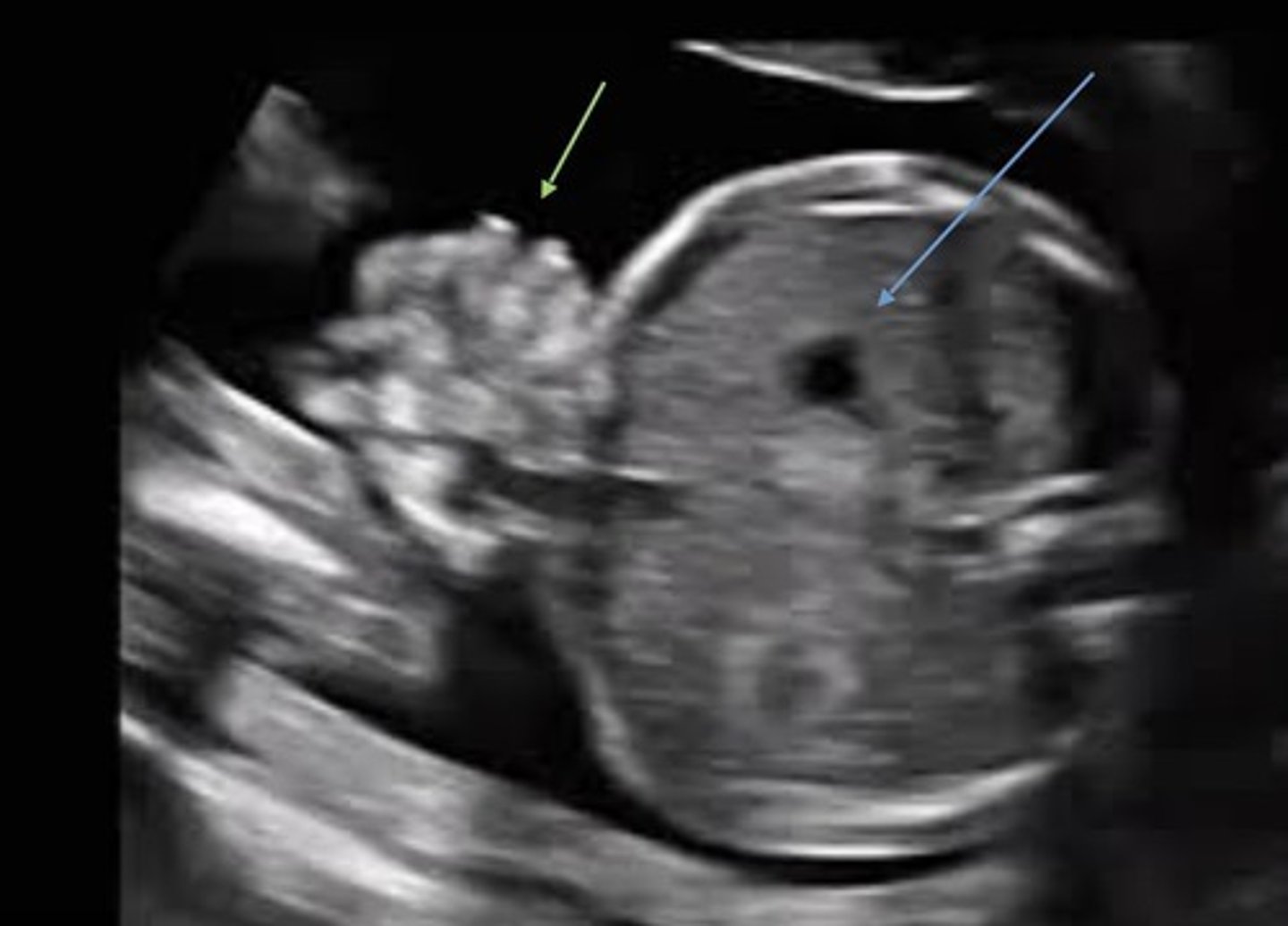
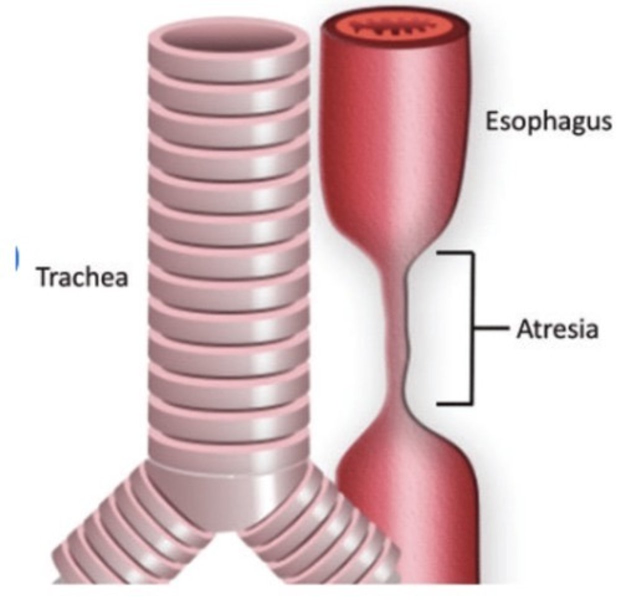
Esophageal Atresia
Stenosis in esophagus
Stomach is not visualized
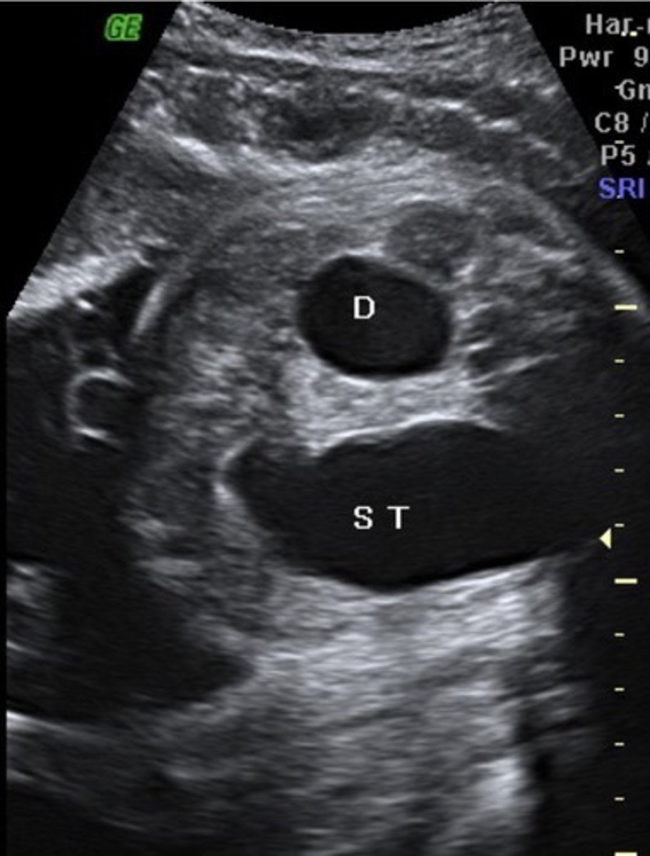
Duodenal Atresia
Fluid reaches duodenum, but cannot move past it
Double Bubble Sign
Enlarged stomach & prox. duodenum
Intestinal Obstruction
Atresia of jejunum and/or ilium
Dilated bowel
Hyperechoic Bowel
Bowel as bright as bone
Classic Potter's Syndrome
Bilateral renal agenesis
Oligohydraminios
Pulmonary hypoplasia
Facial/hand/feet anomalies
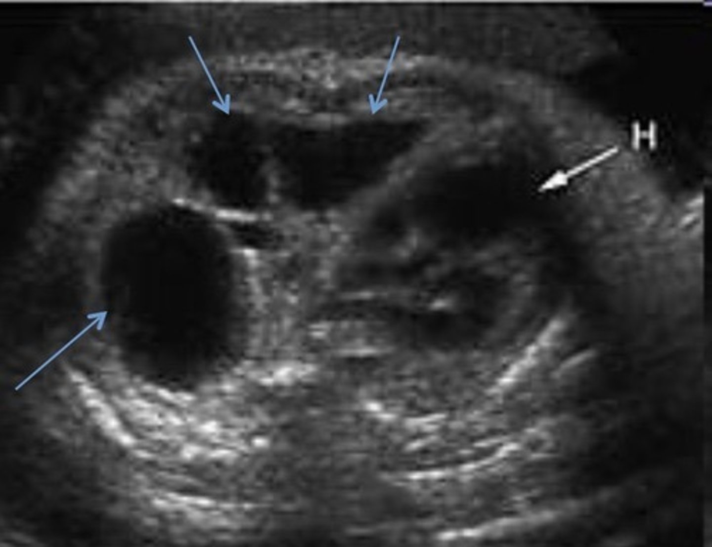
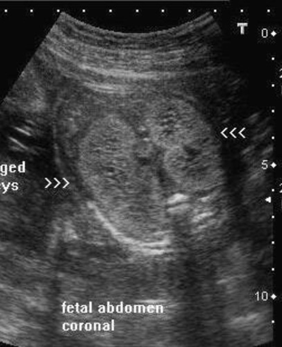
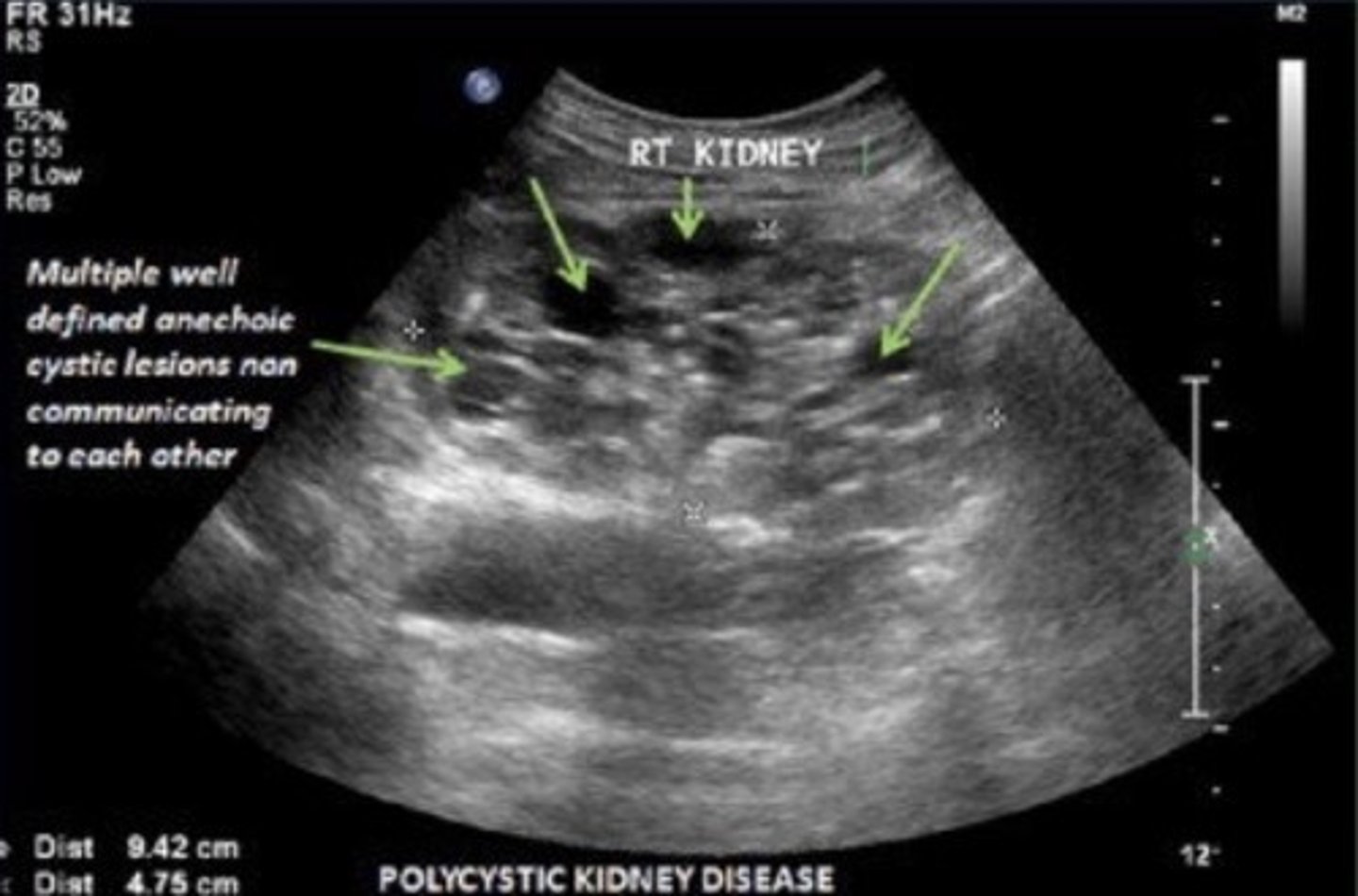
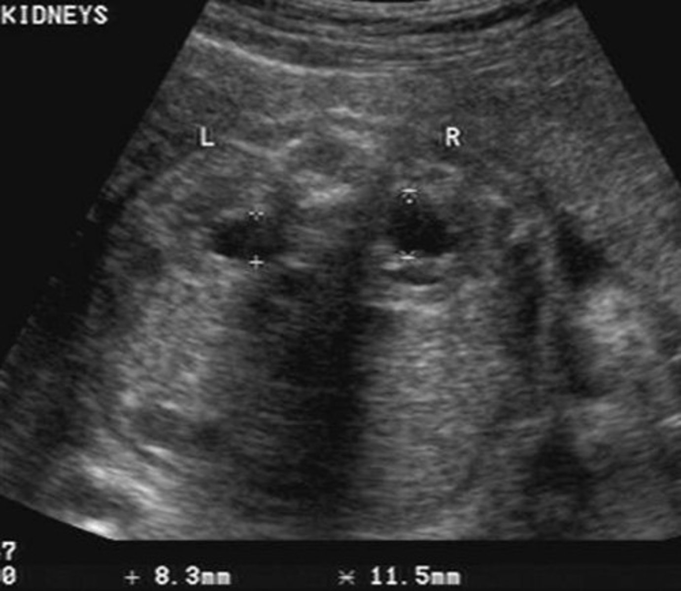
Potter's Syndrome Type I - Polycystic Kidney Disease
Bilateral micro cysts
Bilateral enlarged, echogenic kidneys
Empty bladder
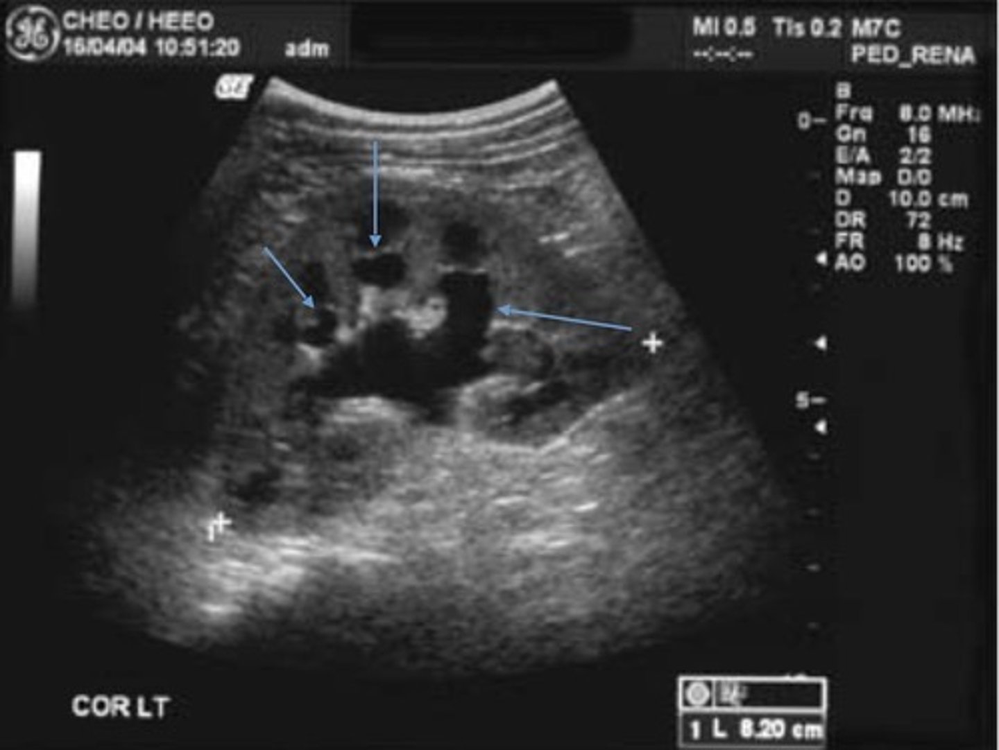
Potter's Syndrome Type II - Multicystic Dysplastic Kidney Disease
Kidney tissue is replaced by cysts - multiple and variable in size
Usually unilateral
Enlarged kidneys
Ill-defined walls & parenchyma/pelvis
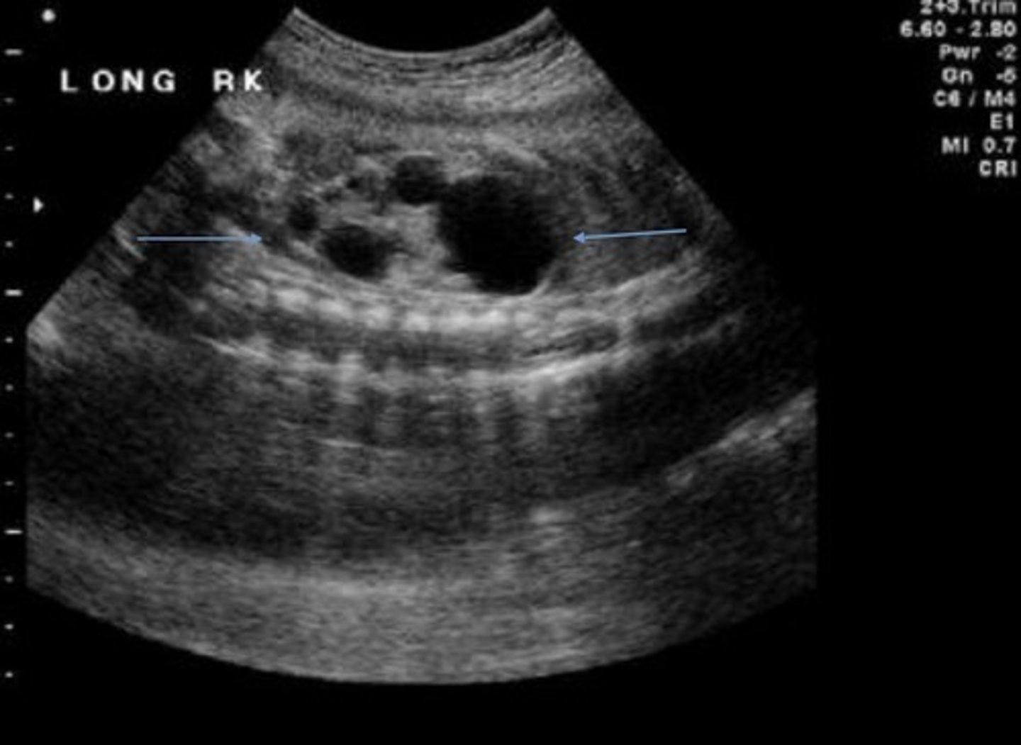
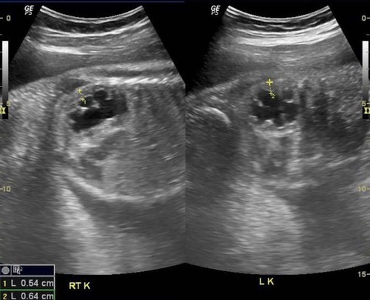
Potter's Syndrome Type III - Autosomal Dominant Polycystic Kidney Disease
Bilateral large cysts
Large kidneys
Hyperechoic parenchyma
Normal bladder/fluid levels
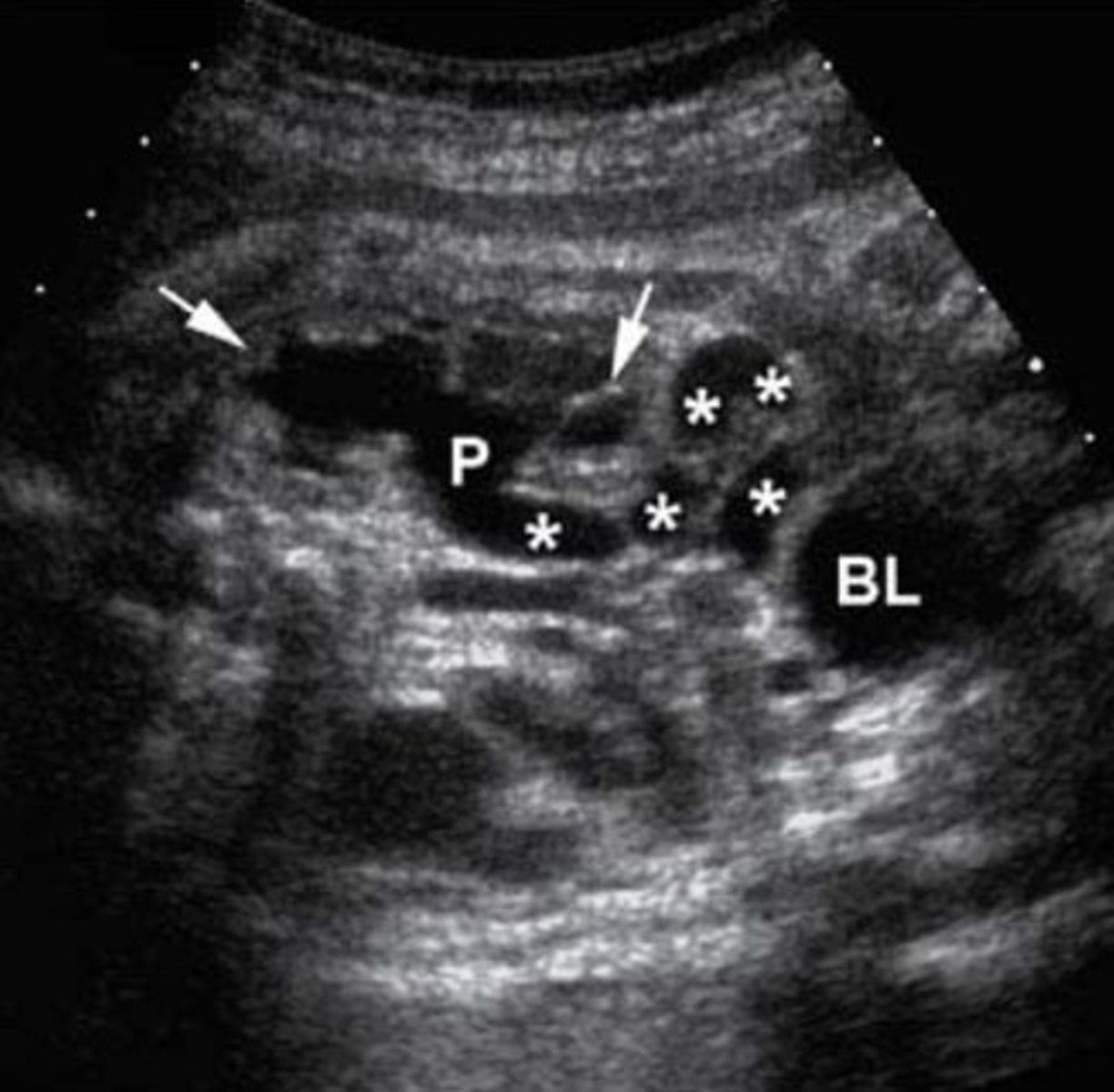
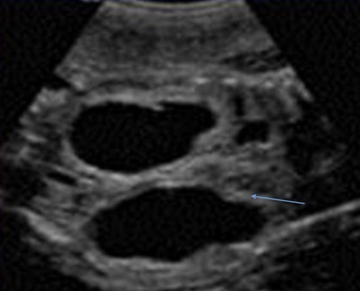
Potter's Syndrome Type IV - Obstructive Cystic Dysplasia
Cortex is replaced with cysts
Dilated pelvis and ureter
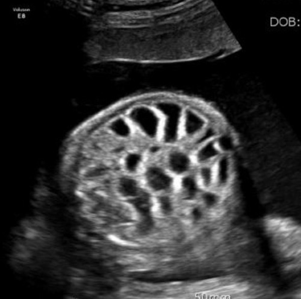
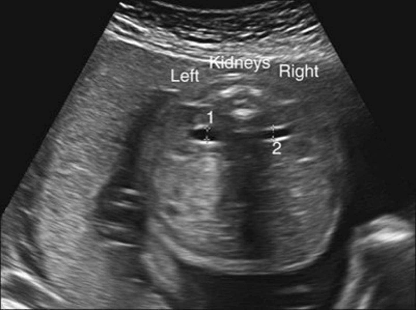
Grade 0 Hydronephrosis
Normal kidney
Grade 1 Hydronephrosis - Pyelectasis
Fluid within renal pelvis, but small enough to be considered insignificant/within normal range
Grade 2 Hydronephrosis
Pelvis is > 10 mm
Few calyces seen
Cortex is normal
Grade 3 Hydronephrosis
Pelvis is > 10 mm
Marked dilation of calyces
Cortex is normal
Grade 4 Hydronephrosis
Pelvis is > 10 mm
Marked dilation of calyces
Mild thinning of cortex
Grade 5 Hydronephrosis
Pelvis is > 10 mm
Marked dilation of calyces
Severe thinning of cortex
Posterior Urethral Valve Obstruction
Only in boys
Causes bilateral hydronephrosis
Thick bladder walls
Keyhole bladder appearance
Prune Belly Syndrome
Triad of symptoms:
Distended abdominal muscles
Obstruction of urinary tract
Cryptorchidism
Distended abdomen
Enlarged bladder
Bilateral hydronephrosis
Oligohydramnios -> hypoplastic lungs
Pathologies that can cause Polyhydramnios
Omphalocele
Esophageal atresia
Duodenal atresia
Midgut Volvulus
Intestinal Obstruction
Mesoblastic Nephroma
Pathologies that can cause Oligohydramnios
Bilateral renal agenesis
Potter's syndrome
Bladder outlet obstruction
Prune belly syndrome
Megalourethra
Reasons for increased hCG levels
Incorrect dates - fetus older than expected
Multiple babies
Trophoblastic/placental disease
Trisomy 21
Reasons for decreased hCG levels
Incorrect dates - fetus younger than expected
Fetal demise
Ectopic pregnancy
Trisomy 18
Reasons for increased AFP levels
Incorrect dates - fetus older than expected
Multiple babies
Placental disease
Leakage from fetus into amniotic fluid via abdominal wall or neural tube defects
Reasons for decreased AFP levels
Incorrect dates - fetus younger than expected
Fetal death
Trisomy 21 or 18
Blockage in fetal urogenital tract
Molar pregnancy
Turner's Syndrome
Absent X chromosome or 46X0/46XXX
Only in females
Mimics Noonan's syndrome (males)
Features of Turner's Syndrome in a Fetus
Horseshoe kidneys
Aortic coarctation
Features of Turner's Syndrome in a Child/Person
Webbed neck
Shield chest
ABNL elbow angle
Trisomy 21
Down syndrome
Extra 21st chromosome
Most common aneuploidy
Low IQ always present
Absent nasal bone
Duodenal atresia
Omphalocele
Echogenic bowel
Sandal gap
Clinodactyly
Trisomy 18
Edward's syndrome
Extra 18th chromosome
Strawberry-shaped head
Chorioid plexus cysts
Septal defects
Clenched fists/talipes/rocker bottom feet
Facial defects
Trisomy 13
Patau syndrome
Extra 13th chromosome
Holoprosencephaly
Cleft lip & cyclopia
Cystic hygroma
Polydactyly
2 vessel cord
Triploidy
Extra chromosome on all sets - 69 total
Apert Snydrome
Craniosynostosis
Hypertelorism
CHARGE Syndrome
Coloboma
Heart defects
FGR
Genital hypoplasia
Ear anomalies
Meckel-Gruber Syndrome
Polycystic kidneys
Encephalocele
Polydactyly
Holt-Oram Syndrome
Radial ray
Cardiac defects -> ASD
Aicardi Syndrome
Agenesis of corpus callosum
Beckwith-Weidemann Syndrome
Overgrowth of tissues & organs
Omphalocele
VACTERL Syndrome
Group of anomalies that sporadically occur together 3 must be present to confirm:
Vertebral defects
Anal atria
Cardiac anomalies
Tracheo-esophageal fistula
Renal anomalies
Limb dysplasia
Limb-Body Wall Complex
Due to amnion rupture or ABNL embryonic folding
Short umbilical cord
Omphalocele
Limb defects
Neural tube defects