Embryology GI
1/79
There's no tags or description
Looks like no tags are added yet.
Name | Mastery | Learn | Test | Matching | Spaced |
|---|
No study sessions yet.
80 Terms
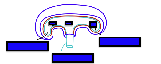
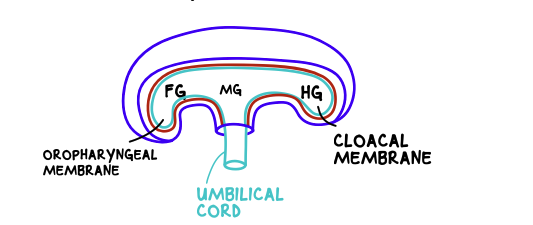
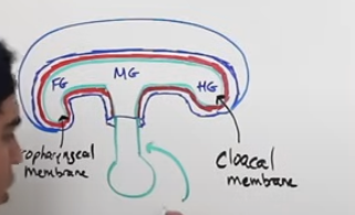
cranial end name
oropharyngeal membrane
caudal end name
cloacal membrane
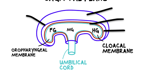
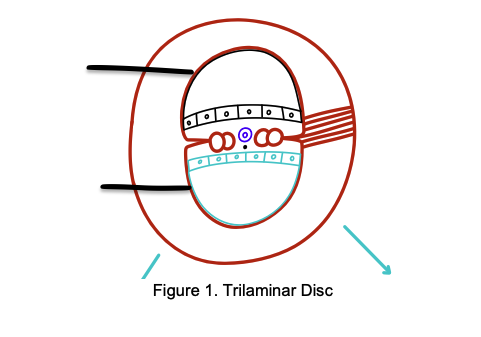
light blue- endoderm
red- mesoderm
dark blue- ectoderm
cavity dark blue- amniotic cavity
4th week what is this duct
vitelline duct
connects the yolk sac to the midgut of the developing embryo
what happens to the vitelline duct in the 6th week
it obliterates leaving only the umblilical cord
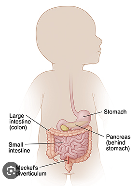
what happens if the vitelline duct doesn’t obliterate
meckel’s diverticulum.
outpouching of the small intestine occurs, vitelline duct forms a ligament between the midgut and the anterior abdominal wall
what is natural herniation
during 6th week, the small intestines will form a loop and herniate through the umbilical cord
because the developing organs within the abdominal cavity grow and push out the small intestines.
do intestines remain herniated
when the abdominal cavity becomes larger, the intestines return back into them during 10th/11th week
what happens if herniation persists past 11th/10th week
o Omphalocele
Results from the failure of the intestinal loop to return inside the abdominal cavity
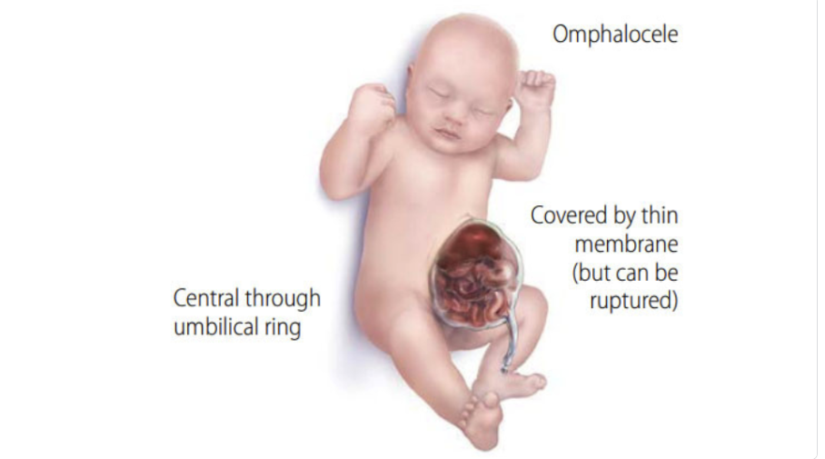
how to detect omphalocele
Can be detected through fetal ultrasound or serum alpha fetoprotein levels of the mother
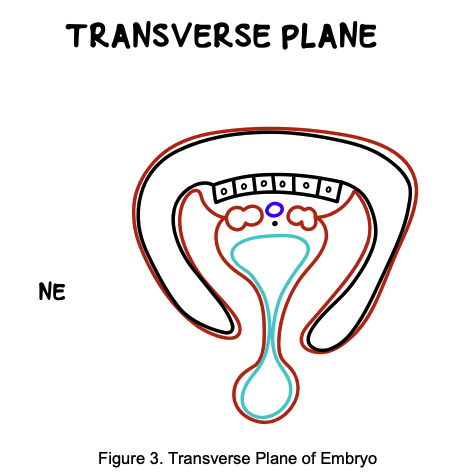
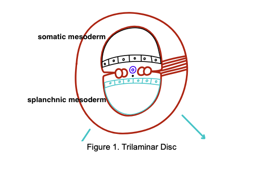
amniotic cavity folds down
splanchnic mesoderm surrounds endoderm, vitelline duct and yolk sac
somatic mesoderm surrounds the amniotic cavity
in the transverse plane what should the lateral folds do
lateral folds of the amniotic cavity fuse
Once the vitelline duct obliterates, the two endoderm folds can merge, forming a closed gut tube
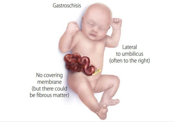
what if the two lateral folds fail to fuse
Gastroschisis
The lateral folds fail to fuse, resulting in the herniation of abdominal contents
The herniated intestines are not covered in peritoneum, which could irritate the abdominal cavity
what forms the visceral peritoneum
splanchnic mesoderm
what forms the parietal peritoneum
somatic mesoderm
what connects the parietal to the visceral peritoneum
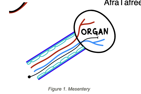
mesentery
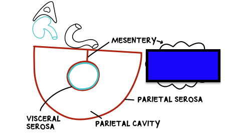
what is a mesentery
is a double-layered serous membrane made up of simple squamous epithelial tissue
Underneath the serous layer is areolar connective tissue (basal layer)
Mesentery is where blood vessels, lymphatic vessels, and nerves can pass through
if an organ has a mesentery and has a visceral and parietal peritoneum, what is it
intra-peritoneal organ
if an organ doesn’t have a mesentery and a visceral and parietal peritoneum
retroperitoneal organ
primary retroperitoneal organ
Organs that develop and remain retroperitoneal—they never had a mesentery or peritoneal covering on their posterior surface.
secondary retroperitoneal organ
Organs that start life intraperitoneal (with a mesentery) but, during gut rotation and fusion in development, lose their dorsal mesentery and become fixed to the posterior abdominal wall.
what ar the primary retroperitoneal organs
-abdominal orta
-inferior vena cava
-kidney
-adrenal glands
-bladder
-ureter
-upper rectum
-esophagus
what are the secondary peritoneal organs
Duodenum (2nd–4th parts)
Pancreas (head, neck, body- not tail)
Ascending colon
Descending colon
what anchors retroperitoneal organs to the posterior abdominal wall
adventitia- a dense fibrous irregular CT
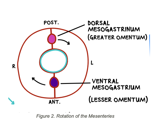
what mesentry is found on foregut
ventral mesogastrium
dorsal mesogastrium
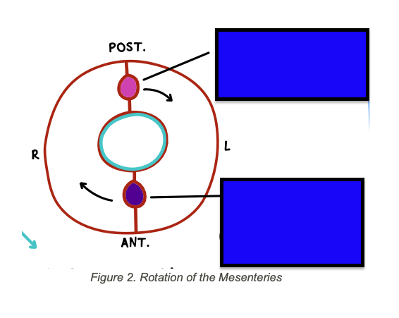
in foregut
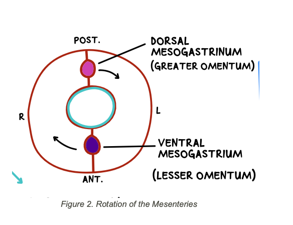
what does the ventral mesogastrium give rise to
will eventually develop into the lesser omentum, which is made up of the:
o Hepatogastric ligament
o Hepatoduodenal ligament
what does the dorsal mesogastrium give rise to
will eventually develop into the greater omentum, which is made up of:
-gastrophrenic ligament
-gastrocolic ligament
-gastrosplenic ligament
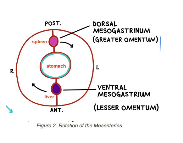
which organ does the ventral mesogastrium help develop
liver
The dorsal mesogastrium will give rise to the (organ)
spleen
why is liver found on right and spleen on left
Further rotation will bring the ventral mesogastrium to the right side and the dorsal mesogastrium to the left side
o This explains why the liver is located on the right side of the body, and the spleen is located on the left side
what does the middle gut tube give rise to
stomach
reference
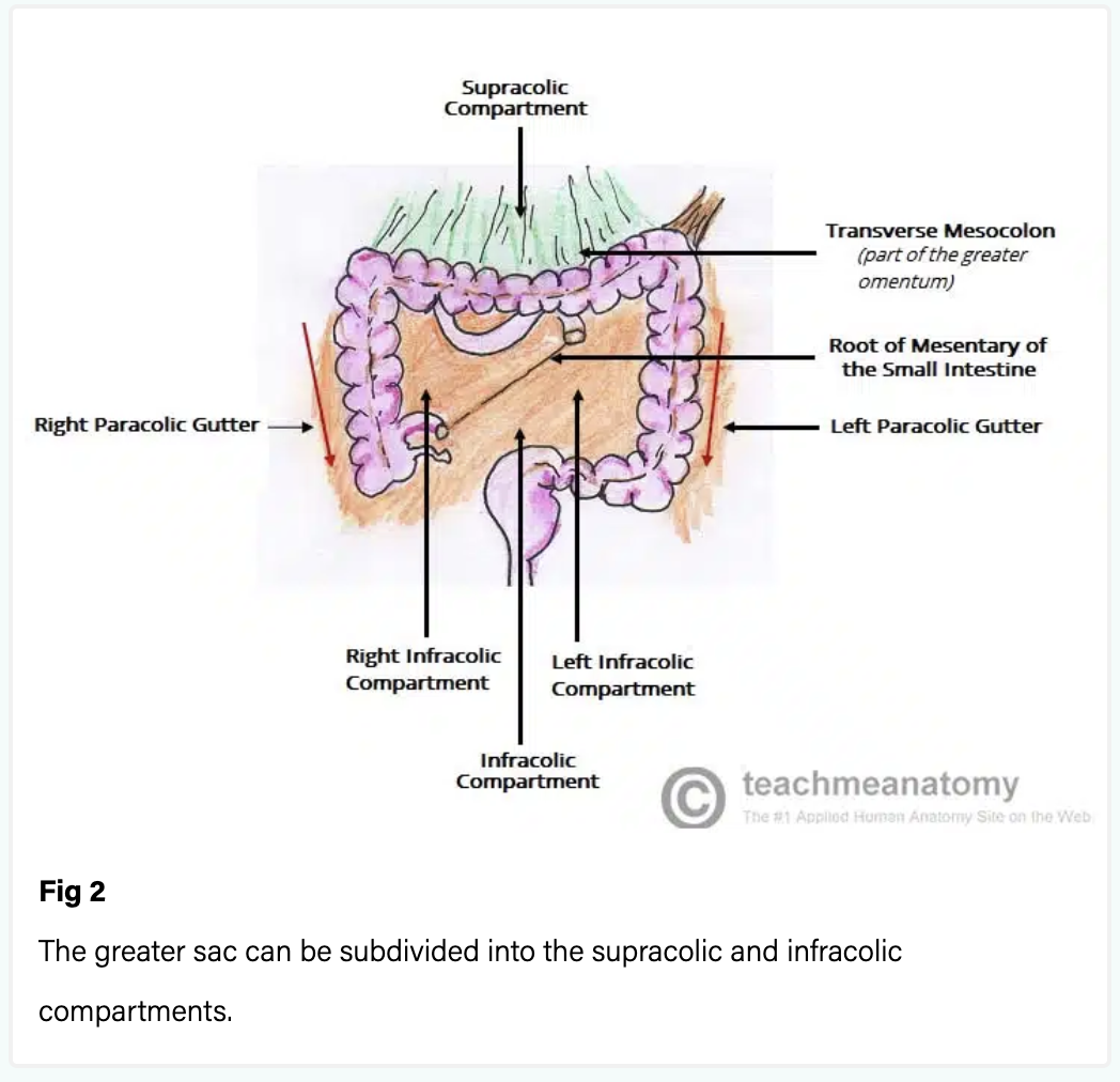
Subdivisions of the Peritoneal Cavity
greater and lesser peritoneal sacs.
Greater Sac is divided by
divided into two compartments by the mesentery of the transverse colon (known as the transverse mesocolon)
divisions of greater sac
Supracolic compartment – lies above the transverse mesocolon and contains the stomach, liver and spleen.
Infracolic compartment – lies below the transverse mesocolon and contains the small intestine, ascending and descending colon. The infracolic compartment is further divided into left and right infracolic spaces by the mesentery of the small intestine.
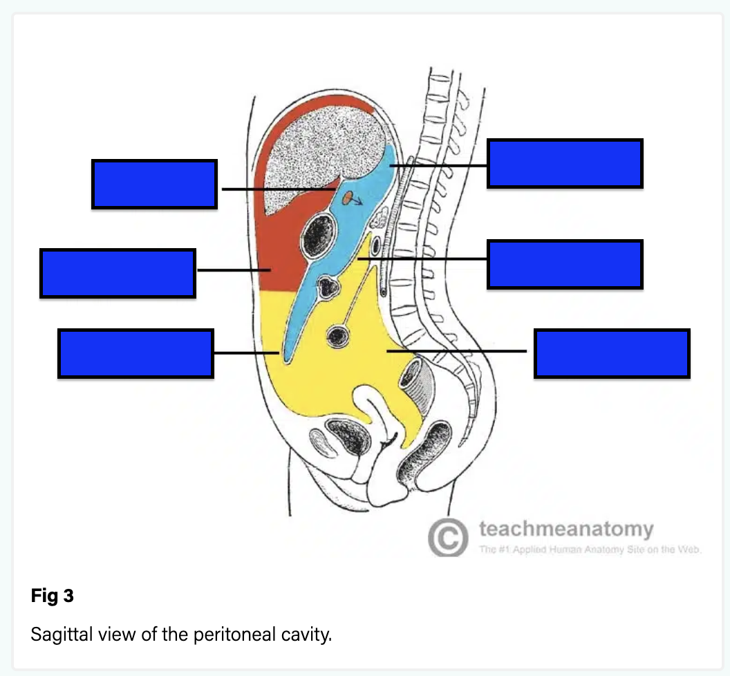
Lesser Sac (Omental Bursa) location
posterior to the stomach and lesser omentum
The omental bursa is connected with the greater sac through
an opening in the omental bursa – the epiploic foramen (of Winslow).
The epiploic foramen is situated posterior to the free edge of the lesser omentum (the hepatoduodenal ligament).

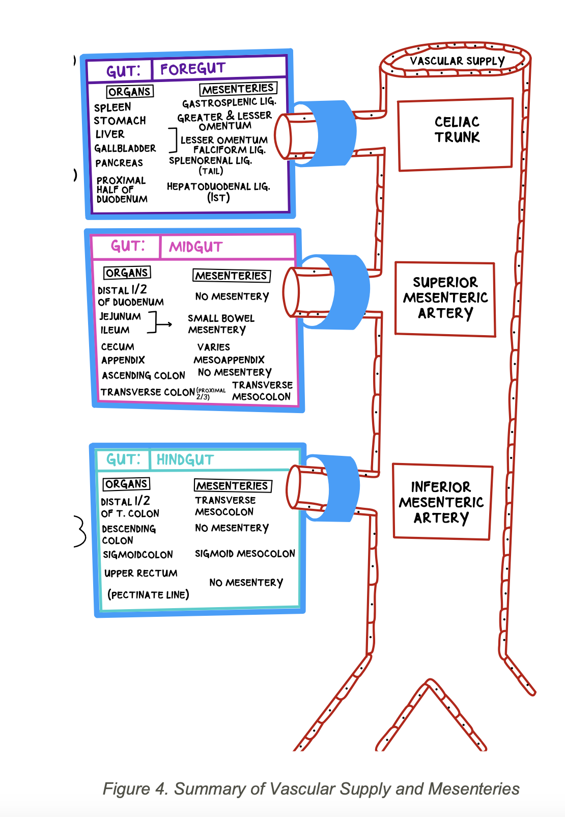
what artery supplies the foregut
the celiac trunk
branches:
-left gastric
-common hepatic: branches into cystic artery (to gallbladder), gastroduodenal artery, superior pancreatic duodenal
-splenic
what organs are formed by foregut
stomach
spleen
liver
gallbladder
proximal duodenum
pancreas
what supplies the stomach
Left gastric a.
what supplies the spleen
Left gastric a.
what supplies the liver
Common hepatic a.
what supplies the gallbladder
Cystic a.
what supplies the Pancreas
Gastroduodenal a.
what supplies the Proximal half of duodenum
Gastroduodenal a.
spleen mesentery
gastrosplenic ligament
stomach mesentery
Greater omentum, Lesser omentum
liver and gall bladder mesentery
lesser omentum including:
o Hepatogastric
o Hepatoduodenal
o Falciform
Pancreas mesentery Head & Body
retroperitoneal; no mesentery
Pancreas mesentery tail
splenorenal ligament (greater omentum)
Proximal half of duodenum mesentery
lesser omentum- Hepatoduodenal ligament
what supplies the midgut
superior mesentric artery
branches of superior mesentric artery
o Intestinal arteries
o Ileo-colic artery
o Right colic artery
o Middle colic artery
organs formed by midgut
-Distal half of Duodenum
-jejunum
-ileum
-Distal Ileum (at the ileo- cecal junction)
-cecum
-appendix
-ascending colon
-Proximal 2/3 of the Transverse colon
intestinal arteries supply
-Distal half of Duodenum
-jejunum
-ileum
Ileo-colic artery suplies
-Distal Ileum (at the ileo- cecal junction)
-cecum
-appendix
Right colic a. supplies
ascending colon
middle colic a. supplies
-Proximal 2/3 of the Transverse colon
-Distal half of Duodenum mesentery
no mesentery
-retroperitoneal organ
Jejunum mesentery
-Small bowel mesentery
-Ligament of Treitz – suspends the fourth part of the duodenum (marks start of jejunum)
Ileum mesentery
-Small bowel mesentery/ mesentery proper
cecum mesentery
Small bowel mesentery; can be retroperitoneal in some
appendix mesentery
mesoappendix
ascending colon mesentery
Retroperitoneal; no mesentery
Proximal 2/3 of the Transverse colon mesentery
Transverse mesocolon
The main vascular supply of the hindgut
inferior mesenteric artery
branches of the inferior mesenteric artery
o Left colic artery
o Sigmoidal arteries
o Superior rectal arteries
organs formed by hindgut
-Distal 1/3 of Transverse Colon
-Descending colon
-Sigmoid colon
-Upper rectum (above the pectinate line)
Left colic a. supplies
-Distal 1/3 of Transverse Colon
-Descending colon
Sigmoidal arteries supplies
-Sigmoid colon
Superior rectal artery supplies
upper rectum (above the pectinate line)
Distal 1/3 of Transverse Colon mesentery
Transverse mesocolon
Descending colon mesentery
Retroperitoneal; no mesentery
Sigmoid colon mesentery
Sigmoid mesocolon
Upper rectum mesentery
Retroperitoneal; no mesentery
reference