Unit 5 Lab Exam flashcards
1/110
Earn XP
Description and Tags
Name | Mastery | Learn | Test | Matching | Spaced | Call with Kai |
|---|
No analytics yet
Send a link to your students to track their progress
111 Terms
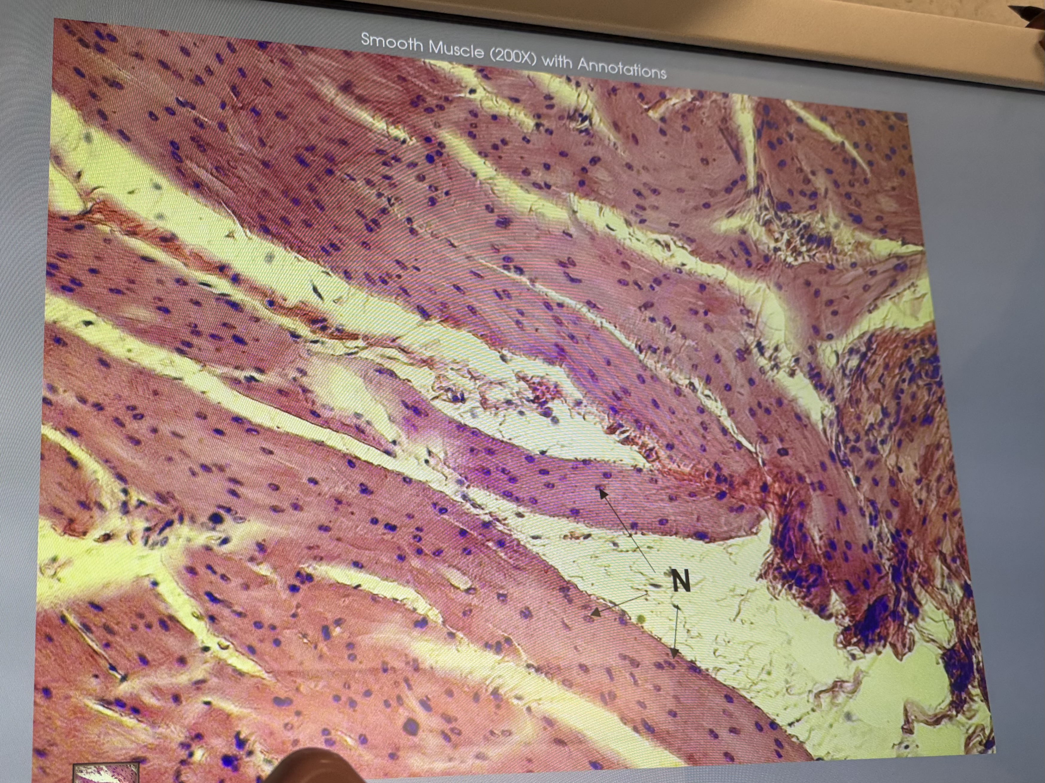
Esophagus (#10937): smooth muscle, shape?, striated or not?, single or multinucleated?
Shape: spindle
Striated: no
Nucleus: single
What kind of ET lines the esophagus?
Non keratinized stratified squamous epithelial tissue
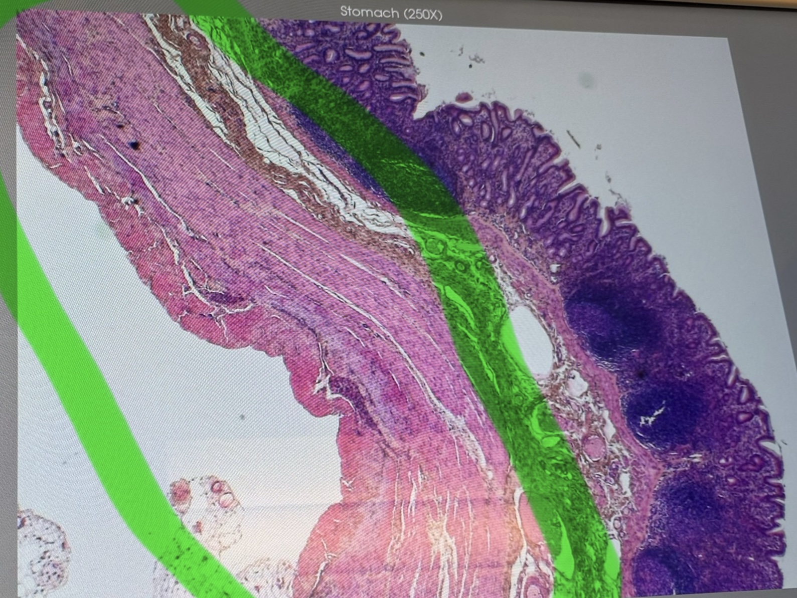
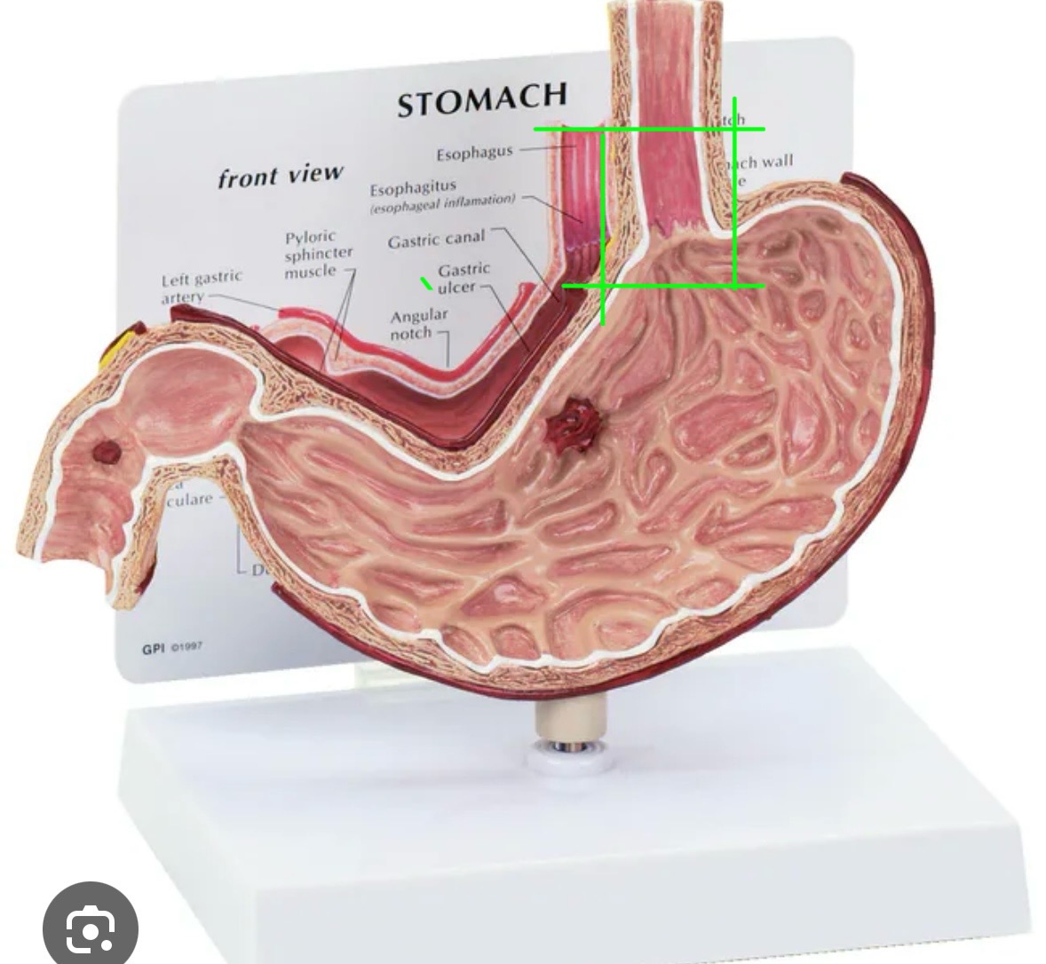
Stomach (#10140): 3 layers of smooth muscle
From outer to innermost: Longitudinal muscle, circular muscle, oblique muscle


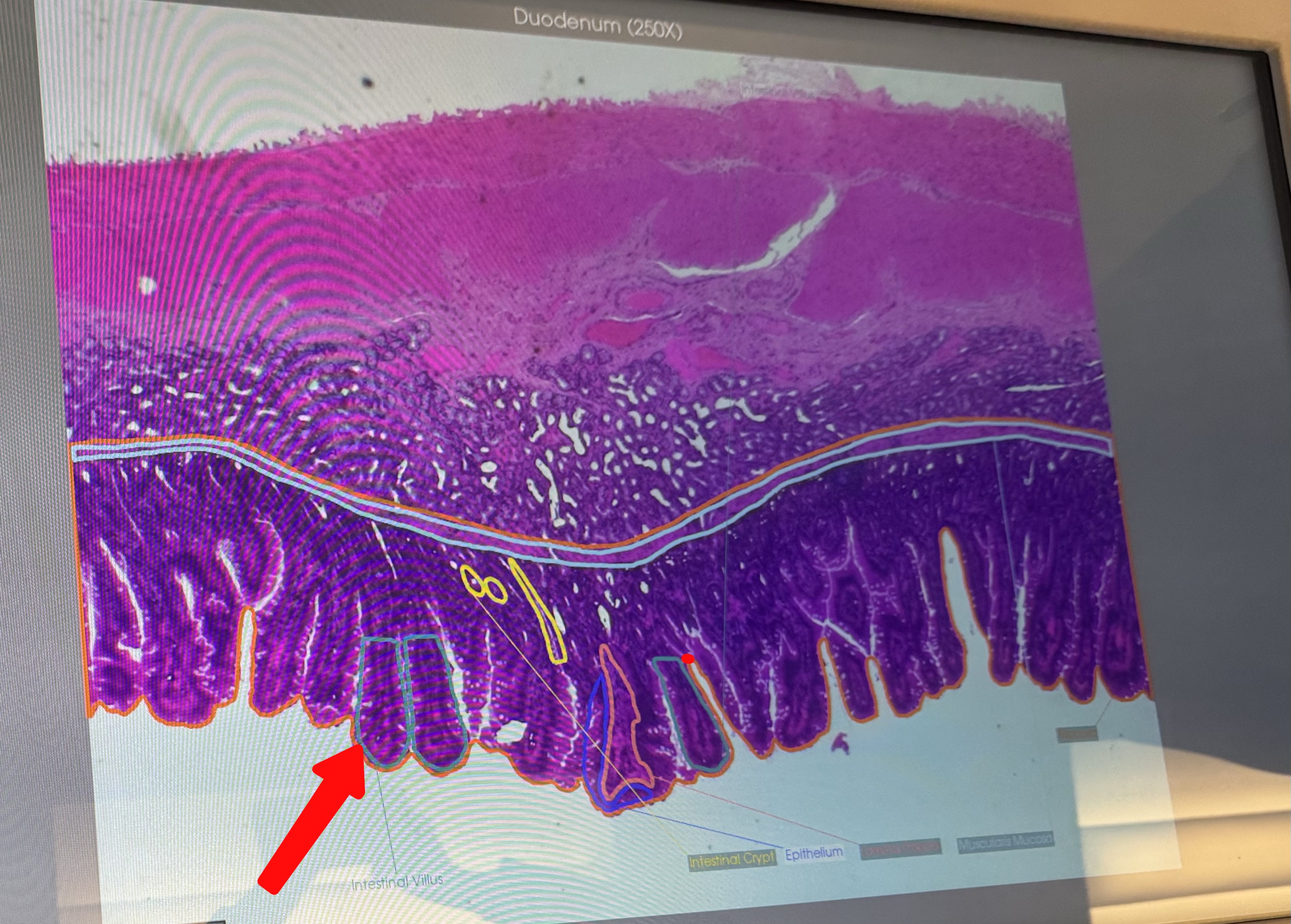
Duodenum (#10152): what is this structure?
villi
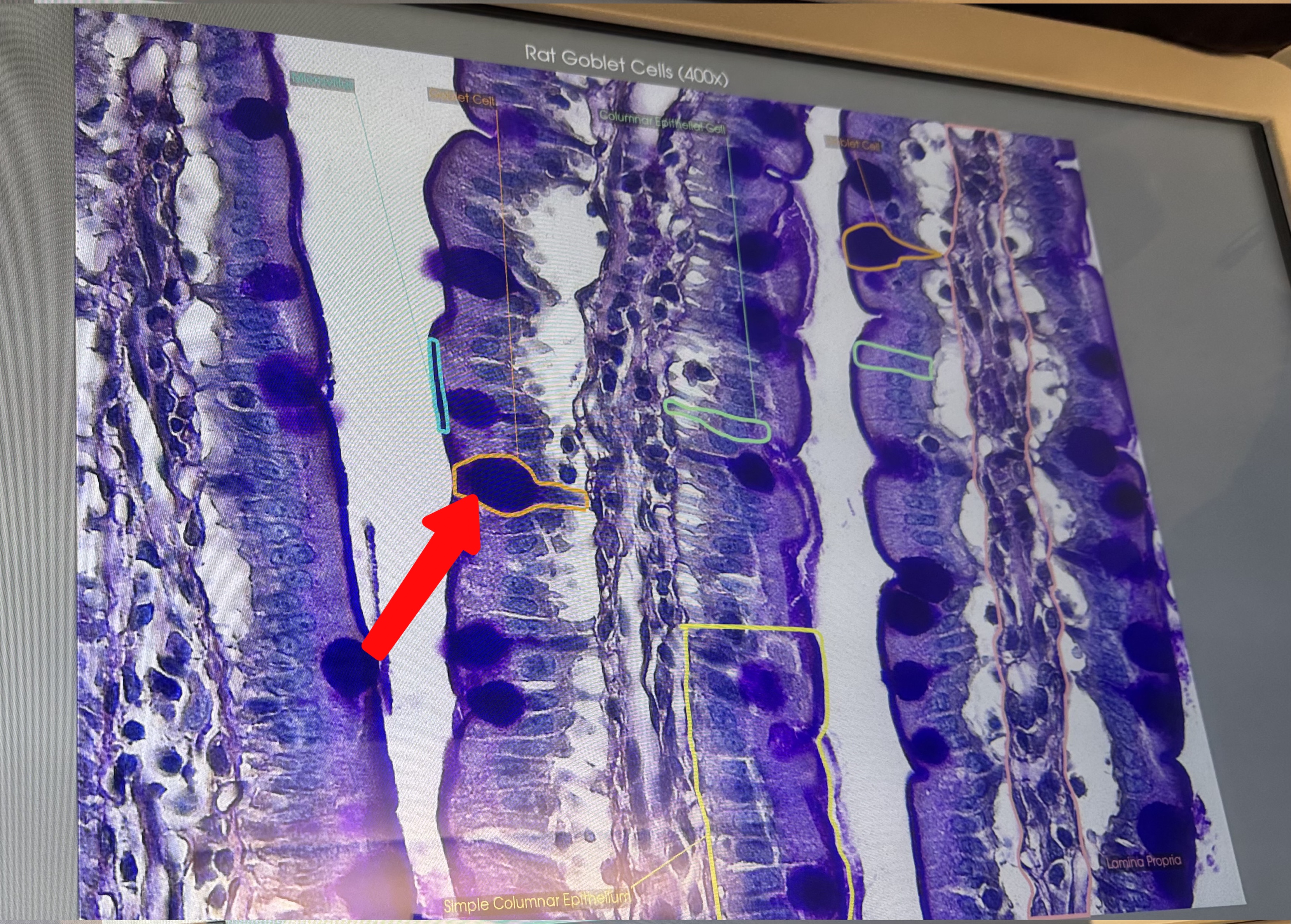
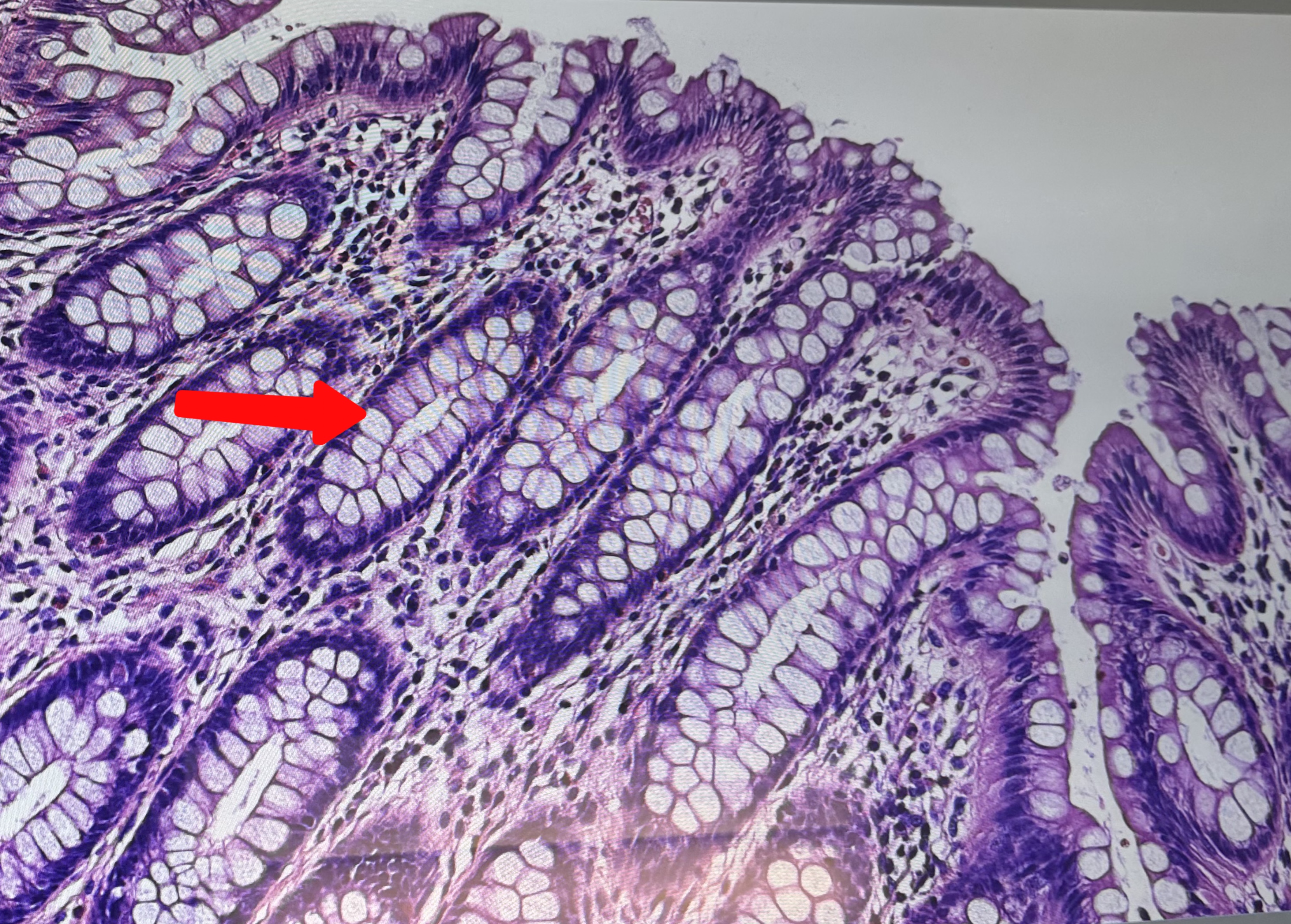
Small intestine 400x (#10068): what is this structure
Goblet cells


Small intestine 400x (#10068): what is this structure
microvilli

Small intestine: what type of ET?
Simple columnar epithelial tissue
Colon: what type of ET?
Simple columnar epithelial tissue
Colon: LOTS of goblet cells, why?
For lubrication for poop
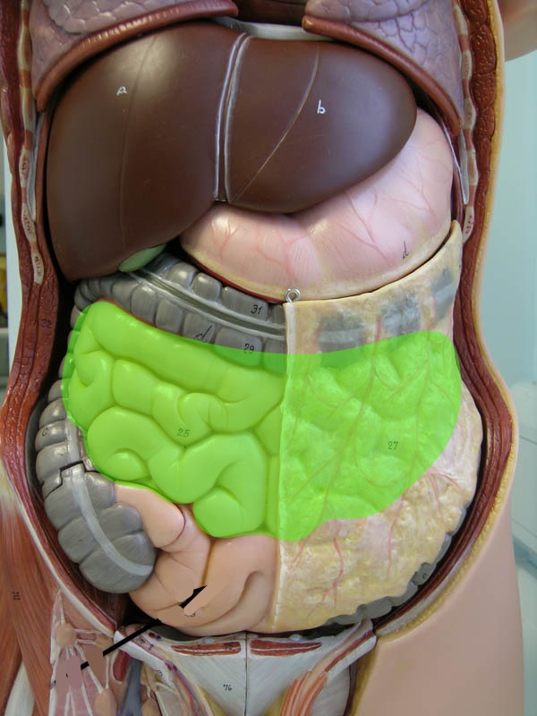
How can you differentiate between the stomach, small intestine, and large intestine?
Amount of goblet cells (mainly colon) presence of villi (mainly small intestine), presence of 3 muscle layers (stomach)
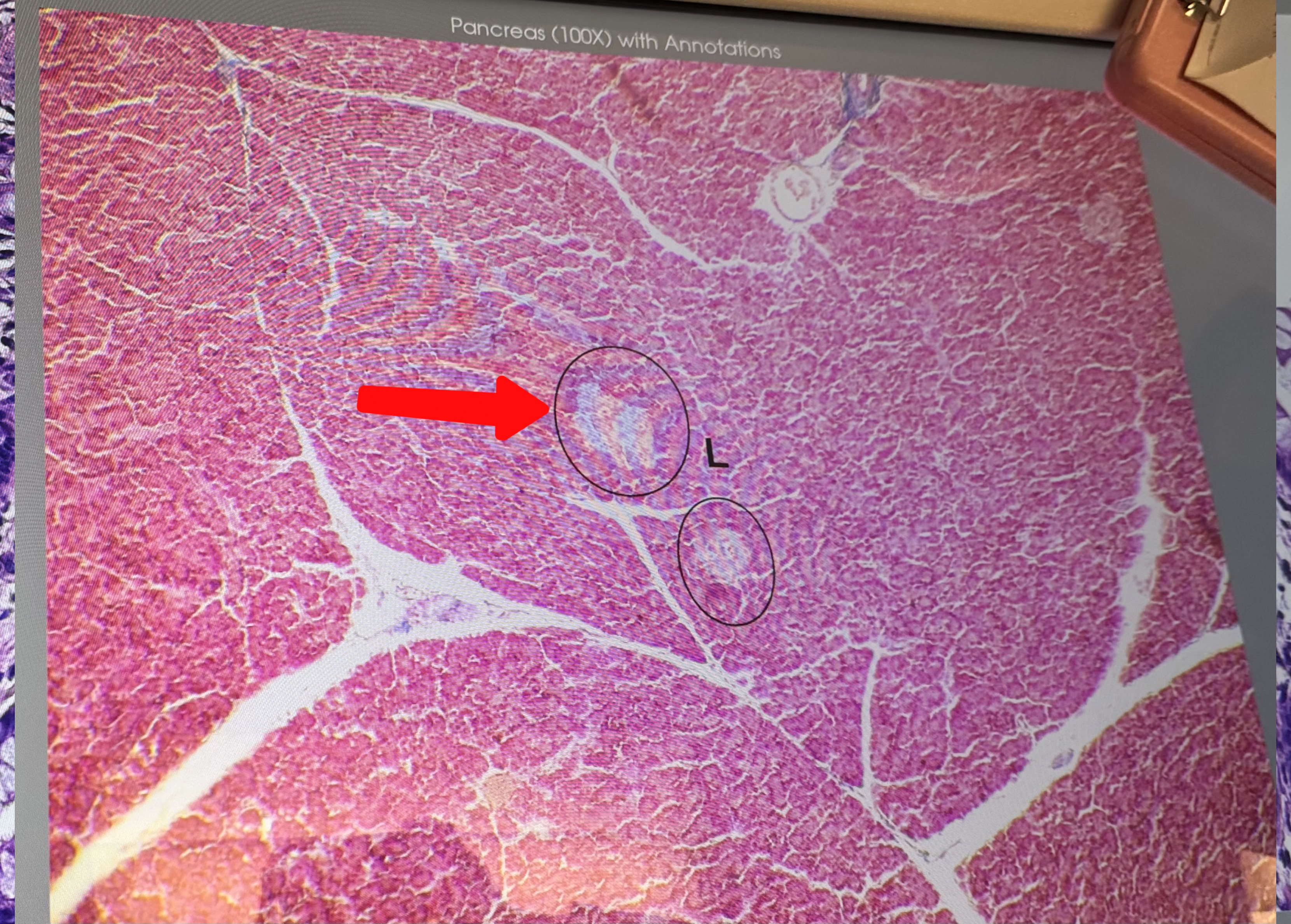
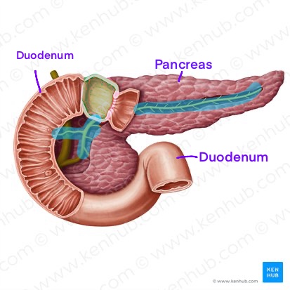
Páncreas: what is this?
Pancreatic islet
Pancreas: pancreatic islets vs. acinar cells--which produces what?
Islets make hormones like insulin and glucagon. acinar cells make enzymes like amylase

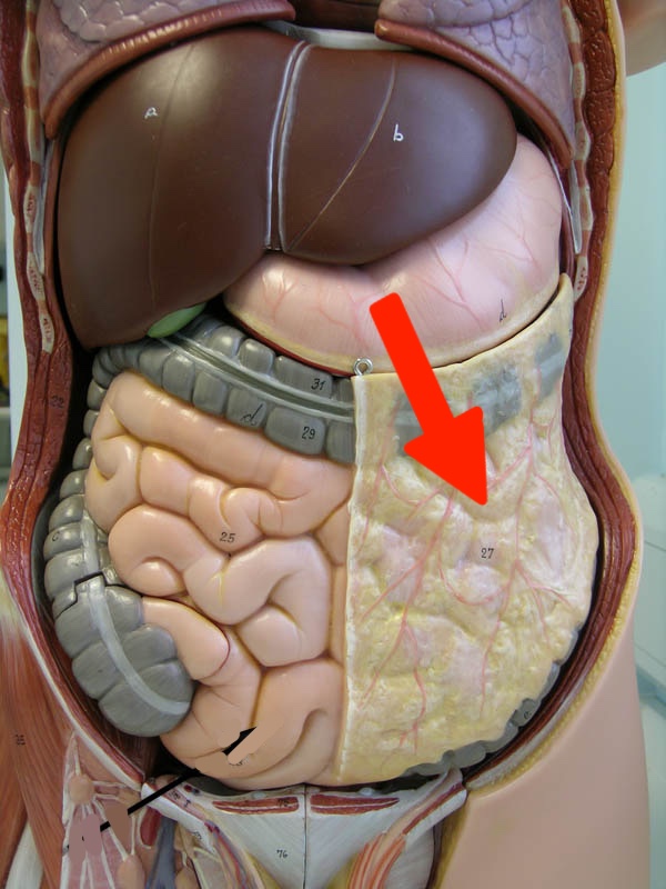
What is this and what is the function?
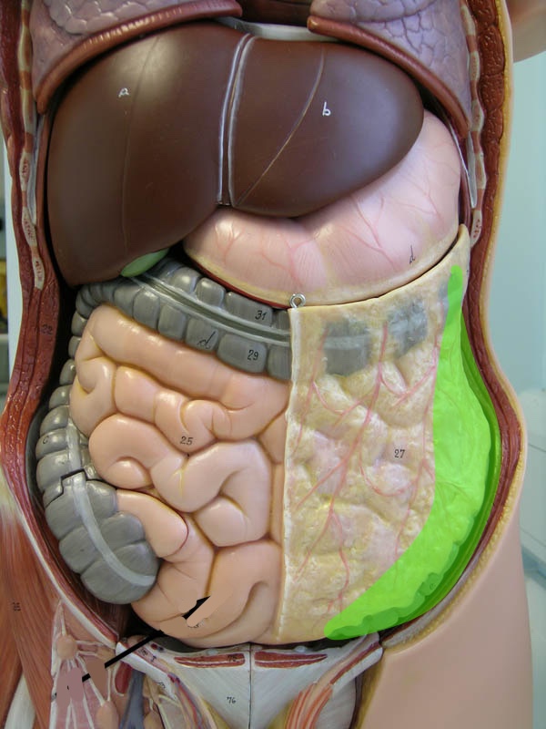
Greater omentum: contains a thick layer of adipose tissue important for energy reserves and insulation to prevent heat loss
What is this and what is the function? ( gray with blood vessels)
Mesentery: Suspends organs in abdominal cavity and contains blood vessels and nerves that supply blood and innervate organs
Esophagus: how do the 2 layers of muscle work together to accomplish peristalsis?
Circular muscle contracts to form bolus and longitudinal muscle contracts to propel bolus forward
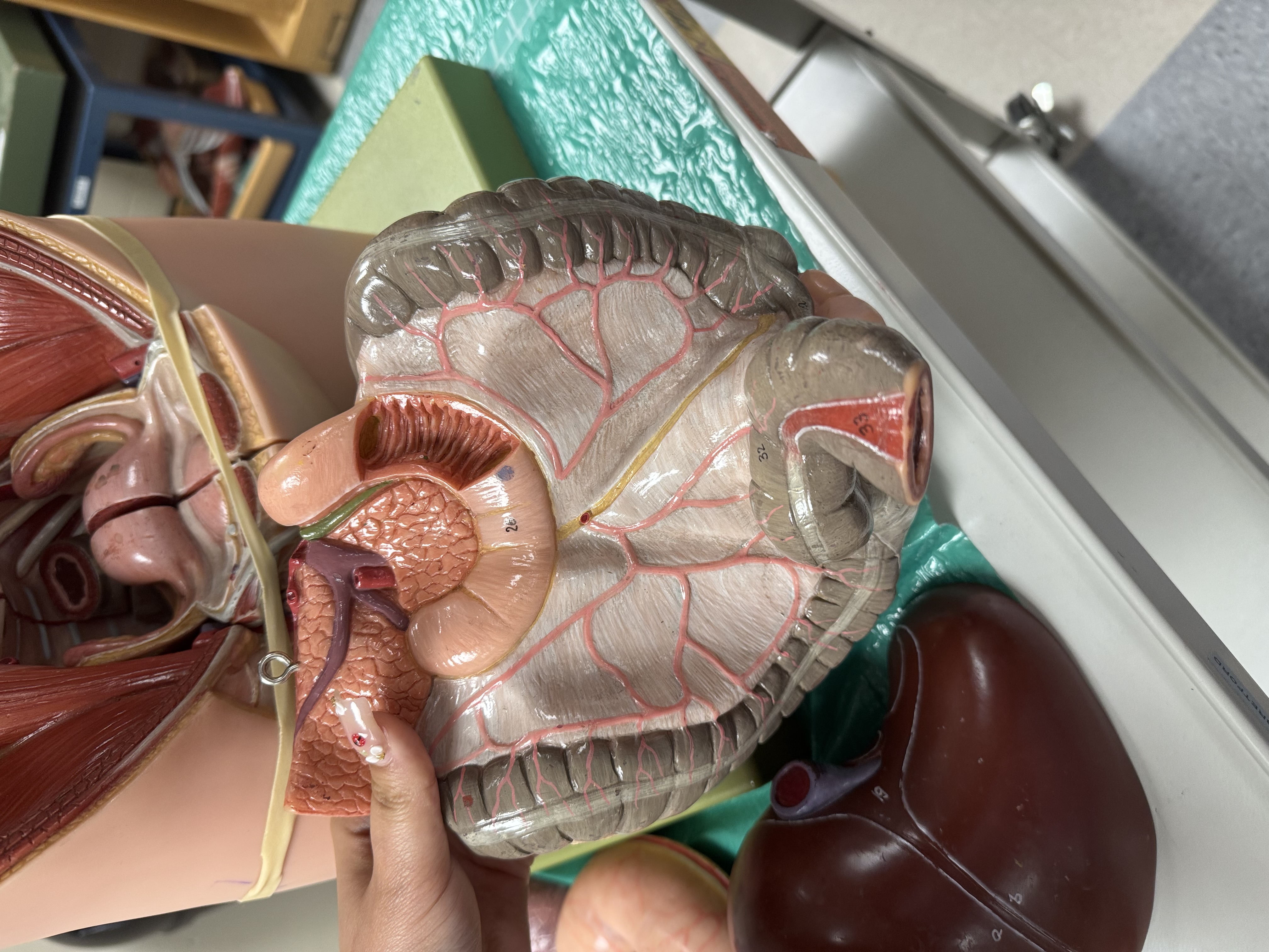
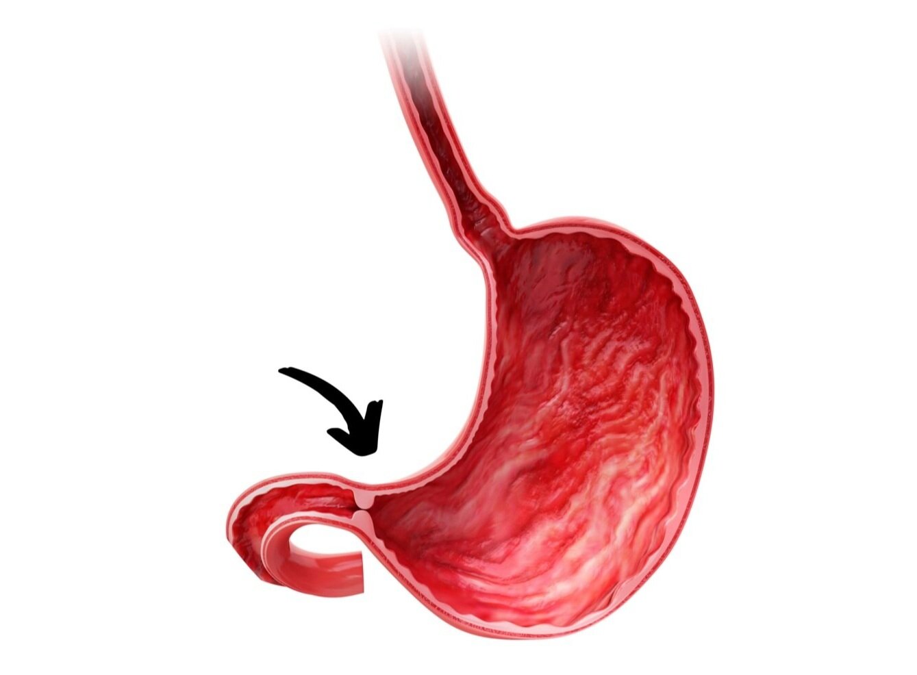
Stomach: notice that a gastric fistula (not normal! Due to her gastric cancer) connect the stomach to the jejunum. Why might this be a super painful condition?
Fistula is an opening and it can be painful because the contents of the stomach leak into the jejunum and can cause alot of irritation because the jejunum doesn’t have the Bruner glands that produce mucus to protect against gastric chyme and acids that the duodenum has
anus: how many sphincters?
2: internal and external anal sphincter
How many sphincters in the colon?
1: ileocecal sphincter
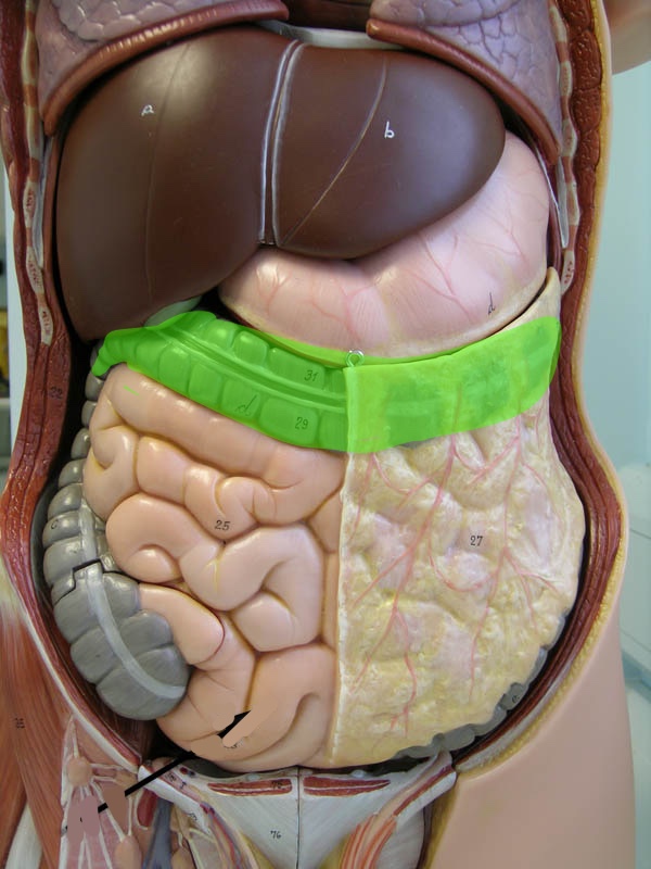
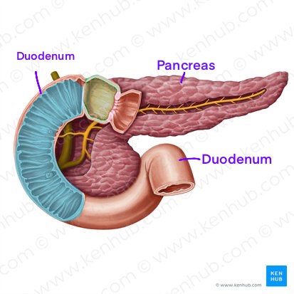
into which organ do the common bile duct and pancreatic duct connect?
Duodenum
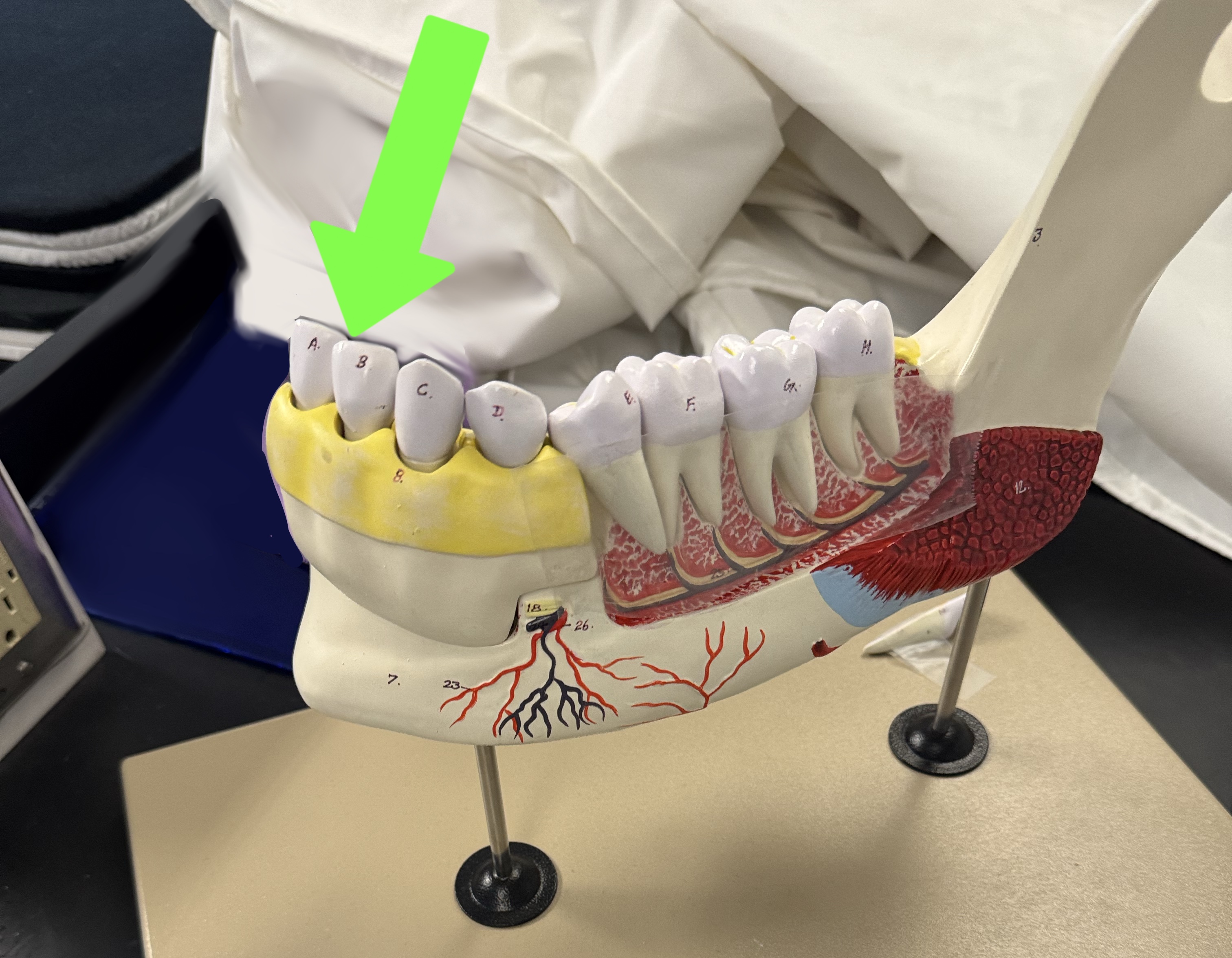
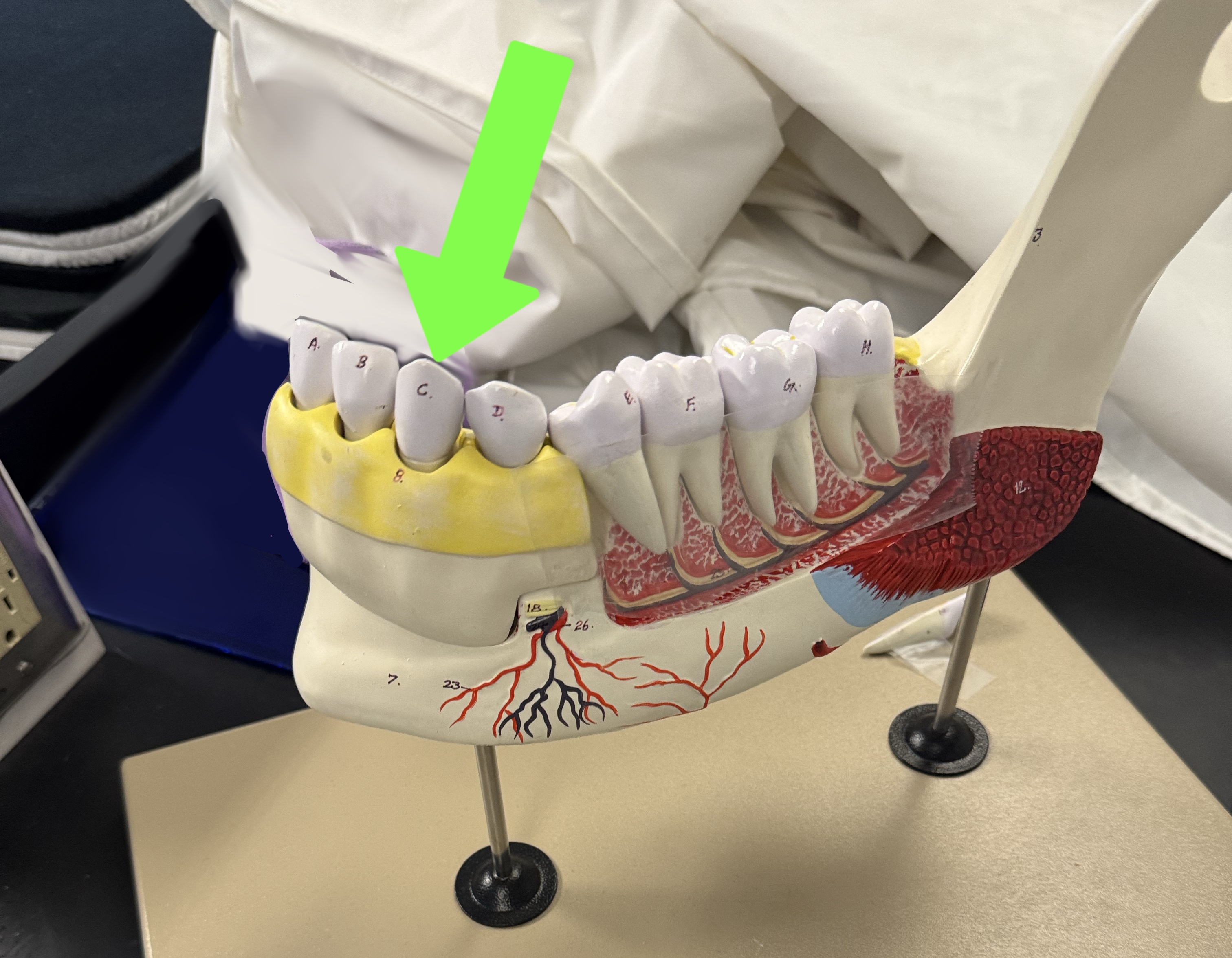
what type of tooth is this
Incisor
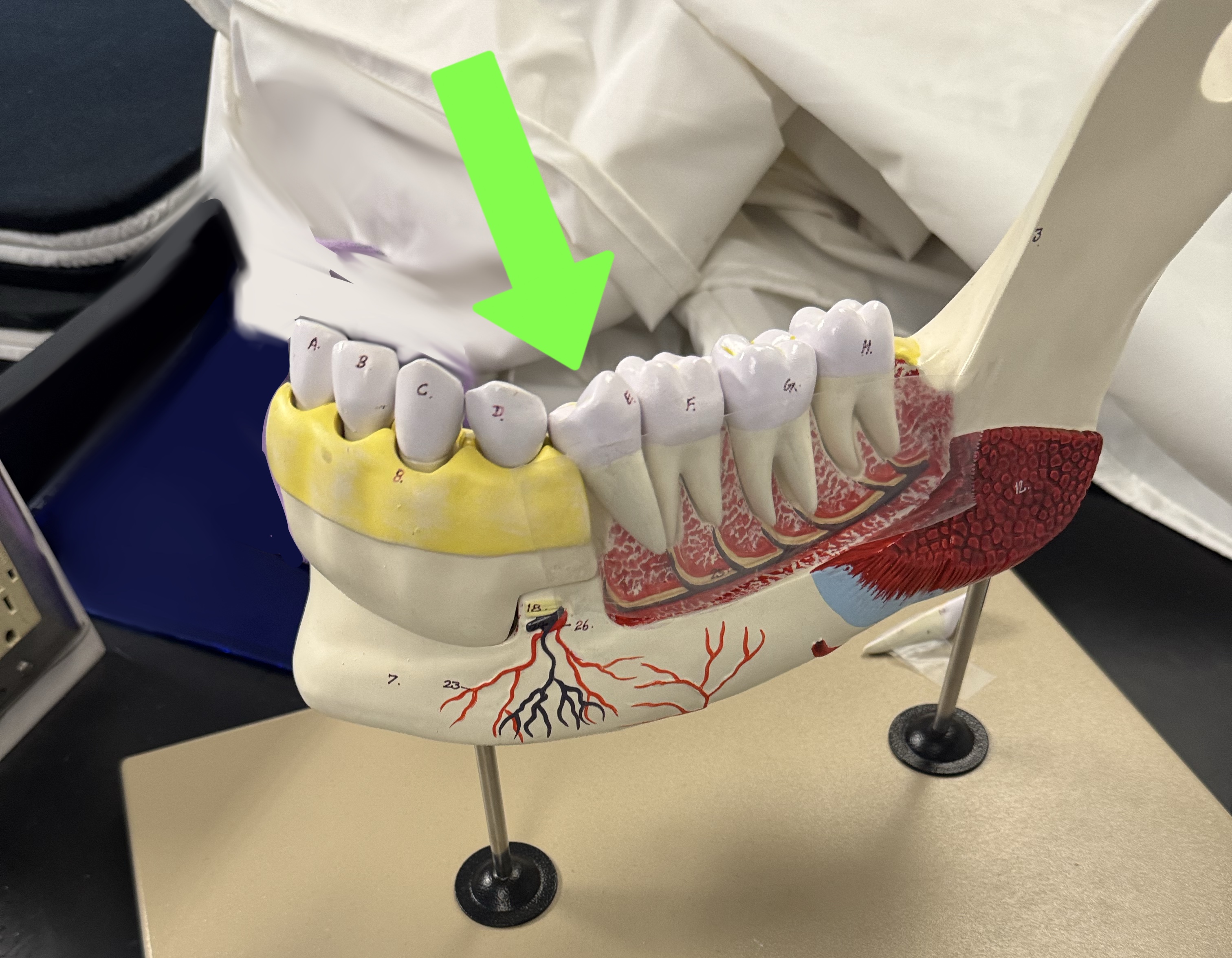
what type of tooth is this
Canine (cuspid)
what type of tooth is this
Premolars (bicuspids)
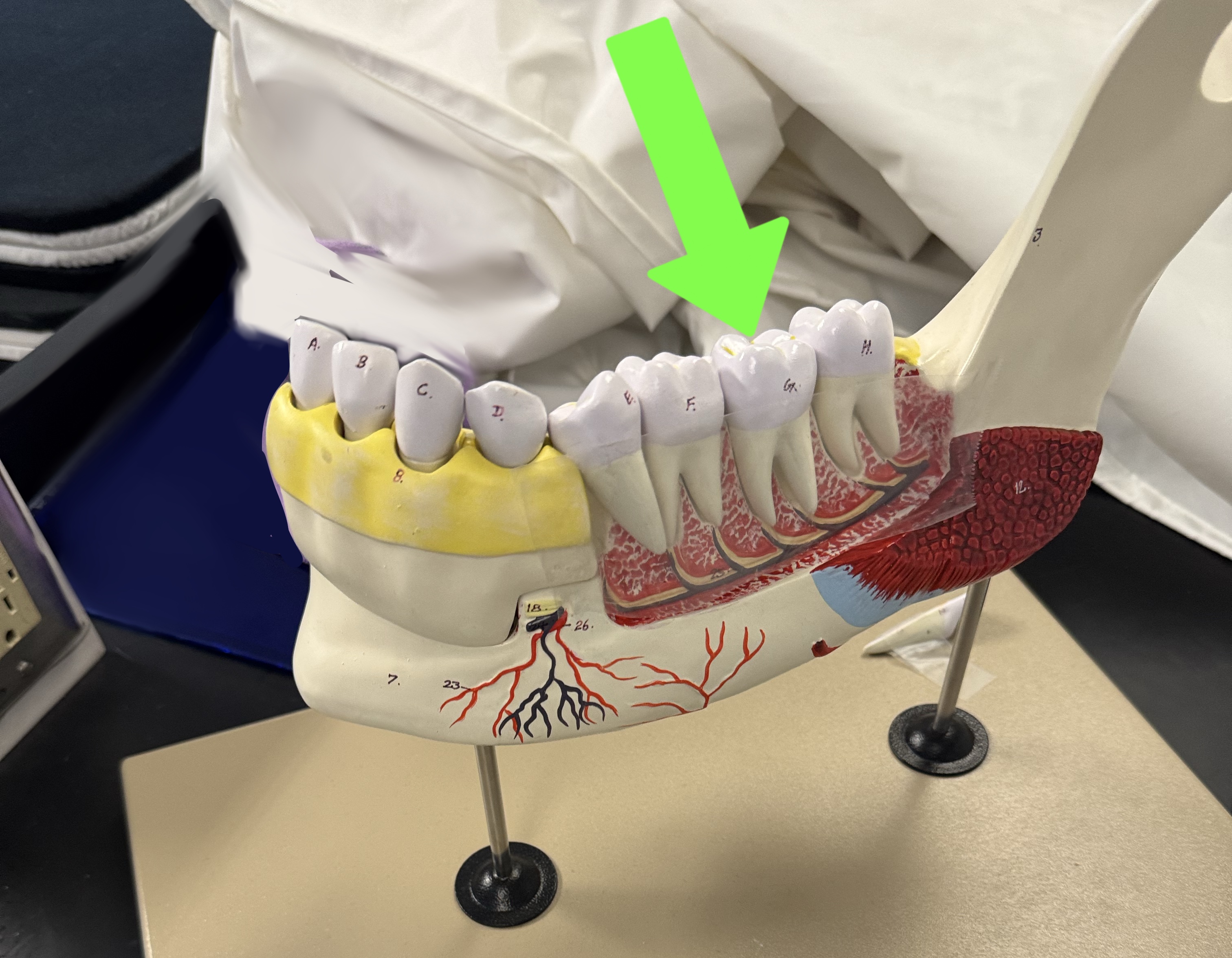
what type of tooth is this
Molars
Which organs do each of these arteries bring blood to? Descending aorta
Abdominal organs
Which organs do each of these arteries bring blood to? hepatic artery
Liver
Which organs do each of these arteries bring blood to? splenic artery
Spleen
Which organs do each of these arteries bring blood to? superior mesenteric artery
Páncreas, duodenum, small intestine, colon
Which organs do each of these arteries bring blood to? inferior mesenteric artery
Colón and rectum
What is the destination of the blood in these veins? Hepatic vein
Inferior vena cava
What is the destination of the blood in these veins? superior mesenteric vein
Liver (hepatic portal vein)
What is the destination of the blood in these veins? splenic vein
Liver (hepatic portal vein)
What is the destination of the blood in these veins? hepatic portal vein
Liver
What is the destination of the blood in these veins? inferior vena cava
Heart (R atrium)
Why does the liver need a hepatic artery if the hepatic portal vein is already bringing nutrient rich blood to the liver?
Because the hepatic artery is to nourish the liver tissue while the vein brings in blood to be filtered
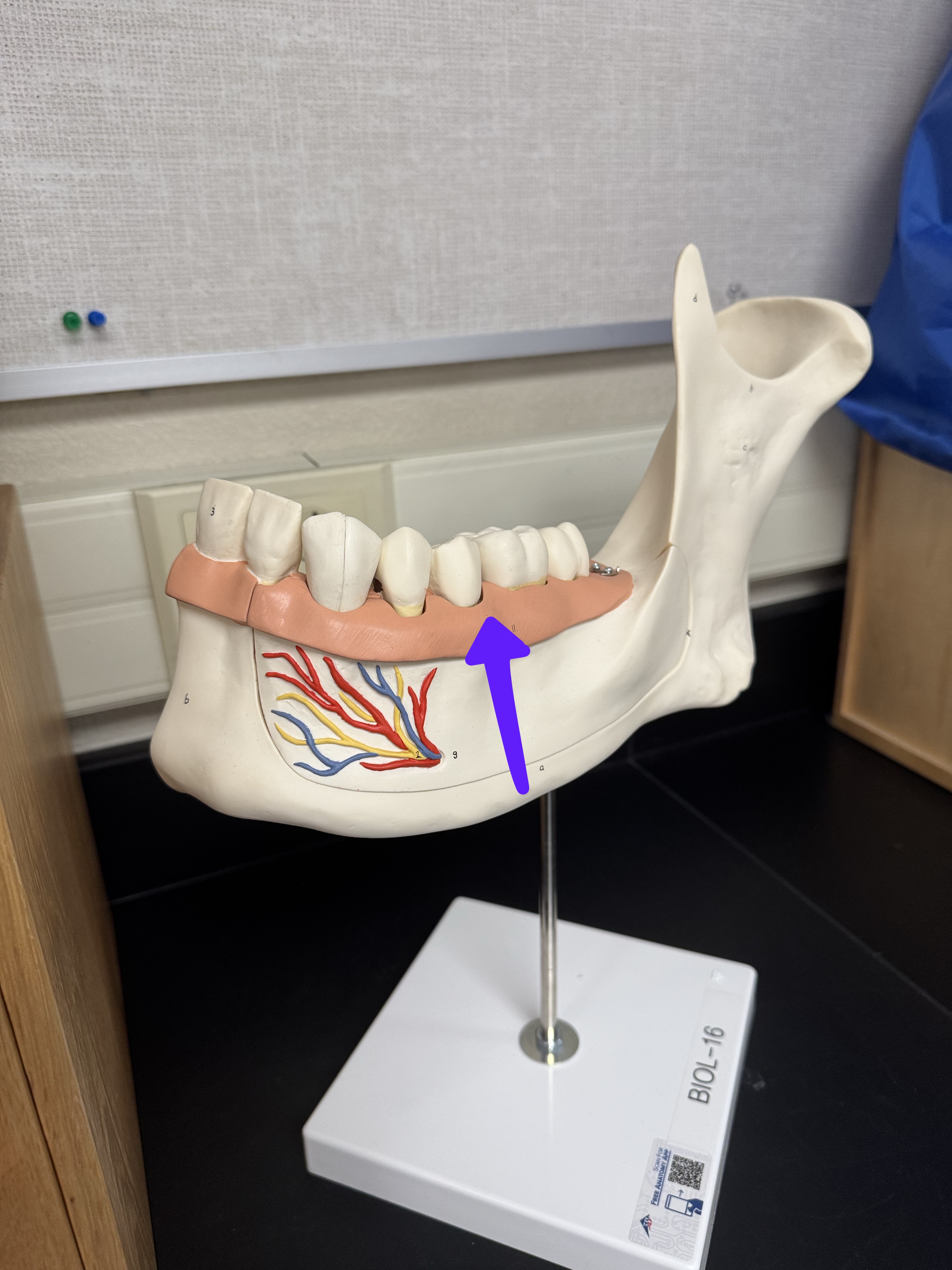
What is this?
Gingiva
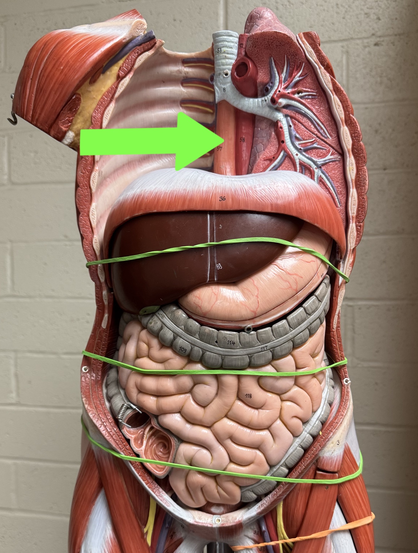
Torso model: what is this?
esophagus
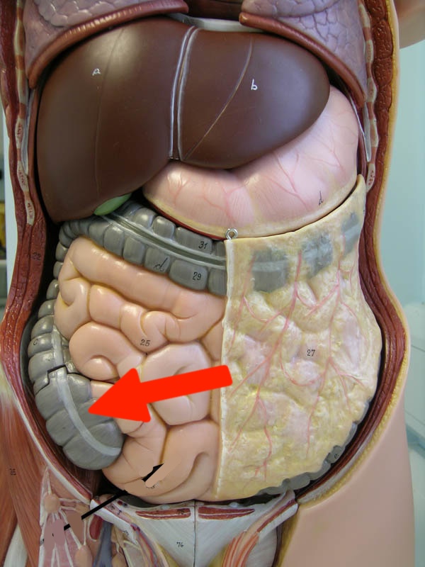
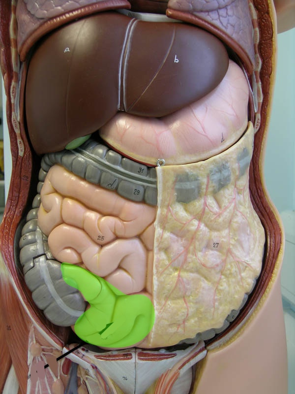
Torso model: what is this?
Cecum
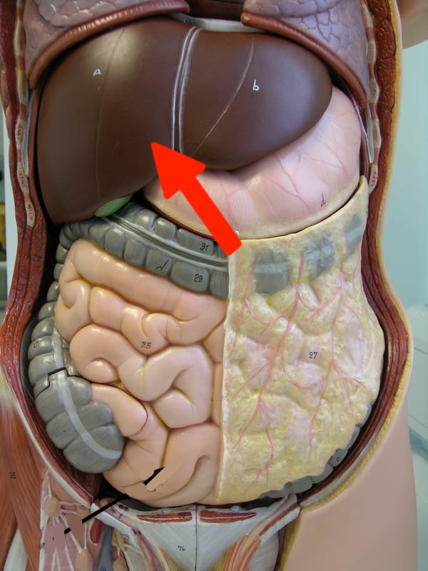
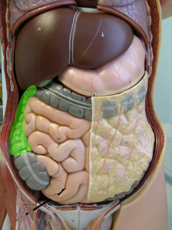
Torso model: what is this?
Liver
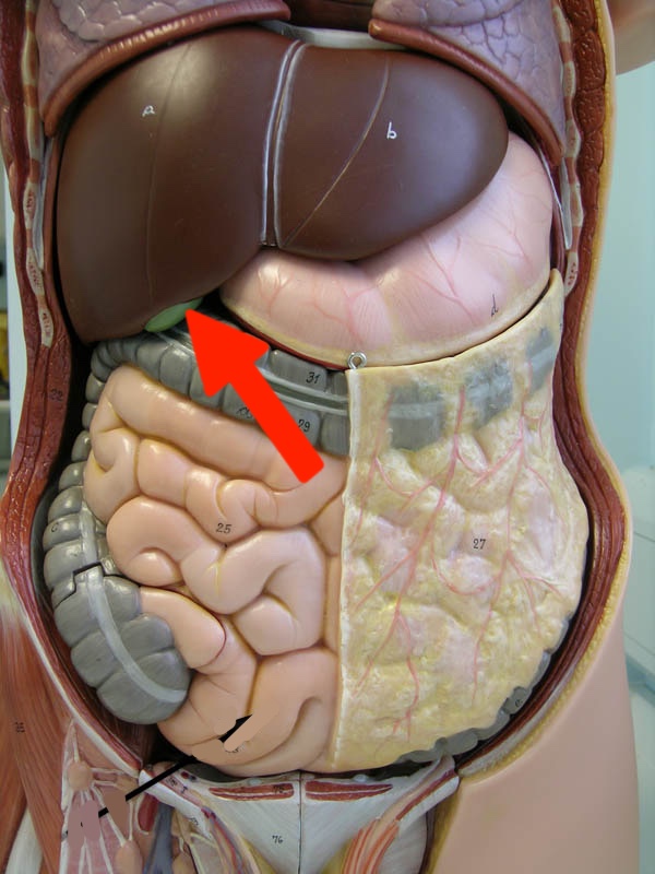
Torso model: what is this?
Gallbladder
Torso model: what is this?
Jejunum
Torso model: what is this?
Ileum
Torso model: what is this?
Ascending colon
Torso model: what is this?
Transverse colon
Torso model: what is this?
Descending colon
What sphincter is this?
Lower esophageal sphincter
what sphincter is this?
pyloric sphincter
What are these circular folds of the small intestine called
Plicae circularis

What are these folds called
Rugae
What is this duct called (highlighted blue)
Pancreatic duct
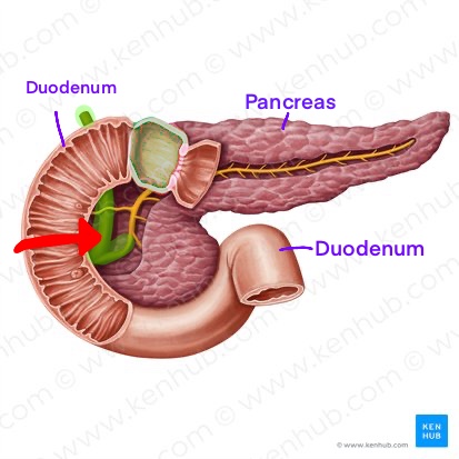
What is this duct called (highlighted green)
Common bile duct
Torso model
Greater omentum
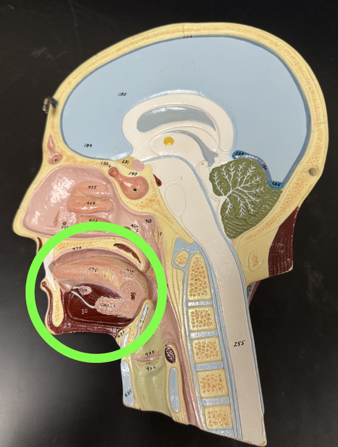
Half head
Oral cavity: tongue
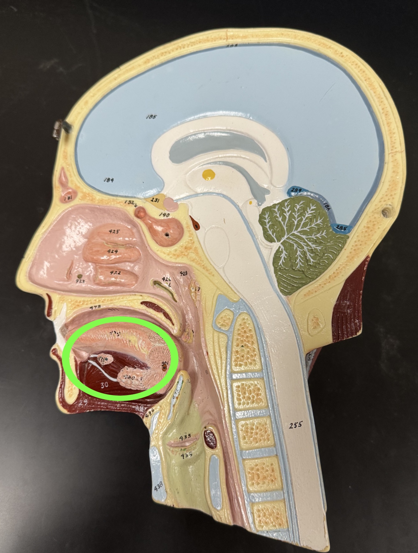
Half head
Tongue
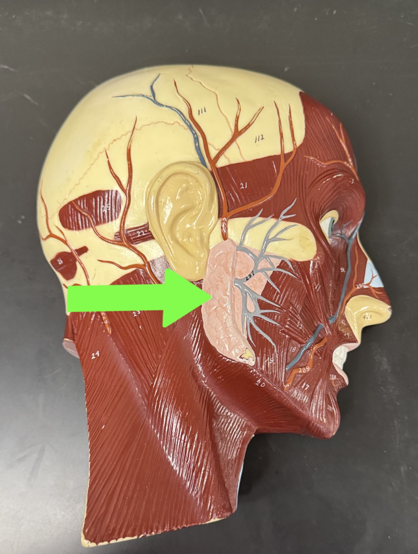
Half head
Salivary glands

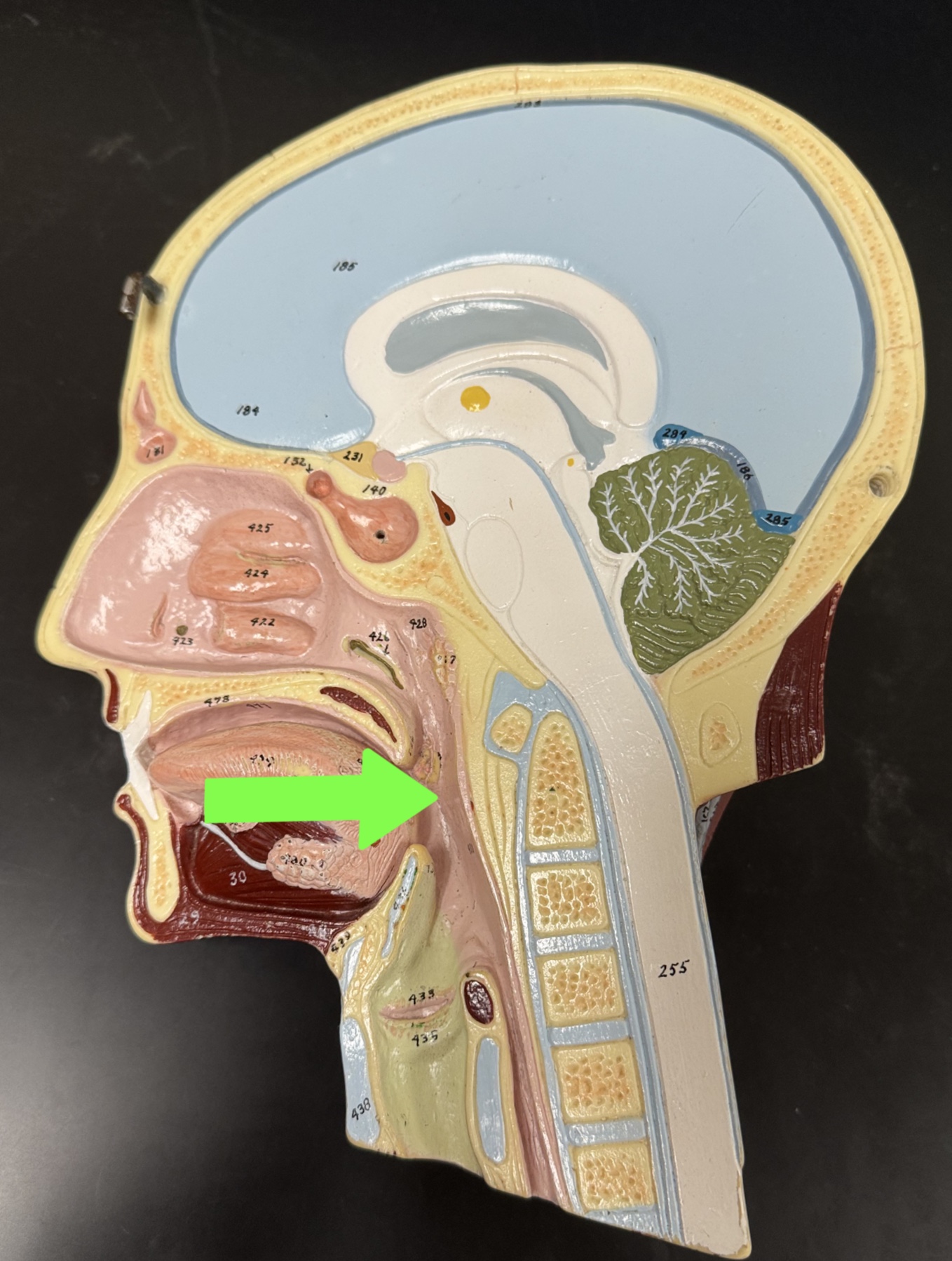
Half head
Esophagus

Half head
Epiglottis
Half head
Pharynx
Rabbit digestive system: Teeth (Adapted for which food? Missing incisors, canines, or molars?)
Has Incisors and molars for herbivore
Missing canines
Rabbit digestive system: How does the rabbit cecum compare to a human cecum?
It is larger
What are three differences between human and rabbit digestive systems?
Rabbit: Larger cecum than human, stomach goes all across abdomen, liver has 5 lobes while humans have 4
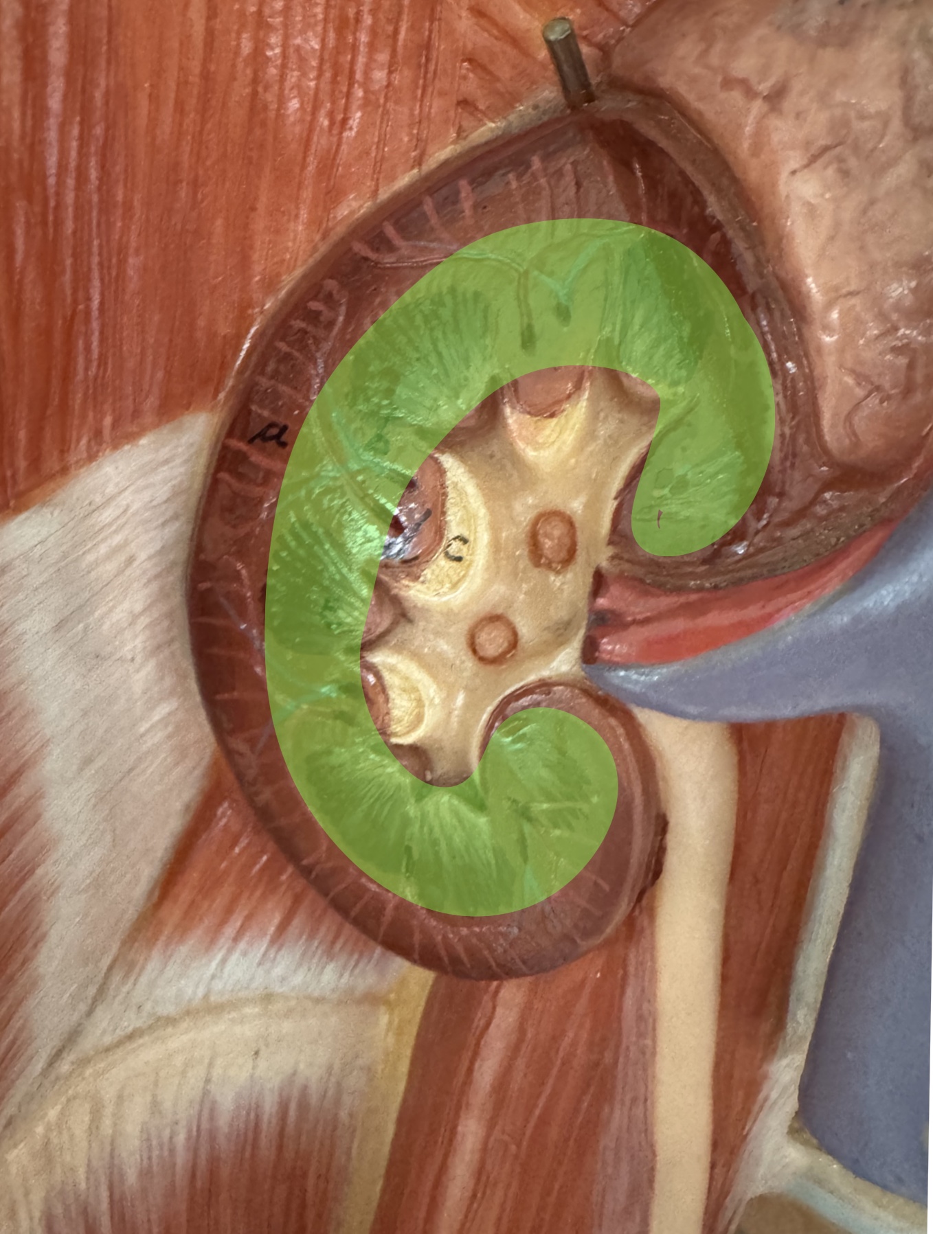
What is the picture of?
Kidney cortex
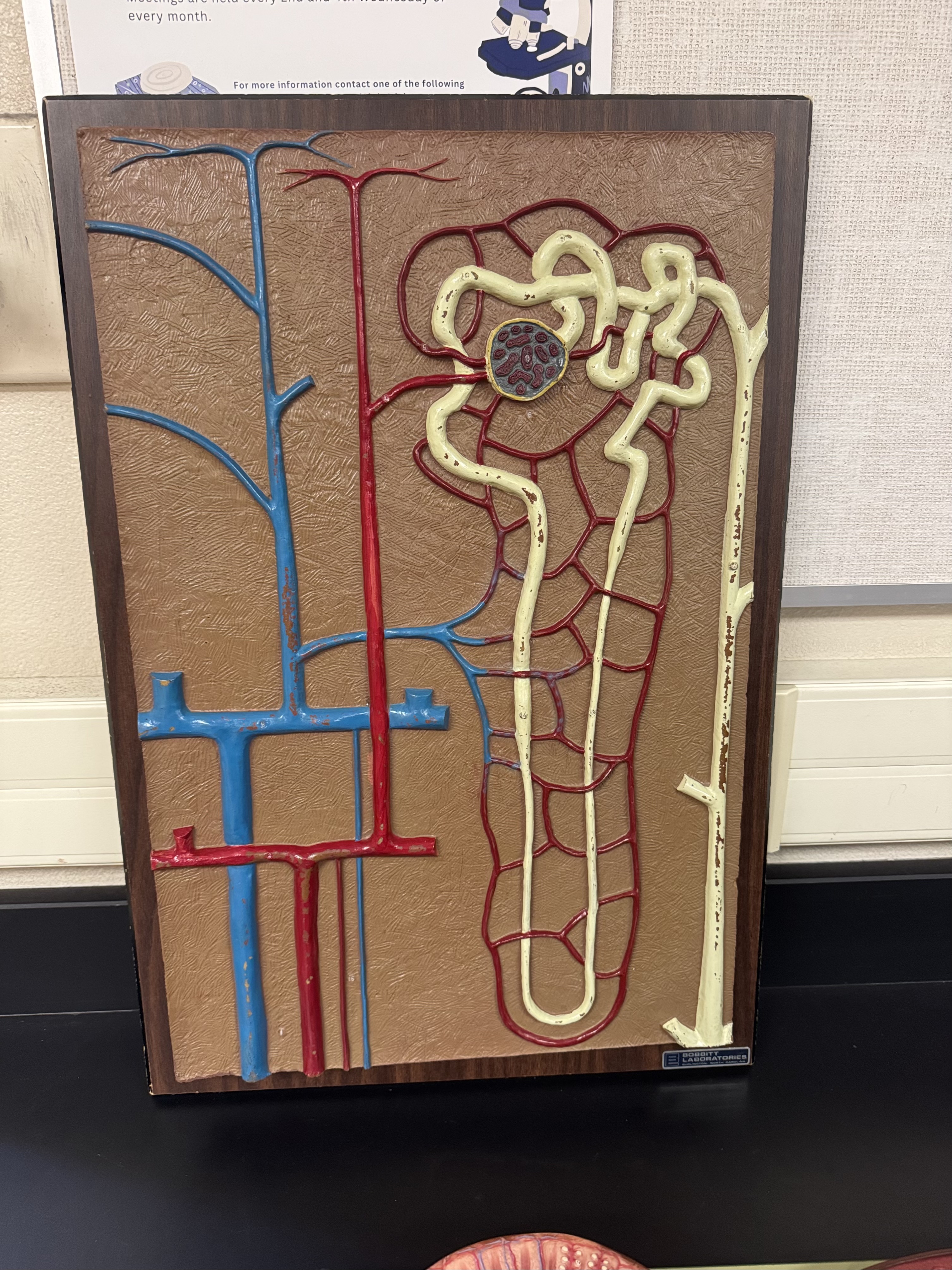
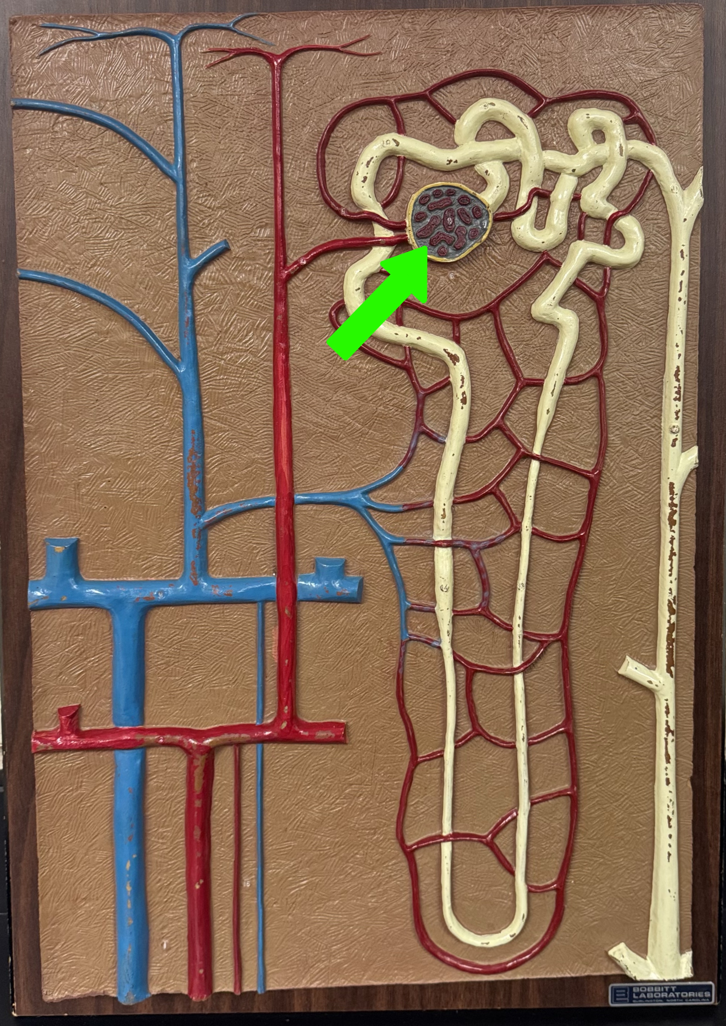
What is this structure
Nephron
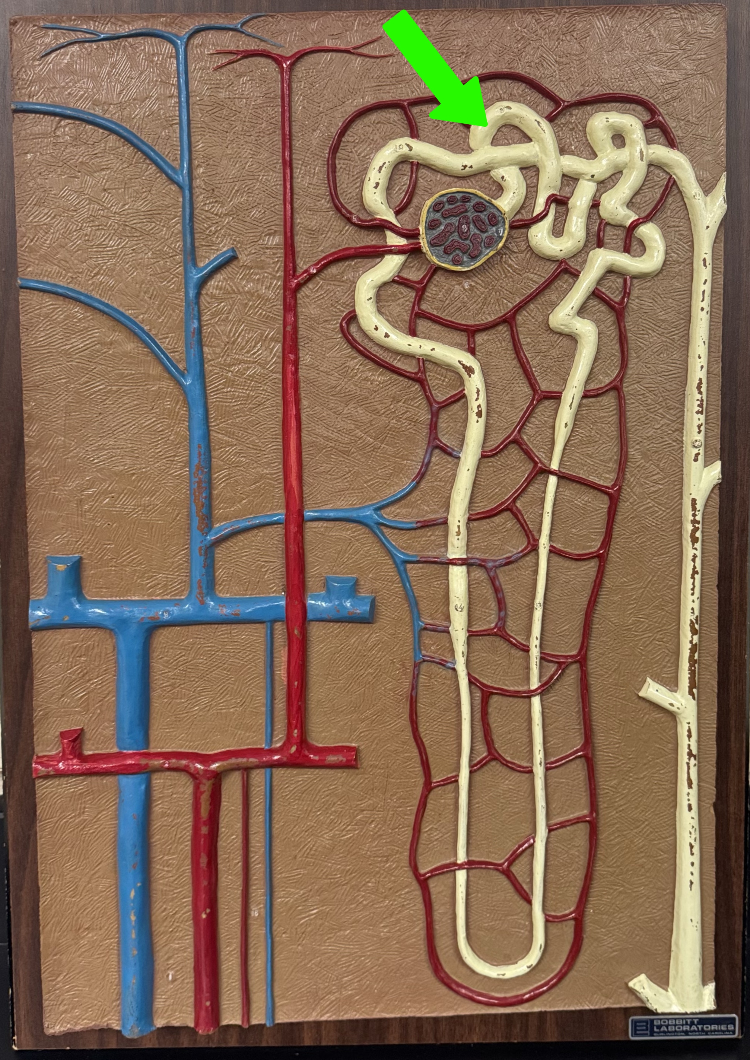
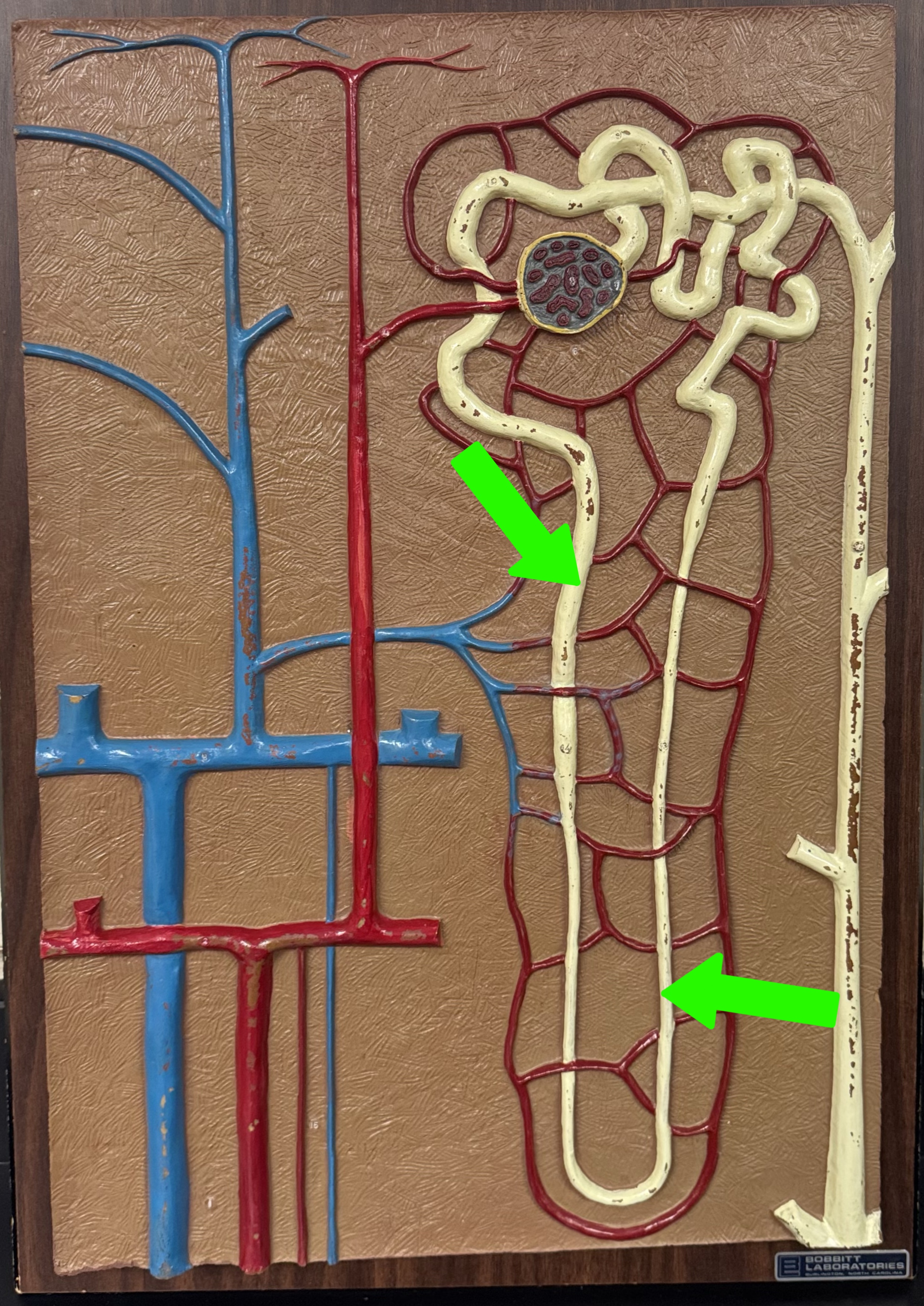
What are these parts of the nephron?
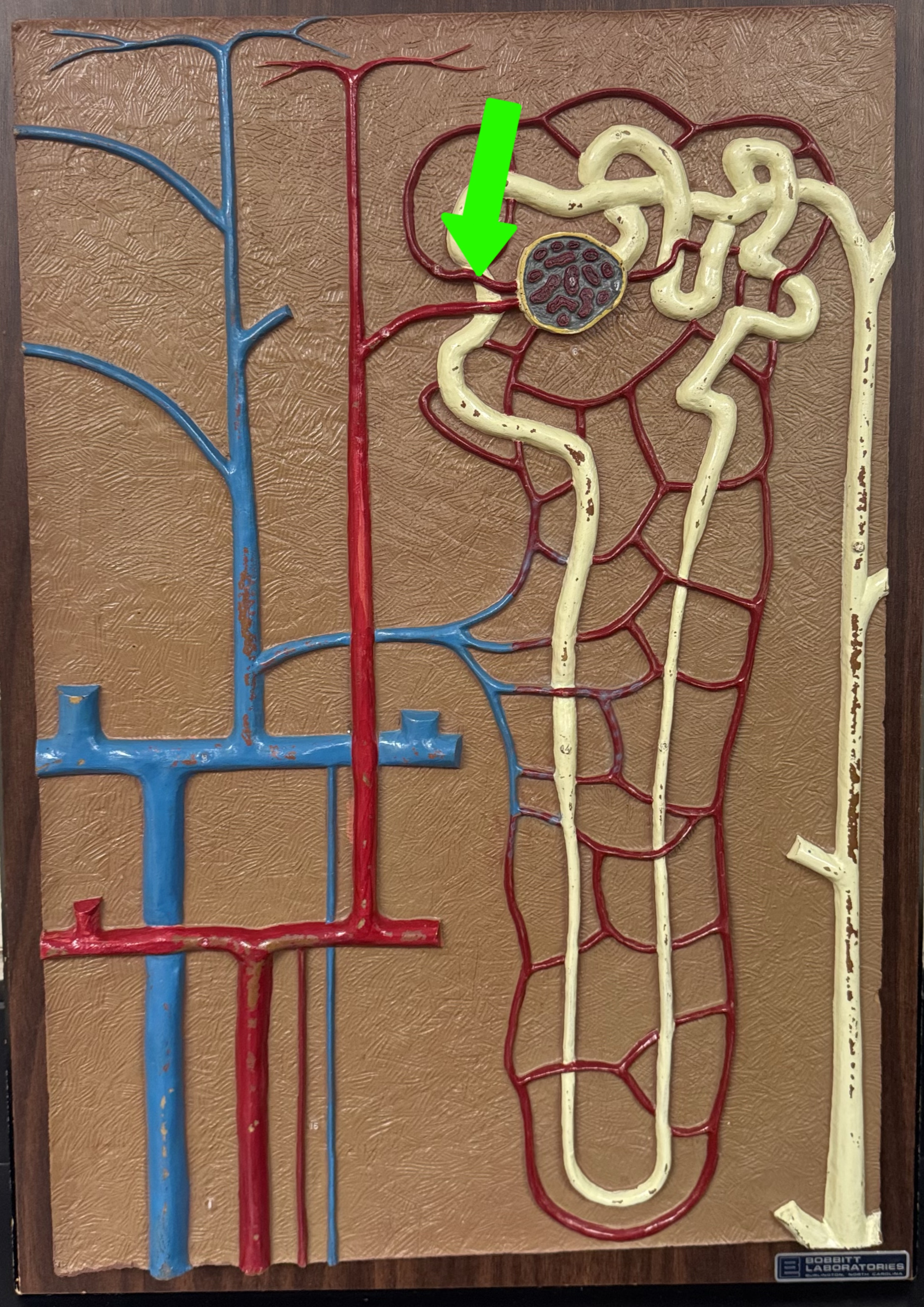
Glomerular capsule with glomerulus
What are these parts of the nephron?
Proximal convoluted tubule
What are these parts of the nephron?
Nephron loop
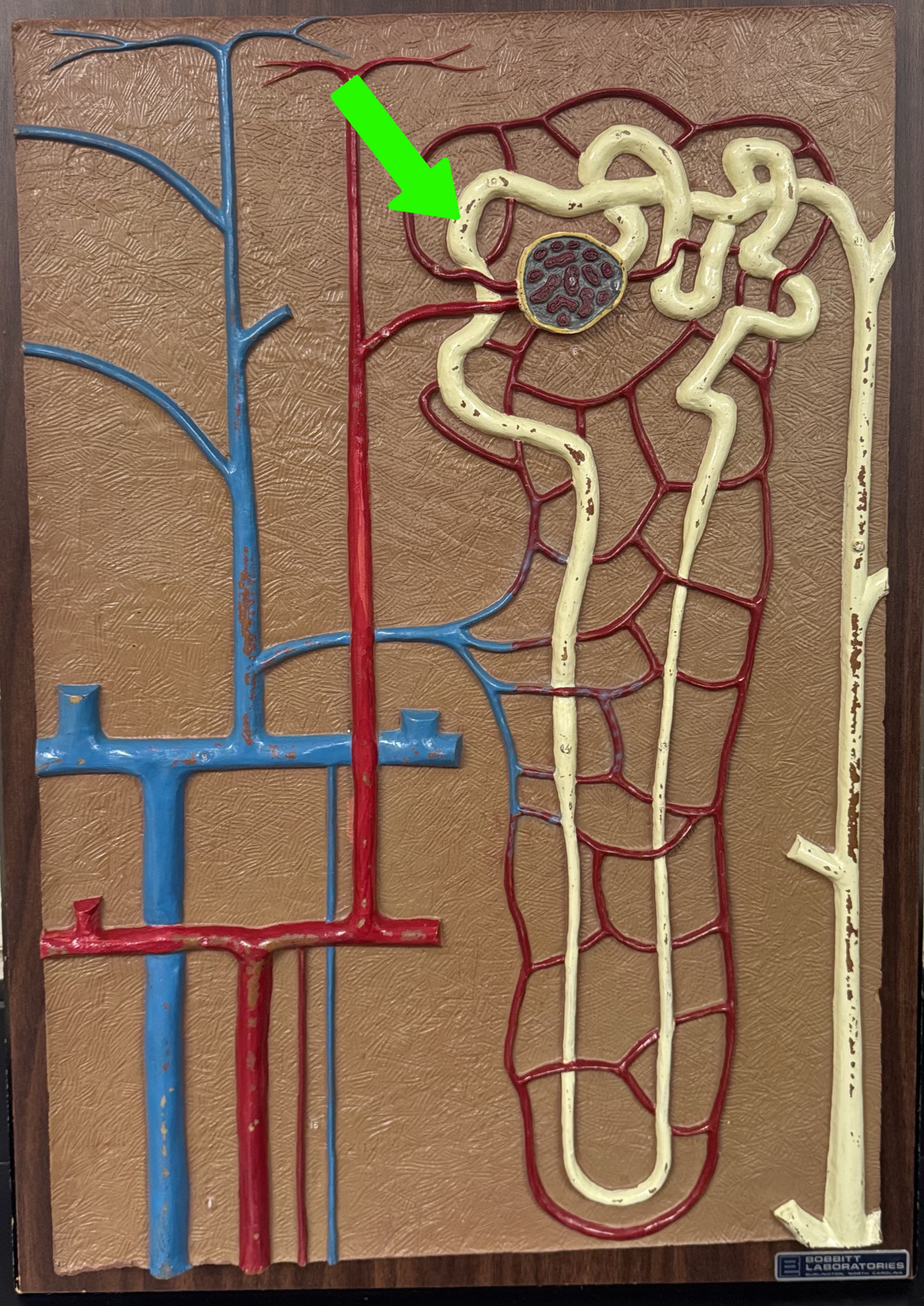
What are these parts of the nephron?
Distal convoluted tubule
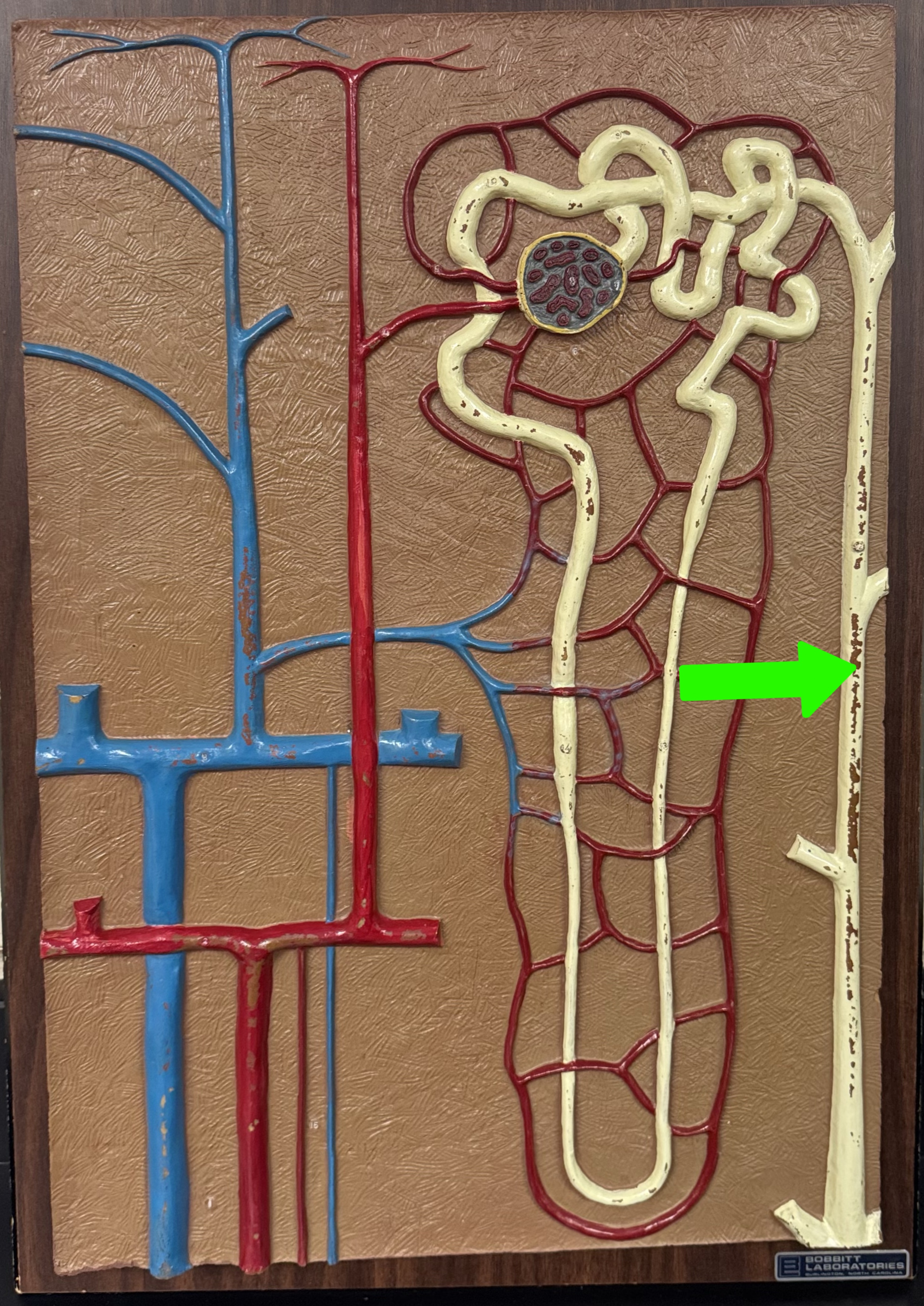
What are these parts of the nephron?
Collecting duct
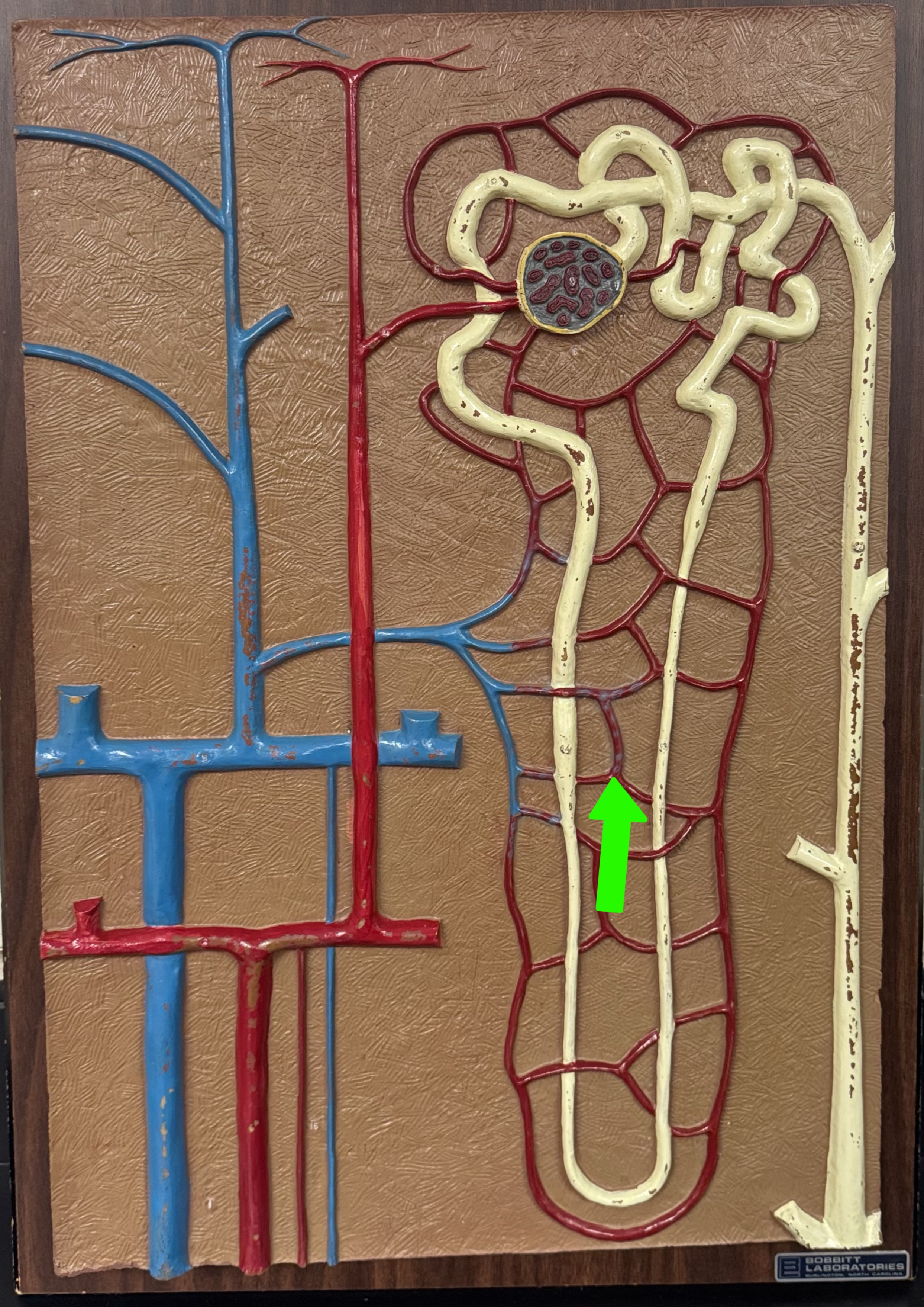
What are these parts of the nephron?
Peritubular capillaries
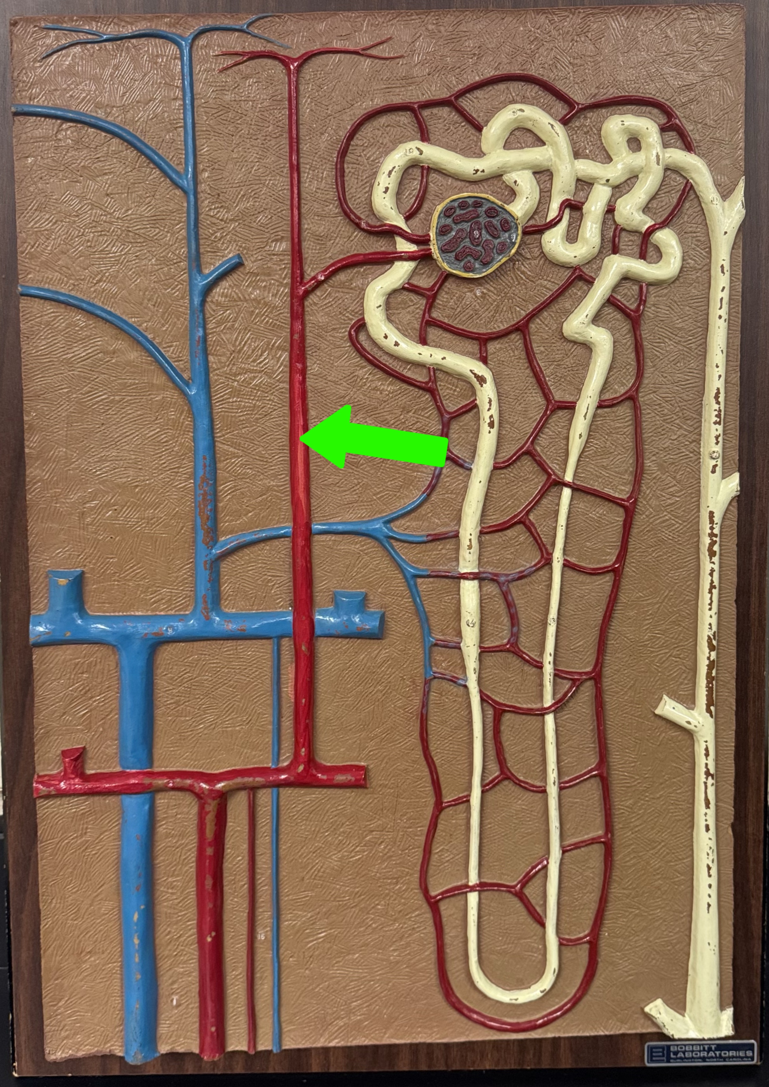
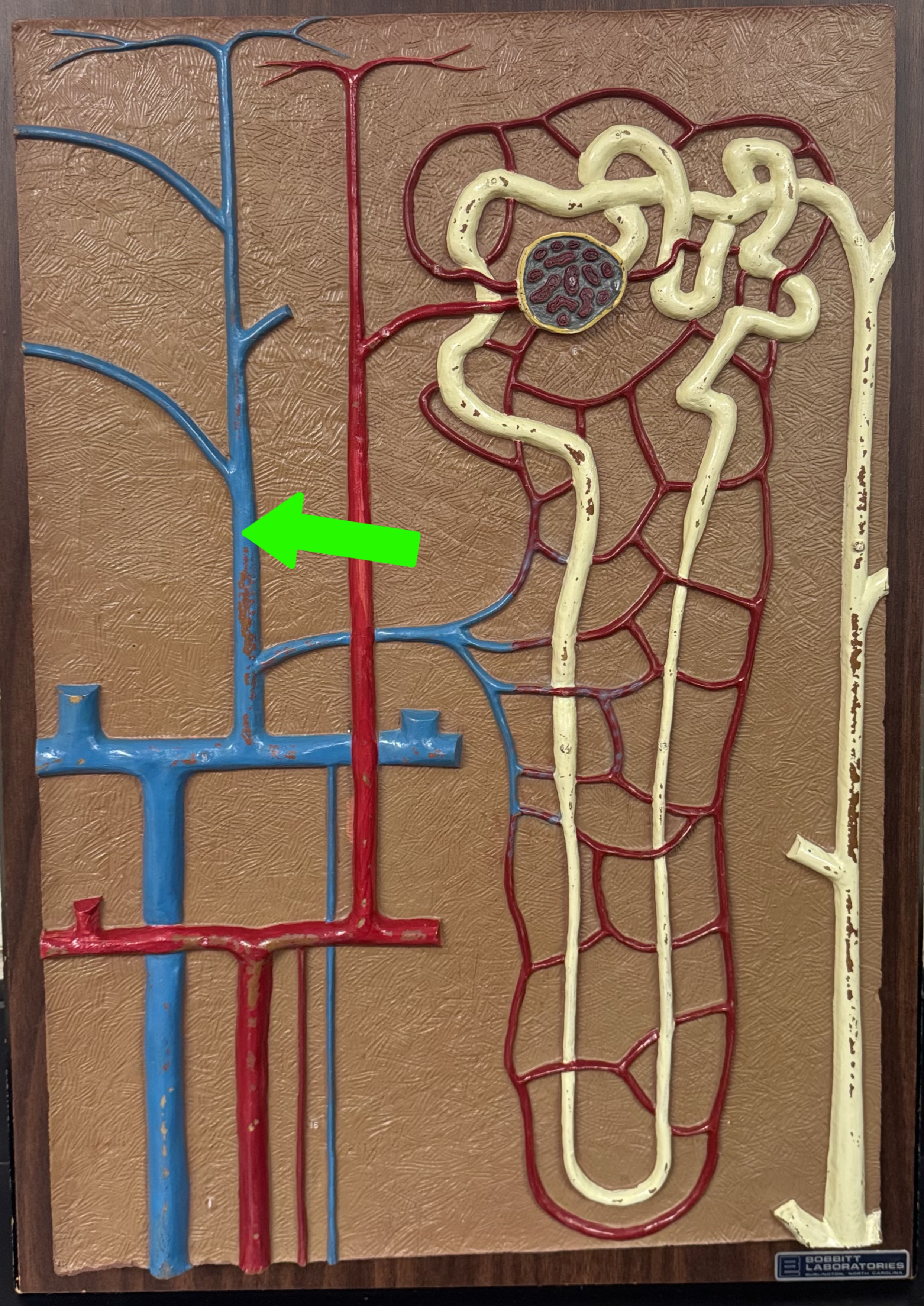
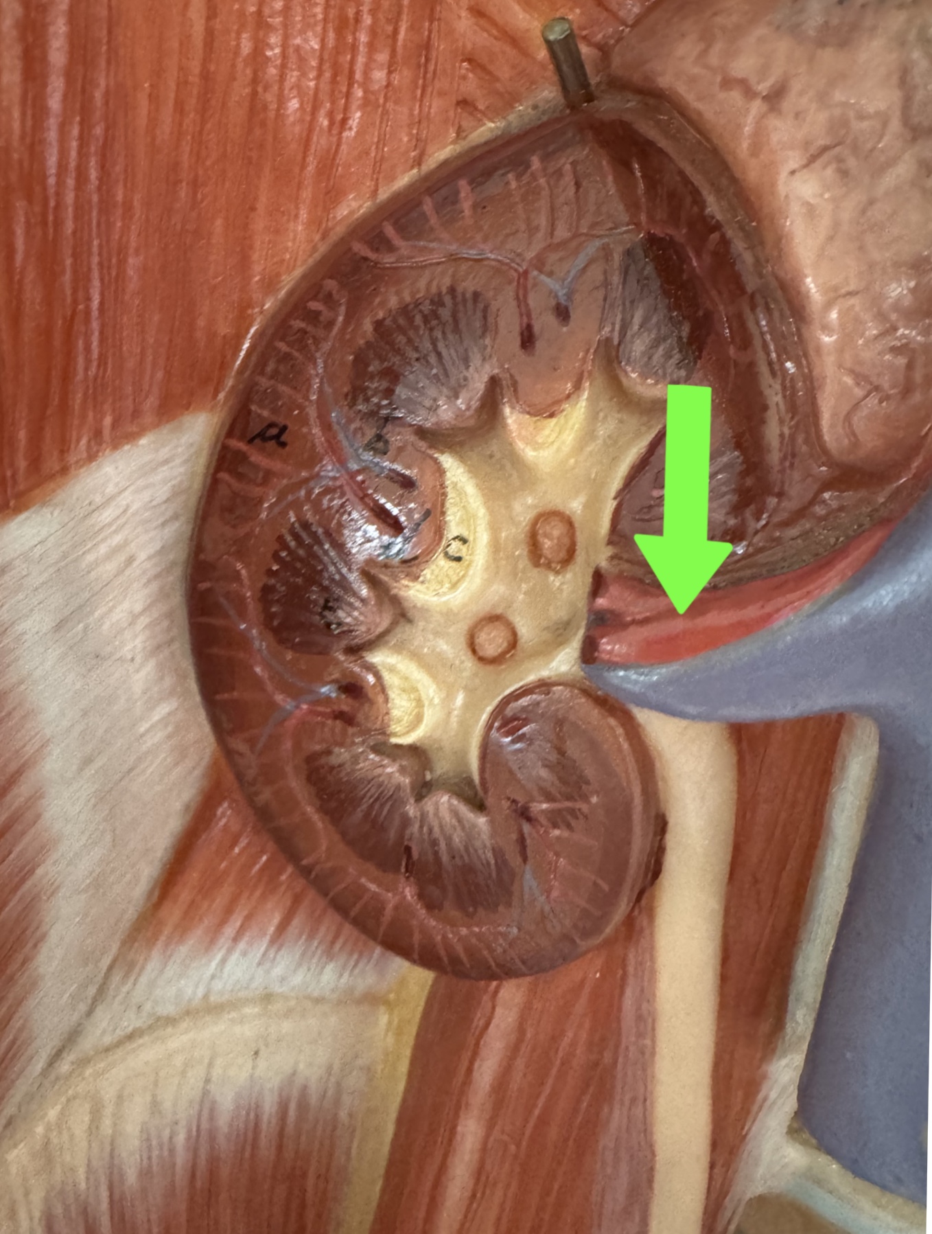
What is this
Renal artery
What are these parts of the nephron?
Renal vein
What are these parts of the nephron?
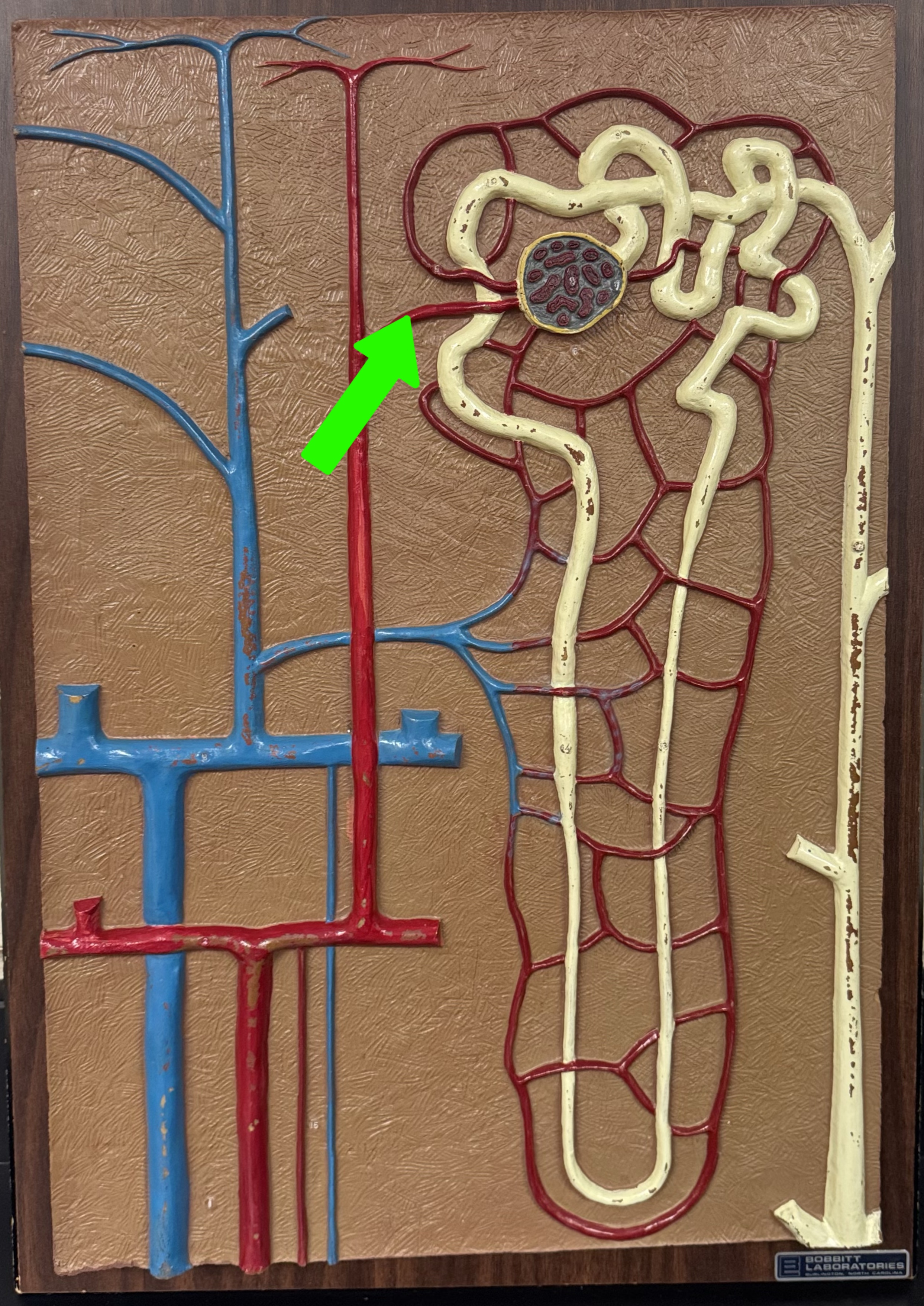
Afferent arteriole
What are these parts of the nephron?
Efferent arteriole
Where does filtration occur? Where does reabsorption and secretion occur?
Filtration: glomerular capsule and glomerulus
Reabsorption: PCT, Nephron loop
Secretion: DCT
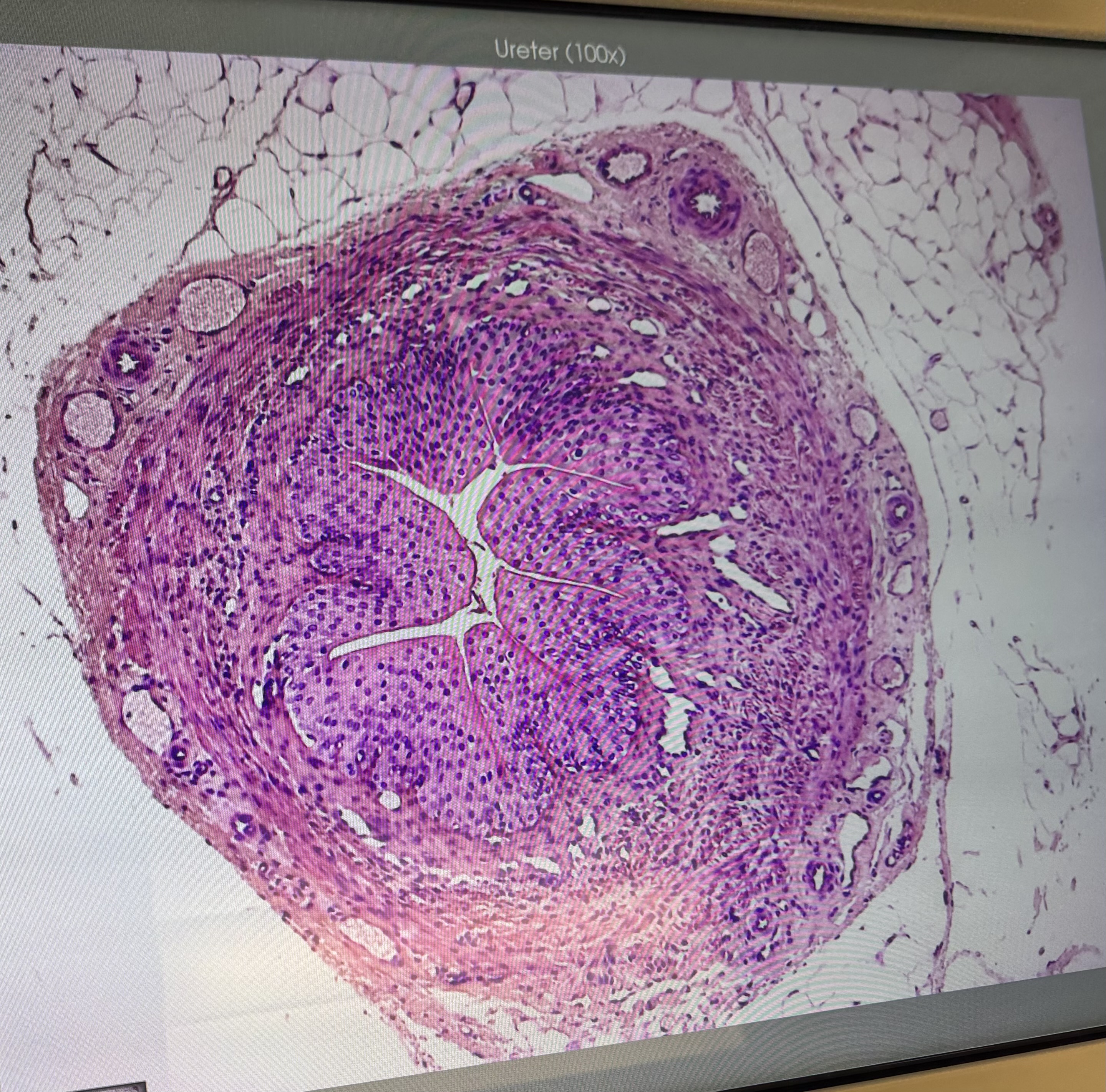
Ureter (#10012): what kind of ET?
Transitional epithelial tissue
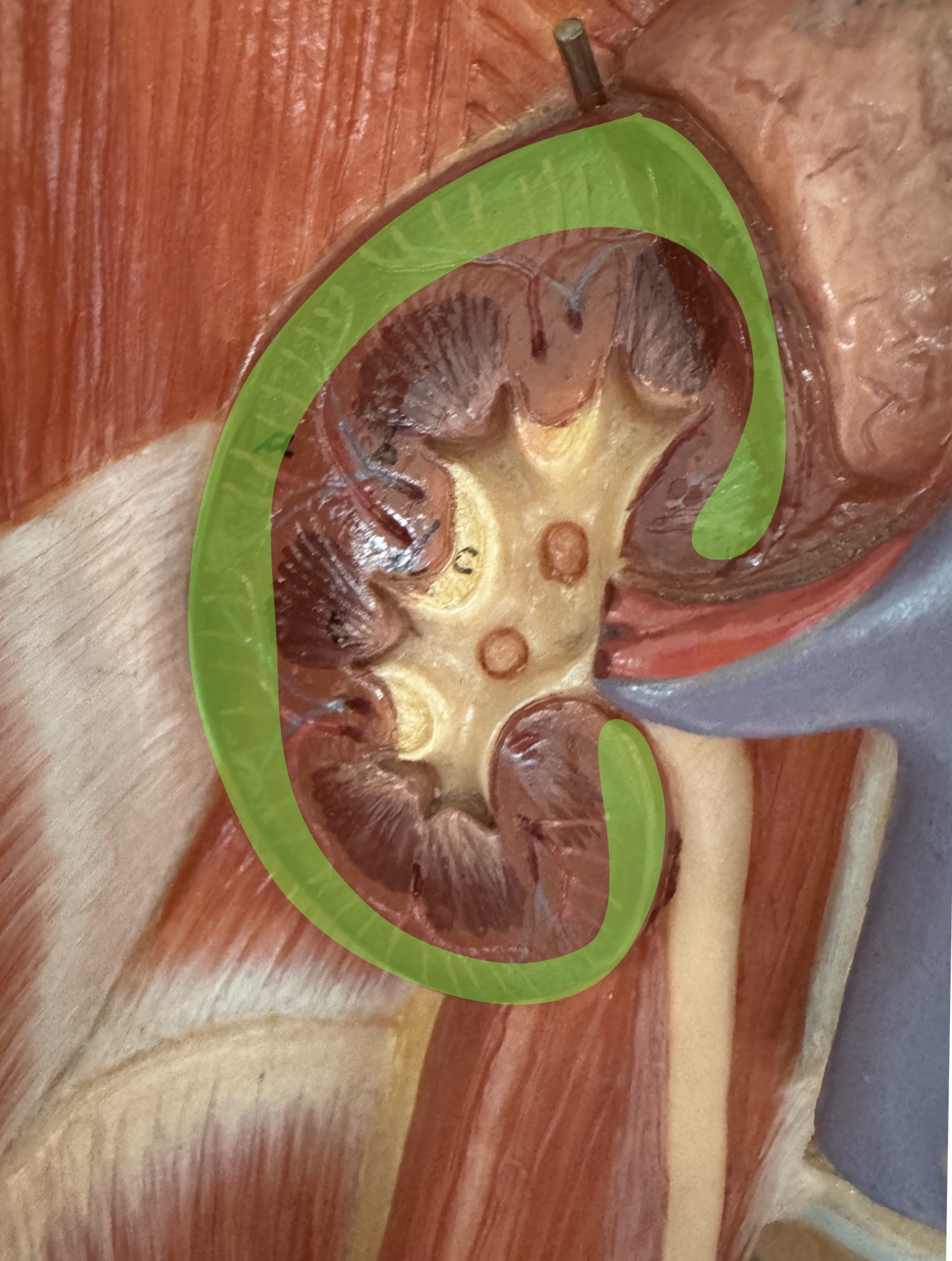
What is this
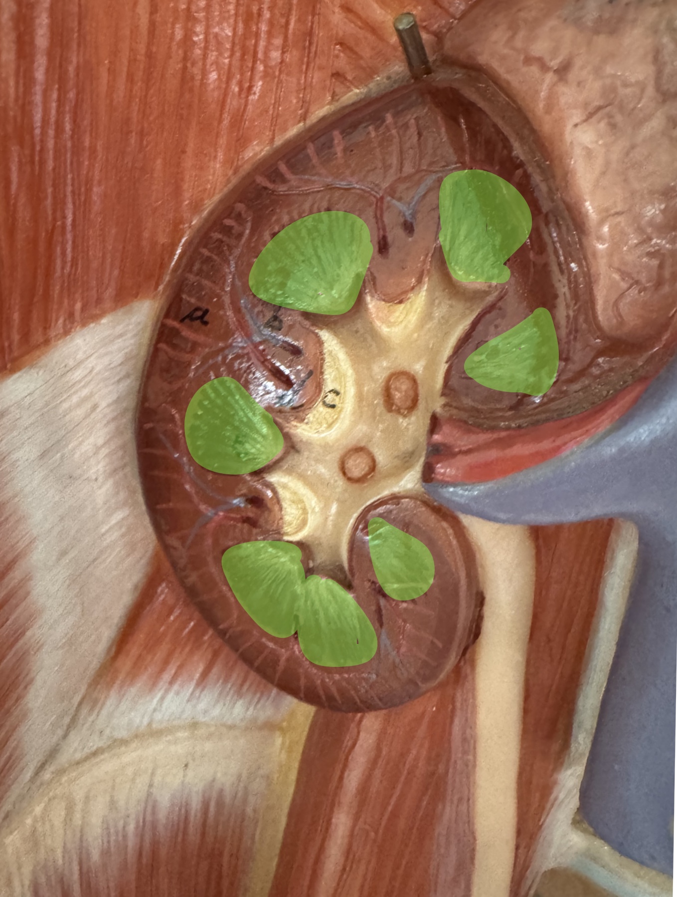
Renal medulla
What is this
Renal pyramids
What is this
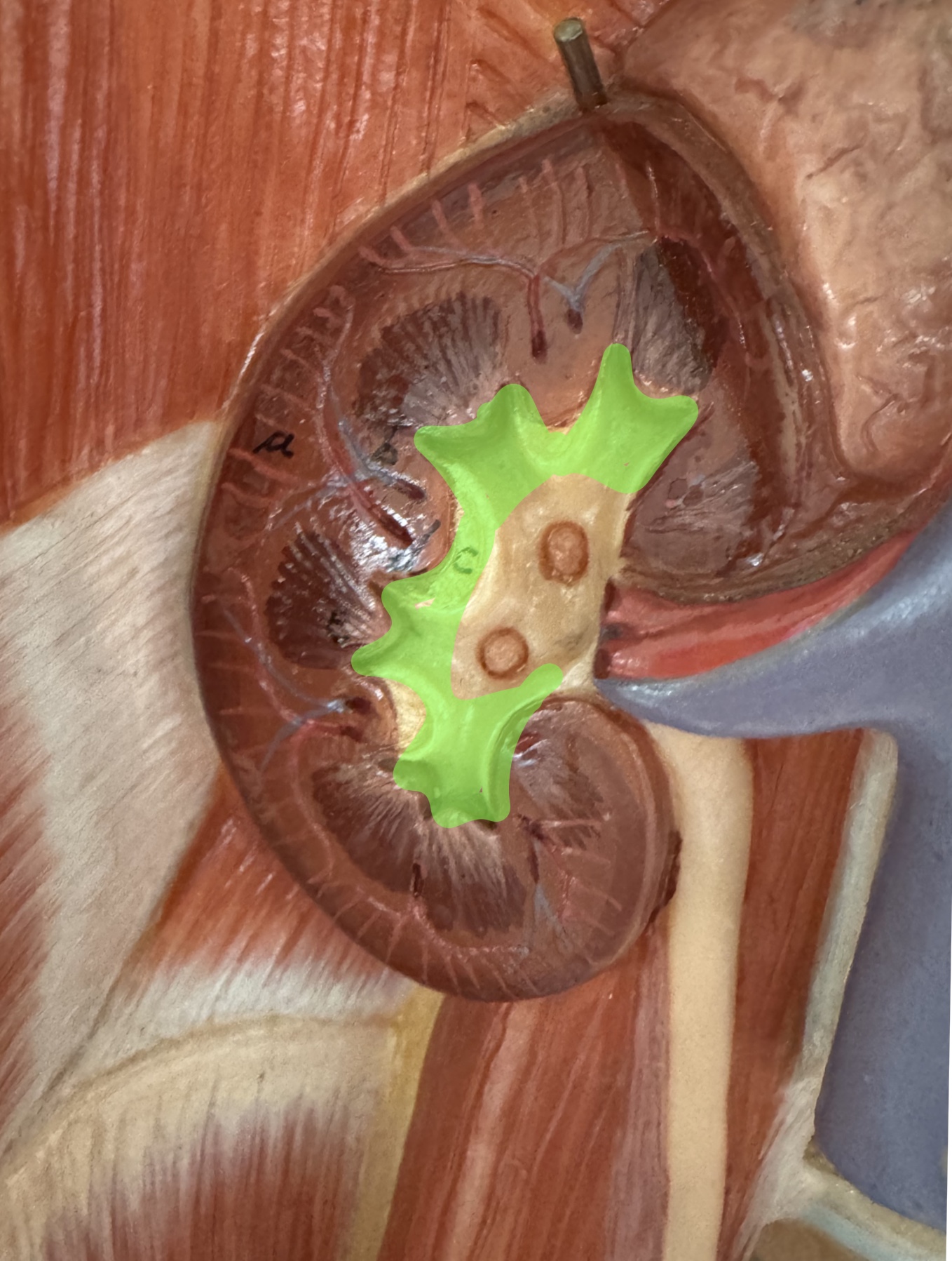
Renal calyces
What is this
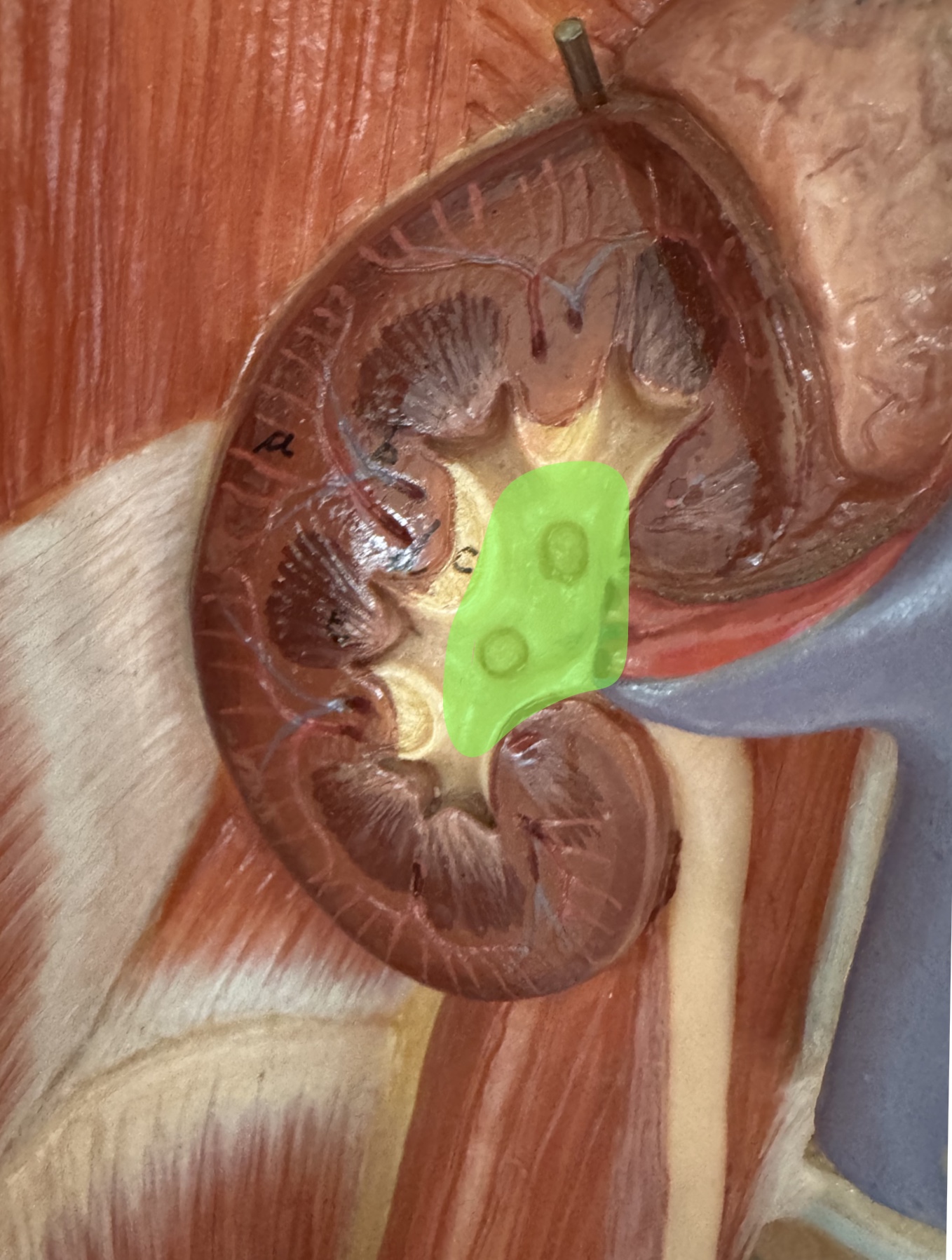
Renal pelvis
What is this
Renal artery
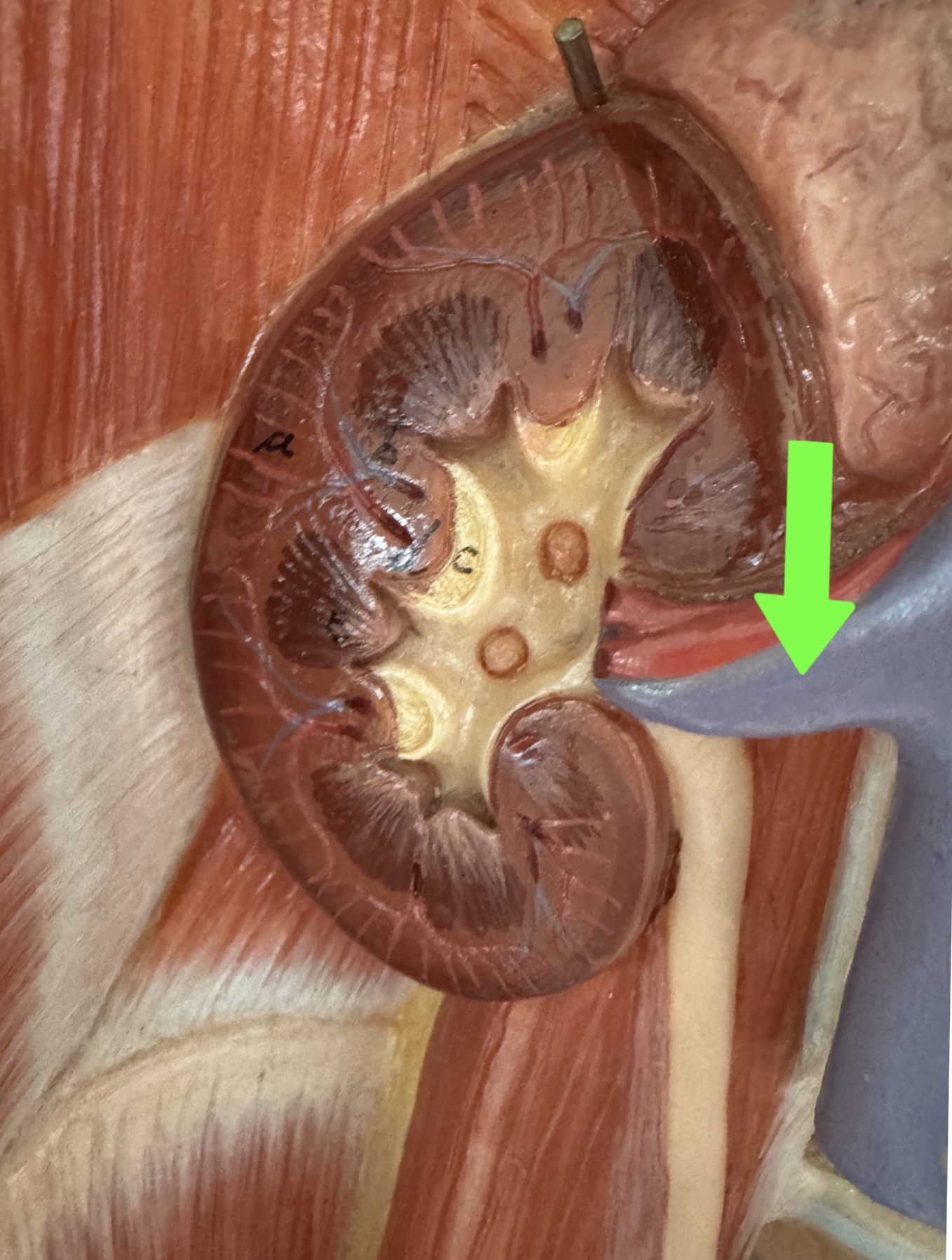
What is this
Renal vein
Vicky Reproductive Cadaver:
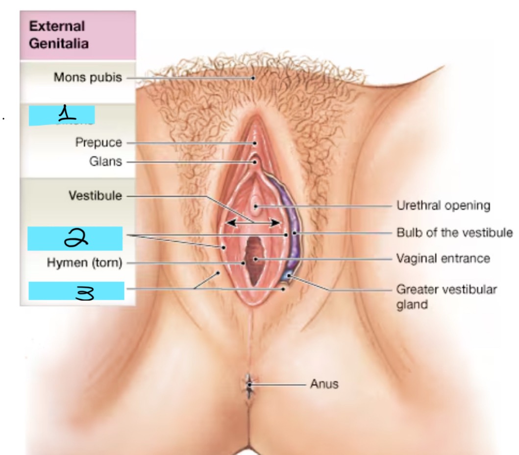
External genitalia:
clitoris
labia minora
Labia majora
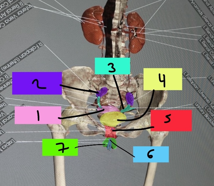
Vicky Reproductive Cadaver: 1-3
Uterus
R ovary
L fallopian/uterine tube

Vicky reproductive cadaver: 4
Urinary bladder

Vicky Reproductive Cadaver: 5-7
Vagina
Clitoris
Labia majora
Note the corpus luteum inside each ovary--what are these formed from and what important hormone do these release?
Ovulated egg; they release progesterone
Where is the vagina in relation to rectum and urethra?
Anterior to rectum and superior to urethra
Where is the uterus in relation to the bladder?
Superior and posterior to it
Turn on pelvic bones and make transparent, can you find the pelvic outlet?
an opening for the anus
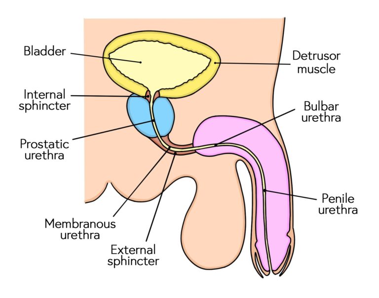
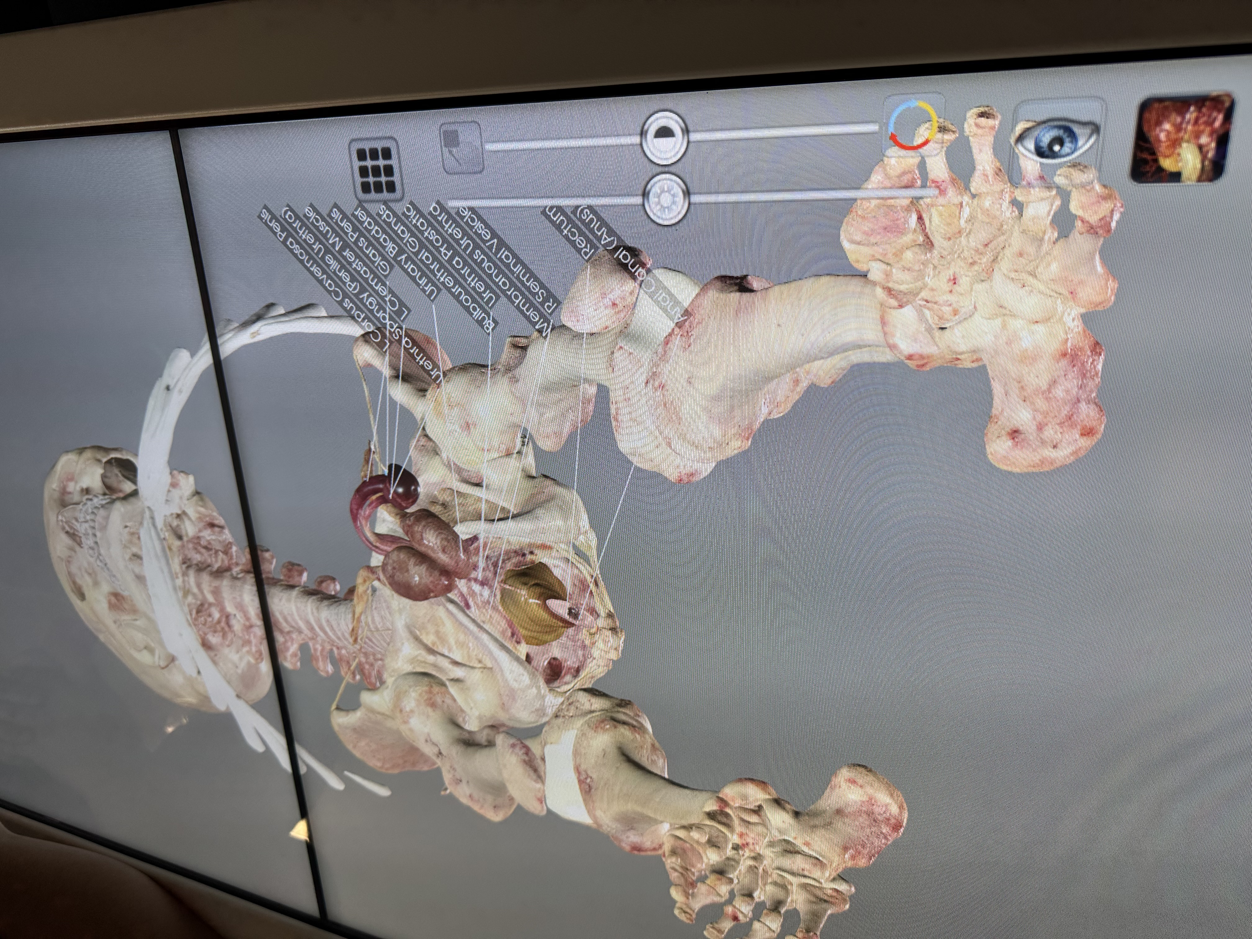
Urethra: note the many regions--which region is a shared pathway for semen and urine?
Penile urethra

Viktor Reproductive Cadaver: 1-3
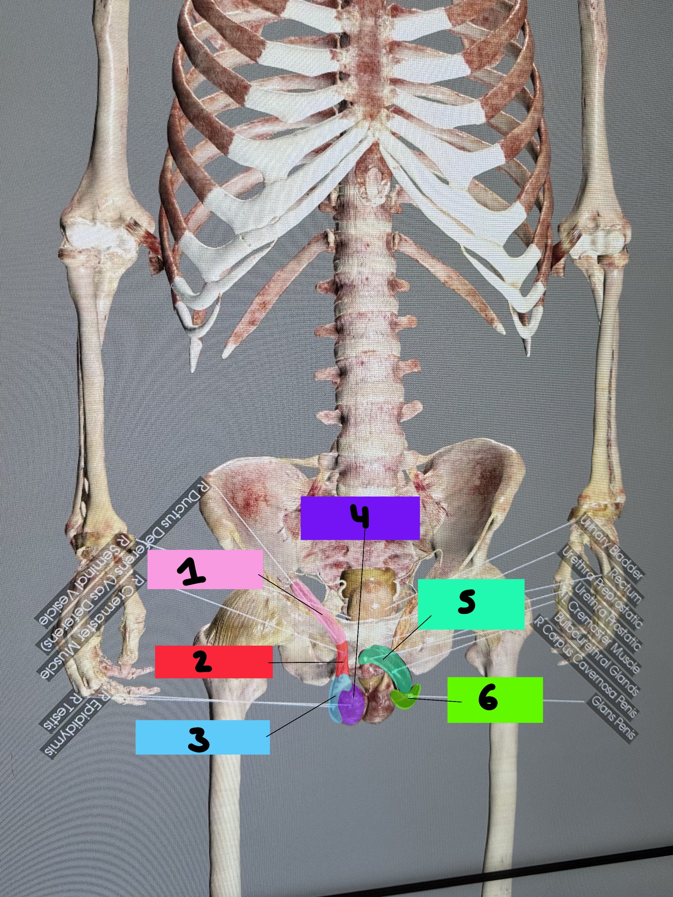
R ductus deferens
R Cremaster muscle
R epididymis

Viktor Reproductive Cadaver: 4-5
R testes
R corpus cavernosa penis
Viktor Reproductive Cadaver: 6
Glans penis

Viktor Reproductive Cadaver: 1-3
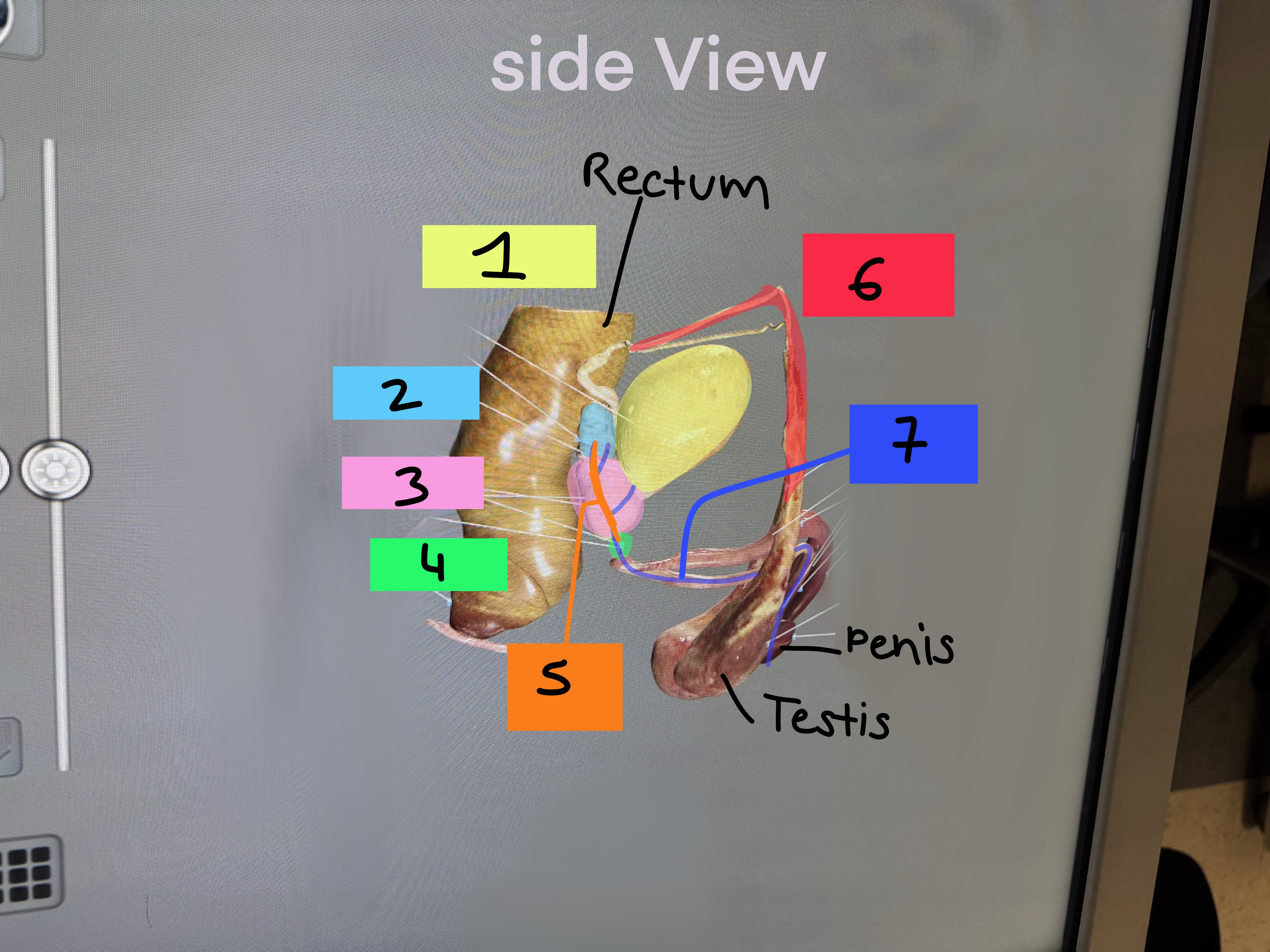
urinary bladder
Seminal gland/vesicle
Prostate gland
Viktor Reproductive Cadaver: 4-7
Bulbourethral gland
Ejaculatory duct
R ductus deferens
Urethra
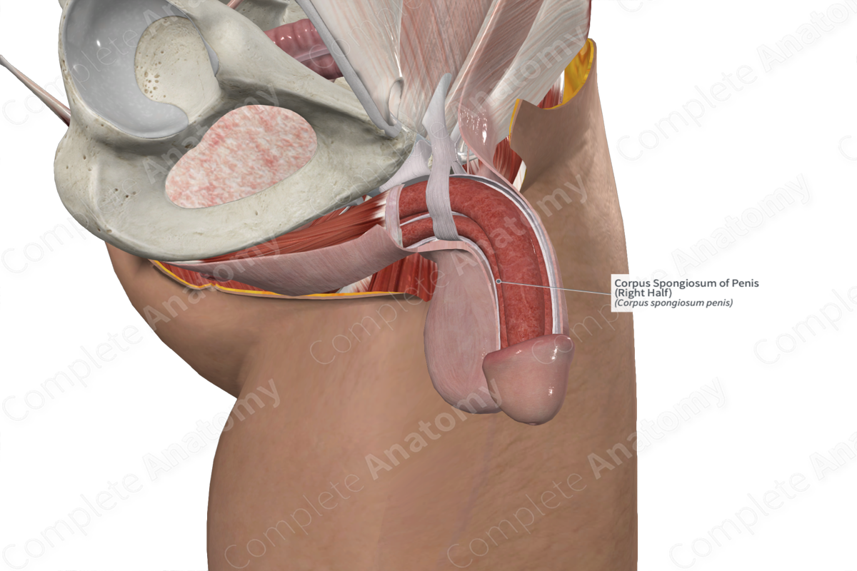
Corpus spongiosum: what is contained inside this erectile tissue?
Urethra
Where is the prostate relative to the urinary bladder?
Below it
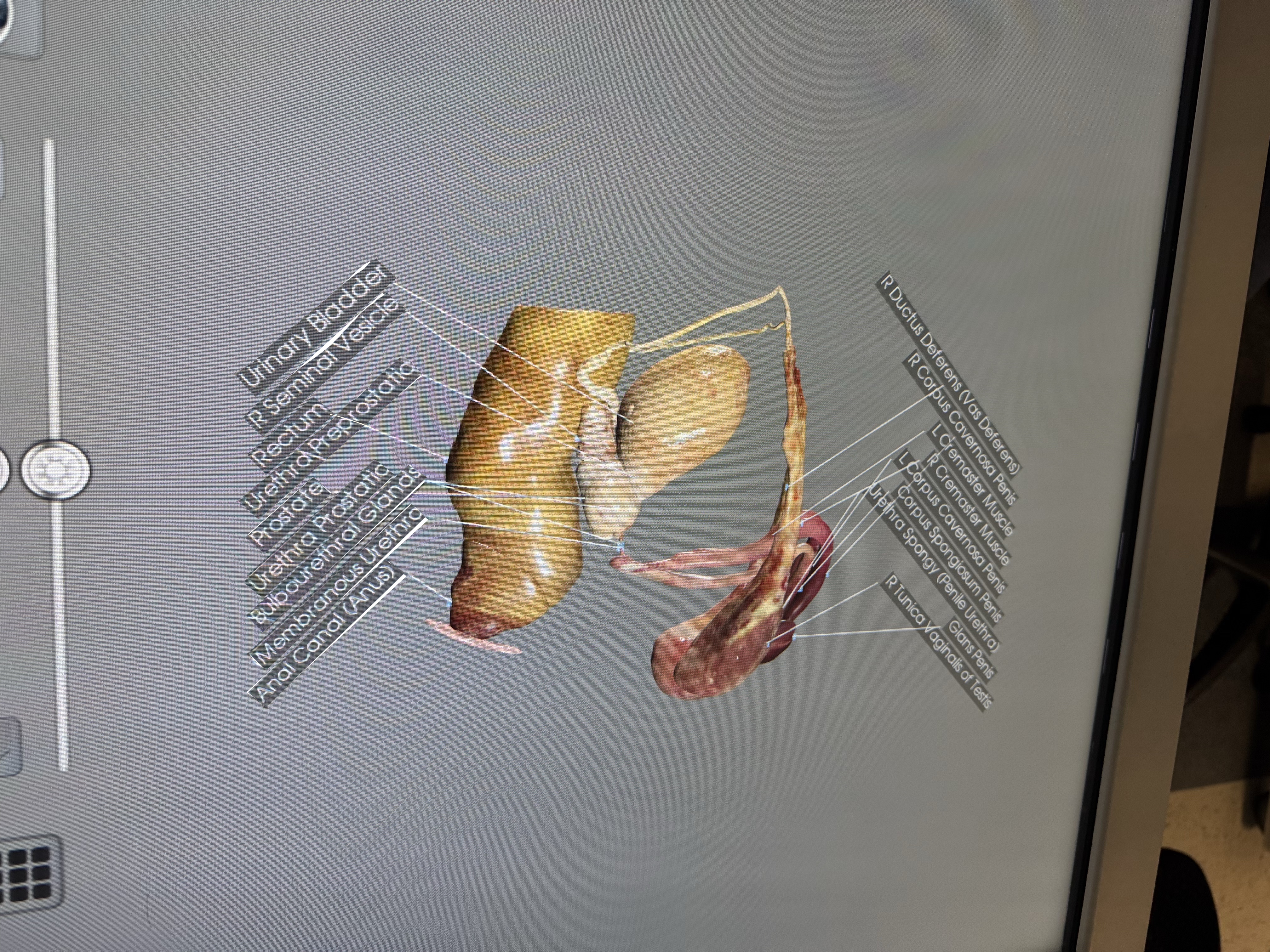
Where is the rectum relative to the seminal vesicles and prostate gland? (think about how a doctor may check a man’s prostate…)
Rectum is Behind it; digital rectal exam to palpate the prostate
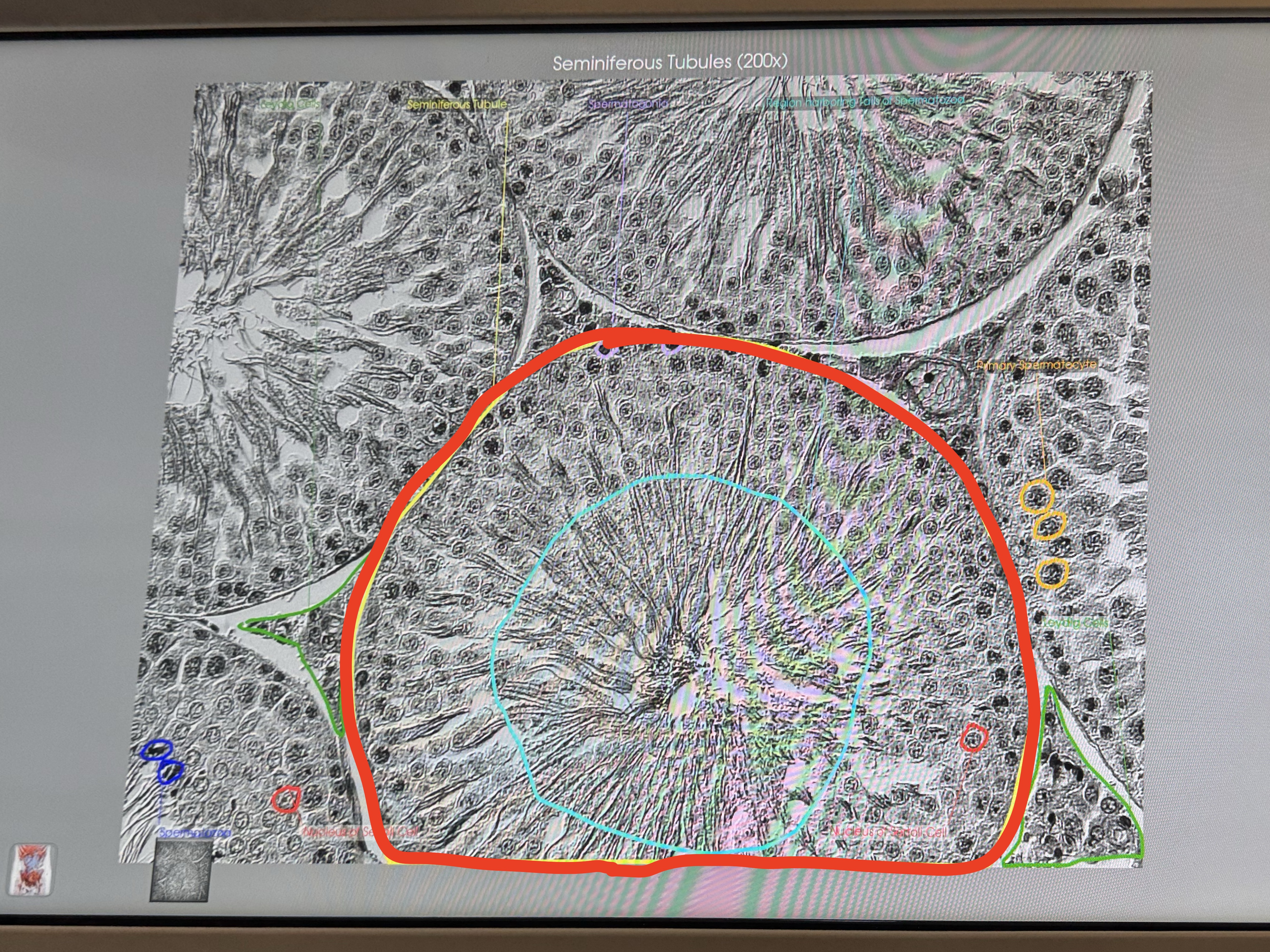
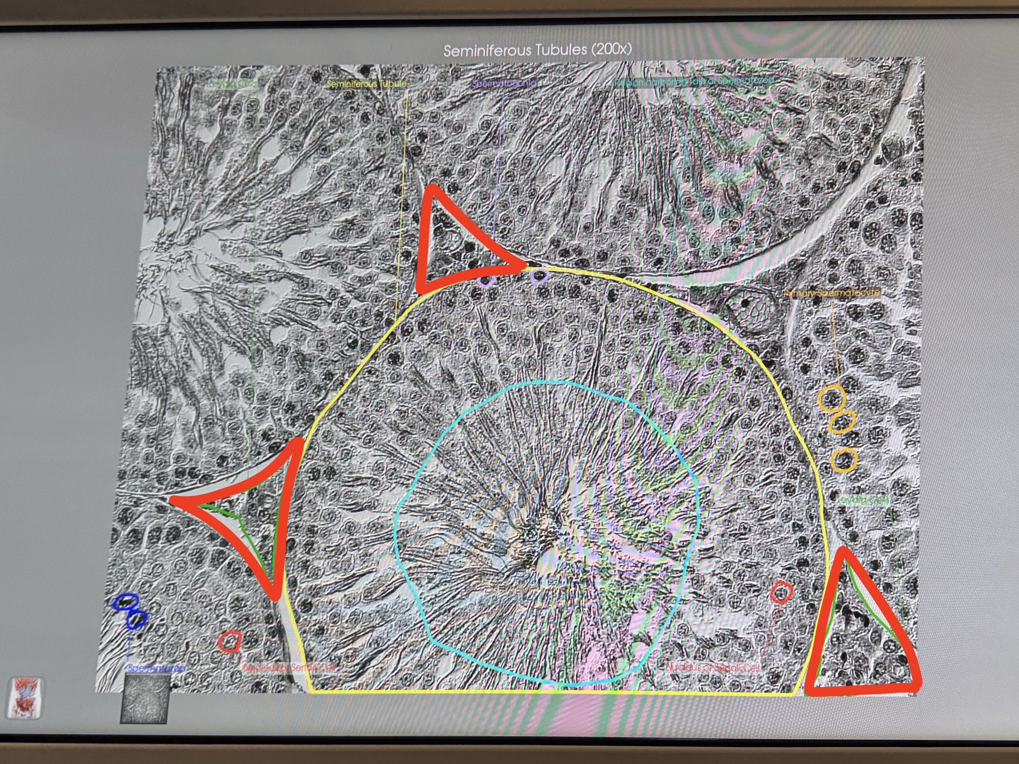
Testis: identity these structures
Seminiferous tubules
What are these?
interstitial endocrine (aka Leydig) cells
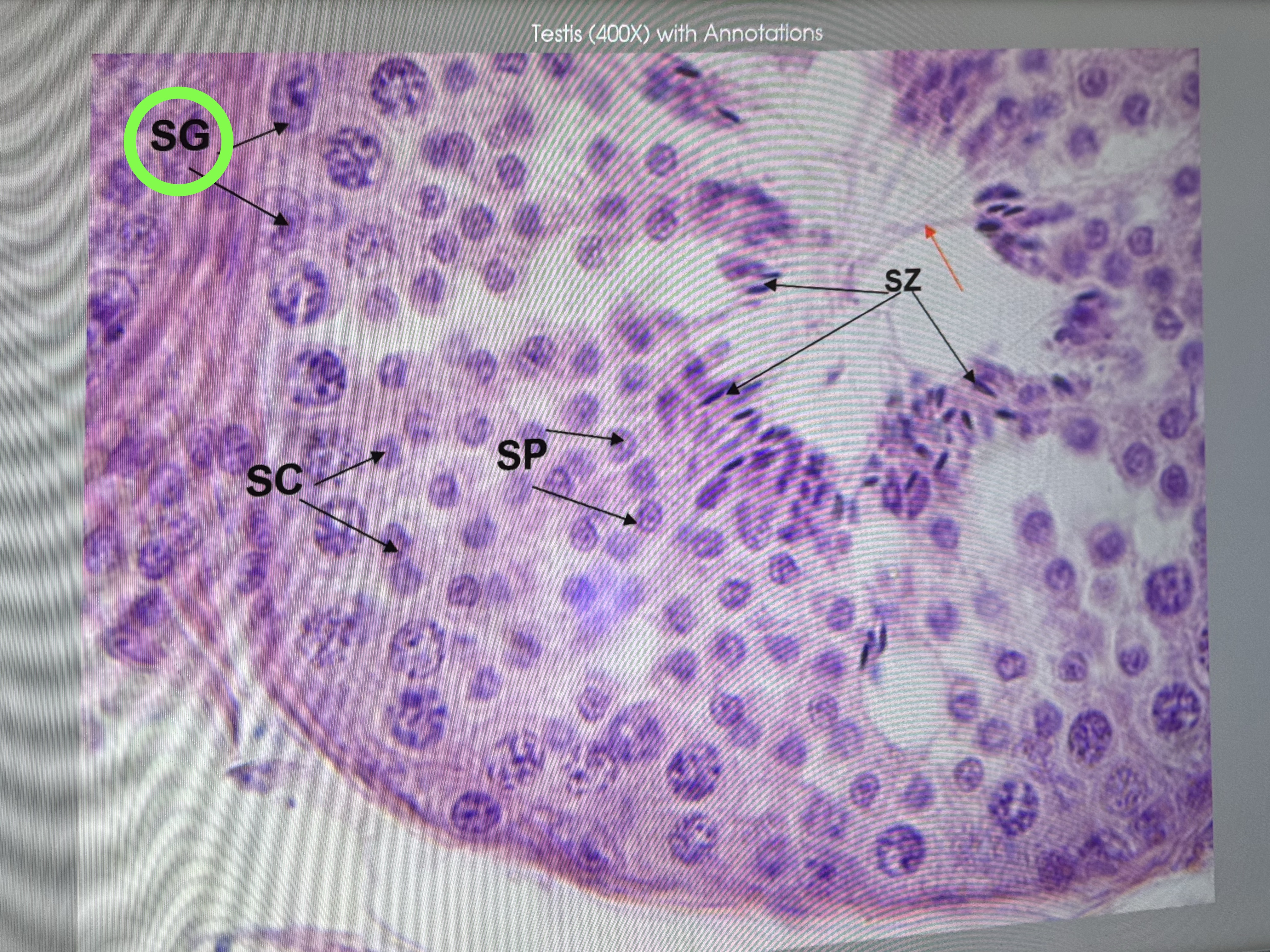
What is this inside the testes
Spermatogonia (sperm stem cells)