BIPN 102 DIGESTIVE SYSTEM — 20 FLASHCARDS Final
1/19
There's no tags or description
Looks like no tags are added yet.
Name | Mastery | Learn | Test | Matching | Spaced | Call with Kai |
|---|
No analytics yet
Send a link to your students to track their progress
20 Terms
What are the major anatomical organs of the digestive system and accessory organs?
Main tract:
Oral cavity
Esophagus
Stomach
Small intestine
Large intestine
Rectum
Accessory organs:
Salivary glands
Liver
Gallbladder
Pancreas
Image suggestion:
Lecture 18 — Slide/Page 4 (full digestive system anatomy diagram)
What are the key sphincters that regulate movement through the GI tract?
Upper esophageal sphincter
Lower esophageal sphincter
Pyloric sphincter
Ileocecal sphincter
Sphincter of Oddi
These regulate movement of food and digestive secretions between compartments.
Image suggestion:
Lecture 18 — Slide/Page 6 (sphincters and organ list)
What are the four fundamental processes of the digestive system?
Motility – movement of food via muscle contraction
Digestion – chemical + mechanical breakdown of food
Secretion – movement of substances from cells into lumen or ECF
Absorption – movement of nutrients from lumen into blood/ECF
Image suggestion:
Lecture 18 — Slide/Page 3 (Functions diagram)
Layers of the GI tract wall (lumen → outside) and their function?
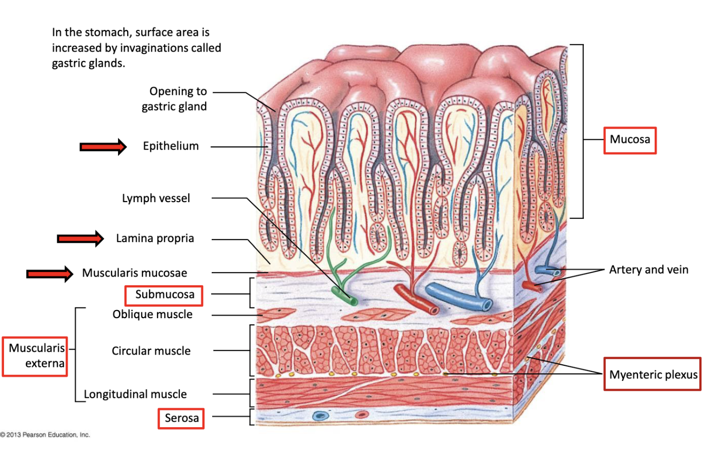
Mucosa — interact with food
(epithelium, lamina propria, muscularis mucosae)Submucosa — support & control
(blood vessels, glands, Meissner plexus)Muscularis externa — move food
(circular + longitudinal muscle, peristalsis)Serosa — protect & reduce friction
Logic:
Interact → Control → Move → ProtectImage suggestion:
Lecture 18 — Slide/Page 8 (GI wall layers diagram)
What are the main functions of motility in the digestive system?
Motility:
Moves food through GI tract
Mechanically mixes food to break it into smaller particles
Image suggestion:
Lecture 19 — Slide/Page 2 (motility diagram)
What are the two major types of GI smooth muscle contractions?
Tonic contractions
Sustained minutes to hours
Found in sphincters
Prevent backward movement
Phasic contractions
Last seconds
Responsible for peristalsis and segmentation
Image suggestion:
Lecture 19 — Slide/Page 4 (types of contraction graphs)
What are the major patterns of GI contraction?
Peristalsis → propels food forward
Segmentation → mixes contents
Migrating Motor Complex (MMC) → fasting “housekeeping” motility pattern
Image suggestion:
Lecture 19 — Slide/Page 5 (patterns list)
What is the Migrating Motor Complex (MMC) and when does it occur?
Cyclic motility pattern during fasting
Clears residual food and bacteria from stomach and intestine
Absence associated with:
gastroparesis
intestinal pseudo-obstruction
bacterial overgrowth
Image suggestion:
Lecture 19 — Slide/Page 6 (MMC diagram)
What do Interstitial Cells of Cajal (ICC) do in the GI tract?
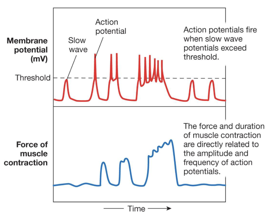
Autorhythmic pacemaker cells
Generate slow-wave potentials
Trigger action potentials when threshold is reached
Control rhythmic smooth muscle contractions
Image suggestion:
Lecture 19 — Slide/Page 3 (slow wave graph)
What are the two major plexuses of the Enteric Nervous System (ENS)?
Myenteric plexus
Controls motility
Submucosal plexus
Controls secretion
!!ENS can function independently of the CNS!!
Image suggestion:
Lecture 19 — Slide/Page 10 (ENS diagram)
What are short vs long digestive reflexes?
Short reflex
Integrated entirely in ENS (enteric nervous system)
Long reflex
Integrated in CNS
Example: cephalic reflex
Image suggestion:
Lecture 19 — Slide/Page 10 (ENS reflex diagram)
What are the three phases of digestion?
Cephalic phase
Gastric phase
Intestinal phase
Each phase regulates digestion through neural and hormonal signals.
Image suggestion:
Lecture 19 — Slide/Page 12 (phase overview)
What occurs during the cephalic phase of digestion?
Triggered by:
sight
smell
thought of food
Mechanism:
Long reflex begins in brain/medulla
Activates vagus nerve
Stimulates gastric secretion and motility
Image suggestion:
Lecture 19 — Slide/Page 13 (cephalic reflex diagram)
What occurs during the gastric phase?
!WHEN FOOD ENTERS THE STOMACH!
Stimulates:
HCl secretion
Pepsinogen secretion → pepsin
Protein digestion
Also includes protective mucus secretion.
Image suggestion:
Lecture 19 — Slide/Page 15 (gastric phase diagram)
What are the six gastric cell types and their functions?
Mucous cells
• secrete protective mucus
• function: protects stomach lining from acid
Parietal cells
• secrete HCl
• secrete intrinsic factor
• function: acid for digestion; intrinsic factor for B12 absorption
Chief cells
• secrete pepsinogen
• function: precursor → pepsin for protein digestion
Enterochromaffin-like (ECL) cells
• release histamine
• function: stimulates parietal cells → ↑ HCl
D cells
• secrete somatostatin
• function: inhibits gastric secretion
G cells (“gassy”)
• secrete gastrin
• function: stimulates acid secretion
Image suggestion:
Lecture 19 — Slide/Page 16 (gastric gland cell diagram)
What hormones regulate the intestinal phase?
CCK (Cholecystokinin)
stimulates pancreatic enzyme secretion
gallbladder contraction
Secretin
stimulates pancreatic bicarbonate secretion
GIP (Glucose Inhibitory Peptide)
stimulates insulin secretion, lowers gastric emptying
GLP-1
stimulates insulin release and promotes satiety, lowering glucagon/gastric function
Image suggestion:
Lecture 20 — Slide/Page 11 (intestinal hormone table)
What is the function of bile and the liver in digestion?
Liver
produces bile
Bile salts
emulsify lipids → increase fat digestion
Gallbladder
stores and concentrates bile
Image suggestion:
Lecture 20 — Slide/Page 12 (bile system diagram)
What are the pancreatic exocrine secretions and cells?
Acinar cells
secrete digestive enzyme zymogens
Duct cells
secrete bicarbonate (HCO₃⁻)
Bicarbonate neutralizes stomach acid in the duodenum.
Image suggestion:
Lecture 20 — Slide/Page 7 (pancreas cell diagram)
How are carbohydrates absorbed in the small intestine?
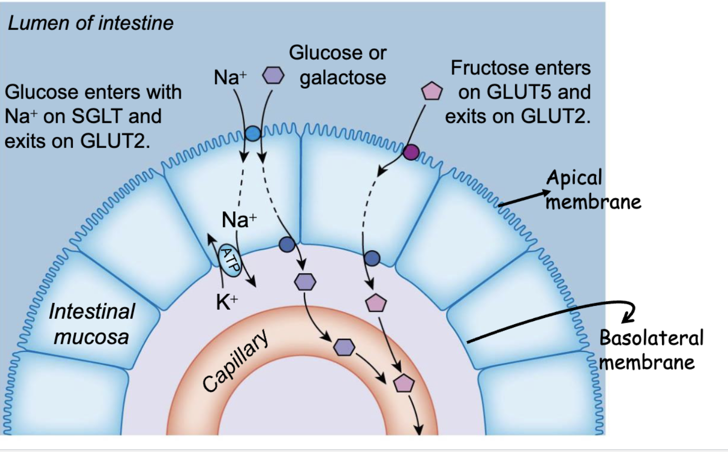
Glucose + galactose
secondary active transport via SGLT
Fructose
facilitated diffusion via GLUT5
All exit enterocytes via GLUT2.
Image suggestion:
Lecture 20b — Slide/Page 3 (carbohydrate absorption diagram)
How are peptides and amino acids absorbed in the intestine?
Amino acids
Na⁺ cotransport
Di- and tripeptides
H⁺ cotransport via PepT1
Inside cell → broken into amino acids → enter blood.
Image suggestion:
Lecture 20b — Slide/Page 6 (peptide absorption diagram)