6. tissues and organs
1/23
There's no tags or description
Looks like no tags are added yet.
Name | Mastery | Learn | Test | Matching | Spaced | Call with Kai |
|---|
No analytics yet
Send a link to your students to track their progress
24 Terms
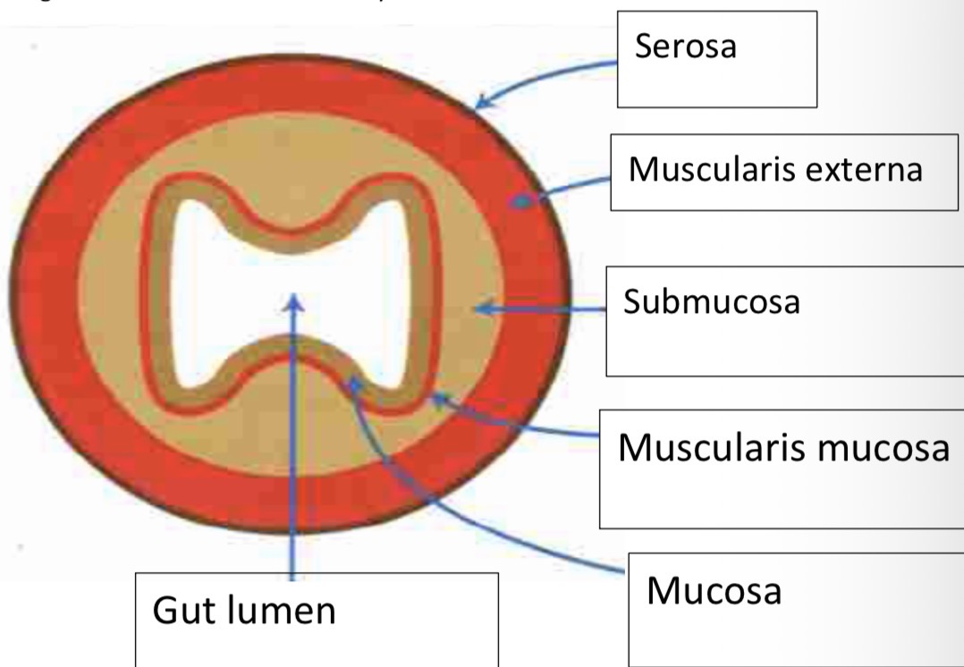
Serosa Structure and function
Outer layer of connective tissue – provides a very thin protective and supportive lining
Muscularis externa Structure and function
Consists of an outer layer of longitutinal muscle – contraction causes pendular movement
And can inner layer of circular muscle – contraction causes local constrictions which causes peristaltic waves to help push food along.
Both muscle help churn and mix the food
Submucosa
Composed of connective tissue and many blood vessels and lymphatic vessels which transport absorbed food products
Muscularis mucosa
Thin layer of muscle. Important in moving the villi to increase contact with the food in the lumen. Small strands of muscle extend into each villus in the mucosa and contract to create a wafting motion.
Mucosa
Layer in contact with the food in gut lumen. Increased surface area due to the presence of villi and microvilli. Villi – finger like extensions of the muscoa layer Micovilli – extensions of the columnar epithelium
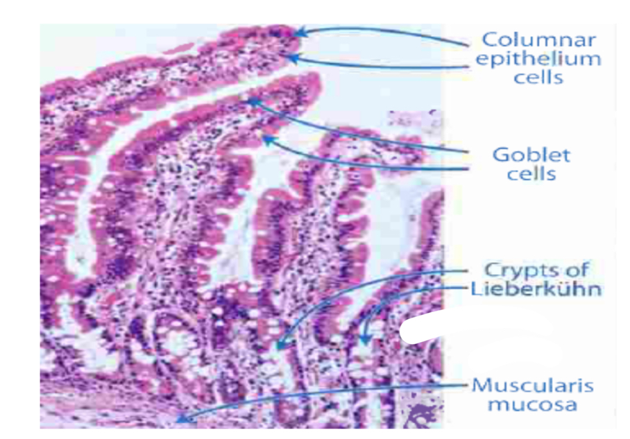
How do columnar epithelial cells have surface area extended
Prescient of numerous microvilli give brush border appearance
Why are the columnar epithelial cells rich in mitochondria?
Produces ATP for active transport of absorbed food molecules
Describe how the products are absorbed into the blood. E.g Glucose, amino acids, lipids.
Involves active transport and diffusion involving protein carrier molecules.
Glucose and amino acids are taken into the epithelial cells by active transport and then diffuse into the blood capillary network.
Products of fat digestion, fatty acids and glycerol are transported by lacteals which are part of the lymphatic system. Move by diffusion
How is pinocytosis involved?
e.g. in babies, antibodies can mass from breast milk into their blood.
Goblet cells location function
Within the columnar epithelial cells
Secrete mucus
Capillaries Location Function
Within the villi
Transport amino acids and glucose
Lacteal Location Function
Within the villi
Transport products of fat digestion/fatty acids and glycerol to lymphatic system
Crypts of LieberkÜhn Location Function
Intestinal glands that lie embedded in the tissue between villi
Cells continually divide to produce new cells that form the columnar epithelium
Paneth cells Location Function
Base of the crypts
Antimicrobial function in protecting their neighbouring actively dividing cells
leaf diagram
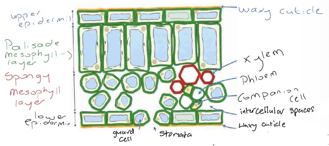
Upper epidermis
Protective layer – no chloroplasts. Covered by a waxy cuticle which prevents water loss and is transparent allowing light to pass through to PML
Palisade mesophyll
Photosynthesising layer. Regularly arranged and tightly packed = increase S.A.
Contains lots of chloroplasts to maximise light absorption for photosynthesis
Spongy mesophyll
Cells loosely and irregularly arranged – Increase S.A for gas exchange. Contain intercellular air spaces that encourage diffusion of gases. Do not photosynthesise
Vascular tissue
Xylem and Phloem.
Lower epidermis
Contains waxy cuticle – thinner than upper. Less transpiration occurs here.
Stomata
Pores. Close at night. Allow gases to enter or leave, including water vapour
Guard cells
Possess chloroplasts. Involved in opening and closing of stomata. When not turgid they close to prevent water loss