Unit 5- Sono Eval Embryo: 48-66
1/28
There's no tags or description
Looks like no tags are added yet.
Name | Mastery | Learn | Test | Matching | Spaced | Call with Kai |
|---|
No analytics yet
Send a link to your students to track their progress
29 Terms
In the embryonic cranium, what is shown in the 6th week of gestation?
Prosencephalon
Mesencephalon
Rhombencephalon
what part of the embryo cranium = forebrain
prosencephalon
what part of the embryo cranium = midbrain
mesencephalon
what part of the embryo cranium = hindbrain
rhombencephalon
What is the Rhombencephalon?
Cystic rhomboid fossa
Imaged routinely from 8-11 weeks
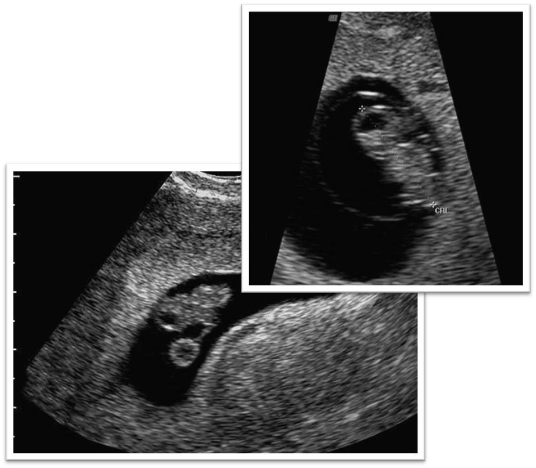
What are these images showing?
Rhombencephalon
What is this image showing?
Rhombencephalon
What is the embryonic cranium?
seen @ 9 weeks gestation
Cerebral hemispheres
Divided by cerebral fill
Lateral ventricles completely fill cerebral vault
Echogenic chorid plexus fills lateral ventricles

What is the purple line
cerebral pole
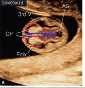
What are the red circles
choroid plexus aka lateral ventricles
what does the choroid plexus produce
spinal fluid
what does the 3rd ventricle do
helps transport spinal fluid
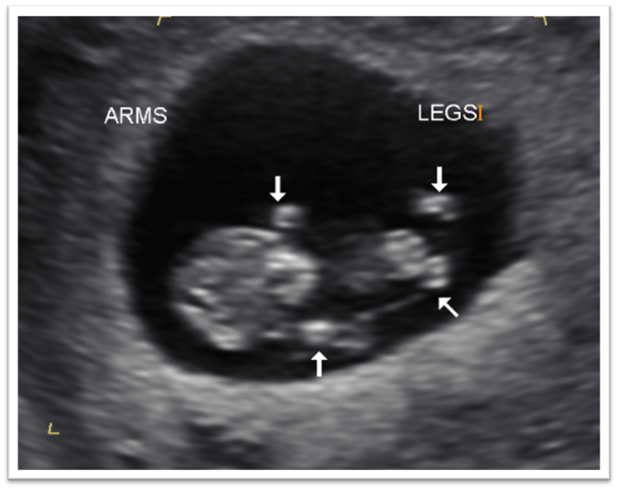
What should be seen on ultrasound at 10-12 weeks?
Head and trunk well defined
Arms and legs formed
Amniotic cavity expanding
Yolk sac usually disappears by 12 weeks
External genitalia appear similar regardless of gender
May see a functional hernia at abdominal cord insertion
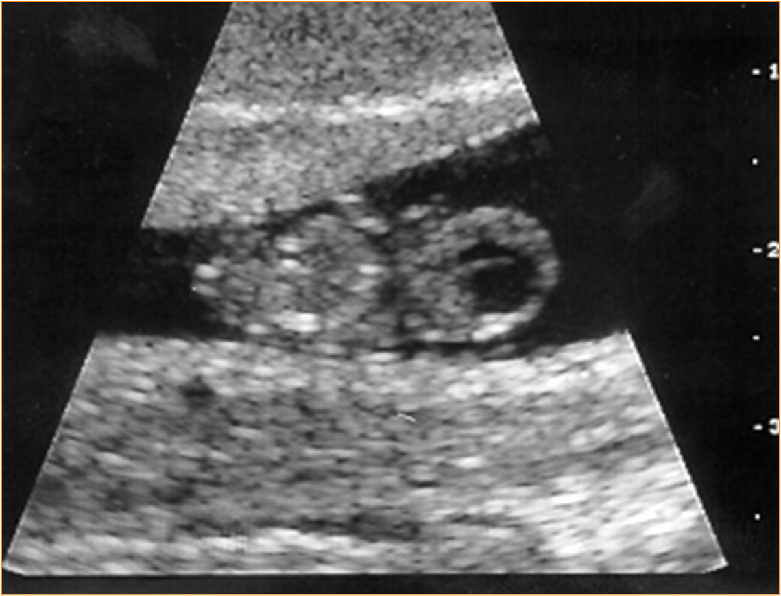
What is this image showing?
Gestation age 9 W 6 D
When does a physiologic herniation of bowel occur?
Bowel normally migrates into the base of the umbilical cord between 8 & 12 menstrual weeks
What is the US of physiologic bowel herniation?
Echogenic mass at base of umbilical cord
Mass should not measure >7mm at any gestational age
Mass should never measure <4mm
When does a physiologic bowel herniation return to abdomen?
Bowel returns to abdominal cavity by 12 menstrual weeks
Echogenic mass no longer visualize
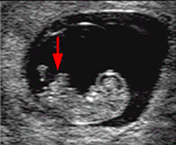
What is this image showing?
Physiologic Herniation of Bowel
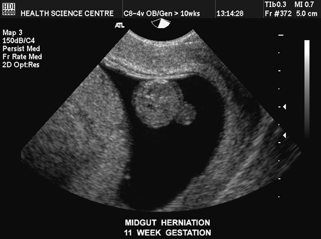
What is this image showing?
Early midgut herniation
What can a physiologic herniation of bowel be confused with?
Omphalocele
Gastroschisis
What is the first organ to function?
Heart
When does the heart start beating?
approximately 35 days (5 weeks)
At what week does the heart have its adult configuration?
End of 8th week
When should cardiac activity always be seen?
49 menstrual(gestational) days - 7 weeks
TV- 5mm CRL (5-6 weeks)
TA- 9mm CRL (7 weeks)
7mm(TV) is discriminatory size
What is the range of heart rates throughout pregnancy? (1st trimester)
90 - 170 bpm
140 bpm average
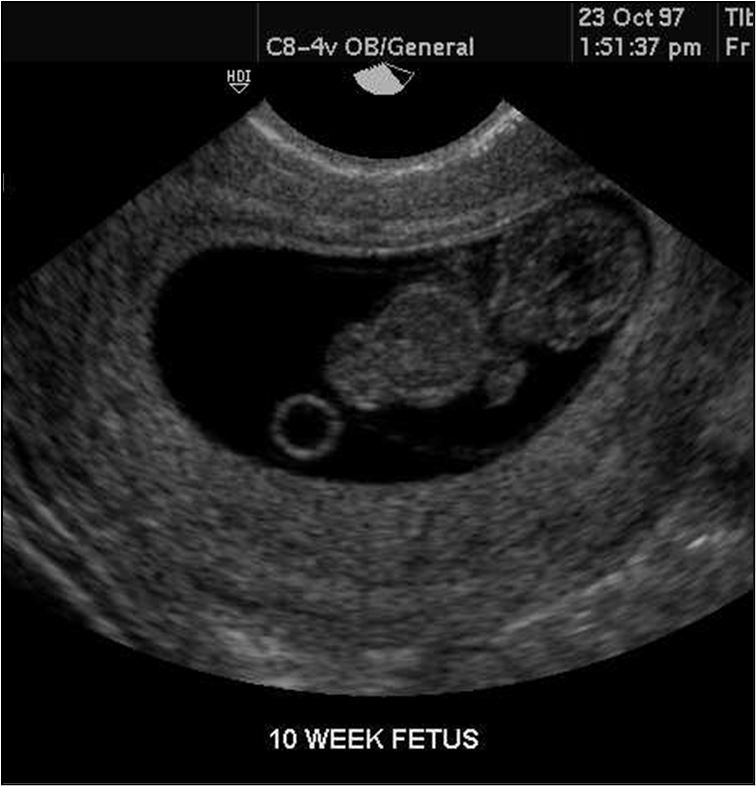
What is this image showing?
10 week fetus/yolk sac
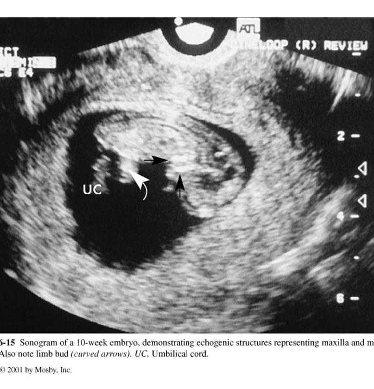
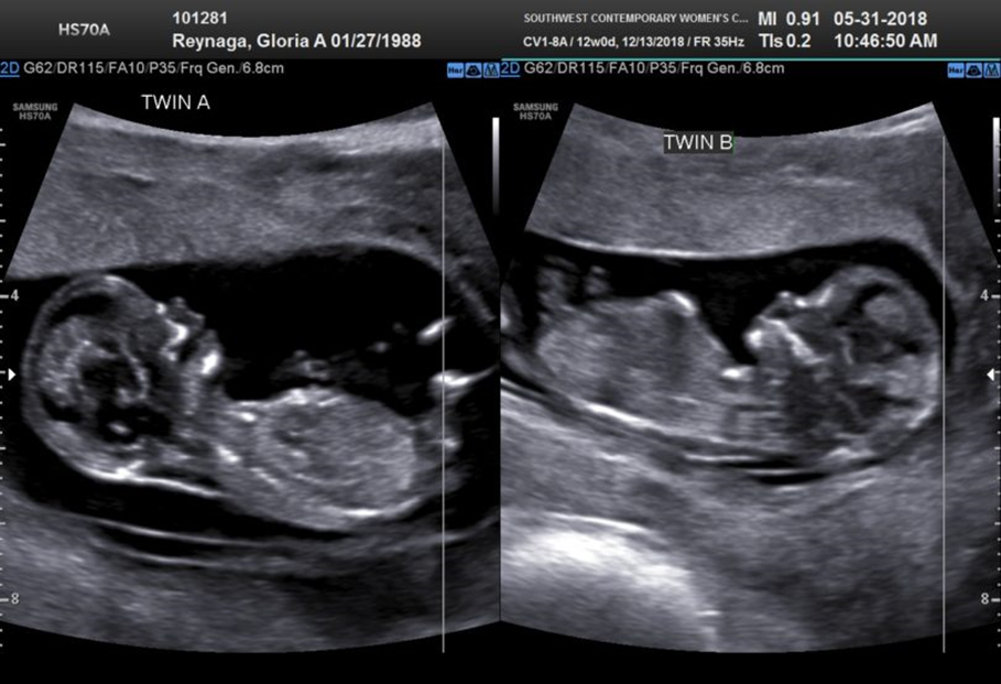
What is this image showing?
10 weeks
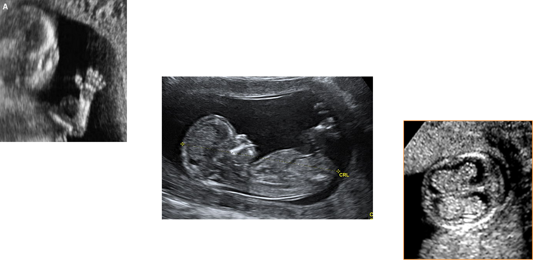
What are these images showing?
12 week fetus
What is this image showing?
12 weeks