NRSC 277 Exam 2
0.0(0)
Card Sorting
1/305
Earn XP
Description and Tags
Last updated 2:47 AM on 3/22/23
Name | Mastery | Learn | Test | Matching | Spaced | Call with Kai |
|---|
No analytics yet
Send a link to your students to track their progress
306 Terms
1
New cards
Gross Anatomy of the Spinal Cord
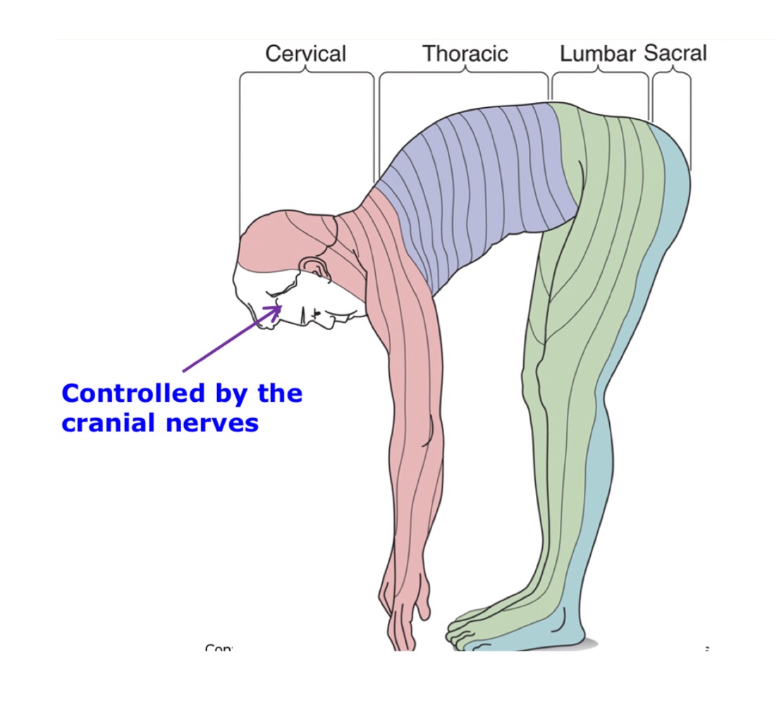
1. Cervical Nerves (8)
2. Thoracic nerves (12)
3. Lumbar nerves (5)
4. Sacral nerves (5)
5. Coccygeal Nerve
2
New cards
\
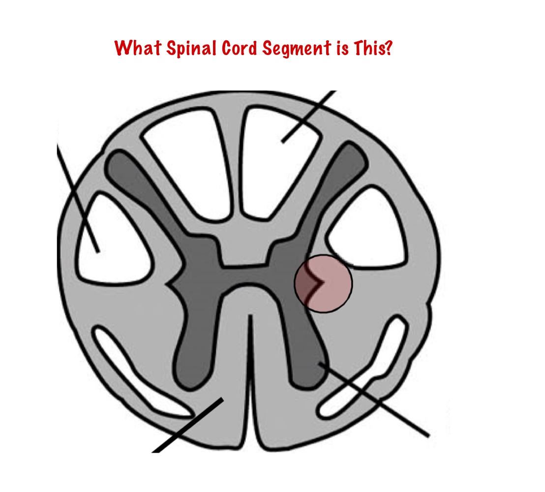
Lumbar
3
New cards
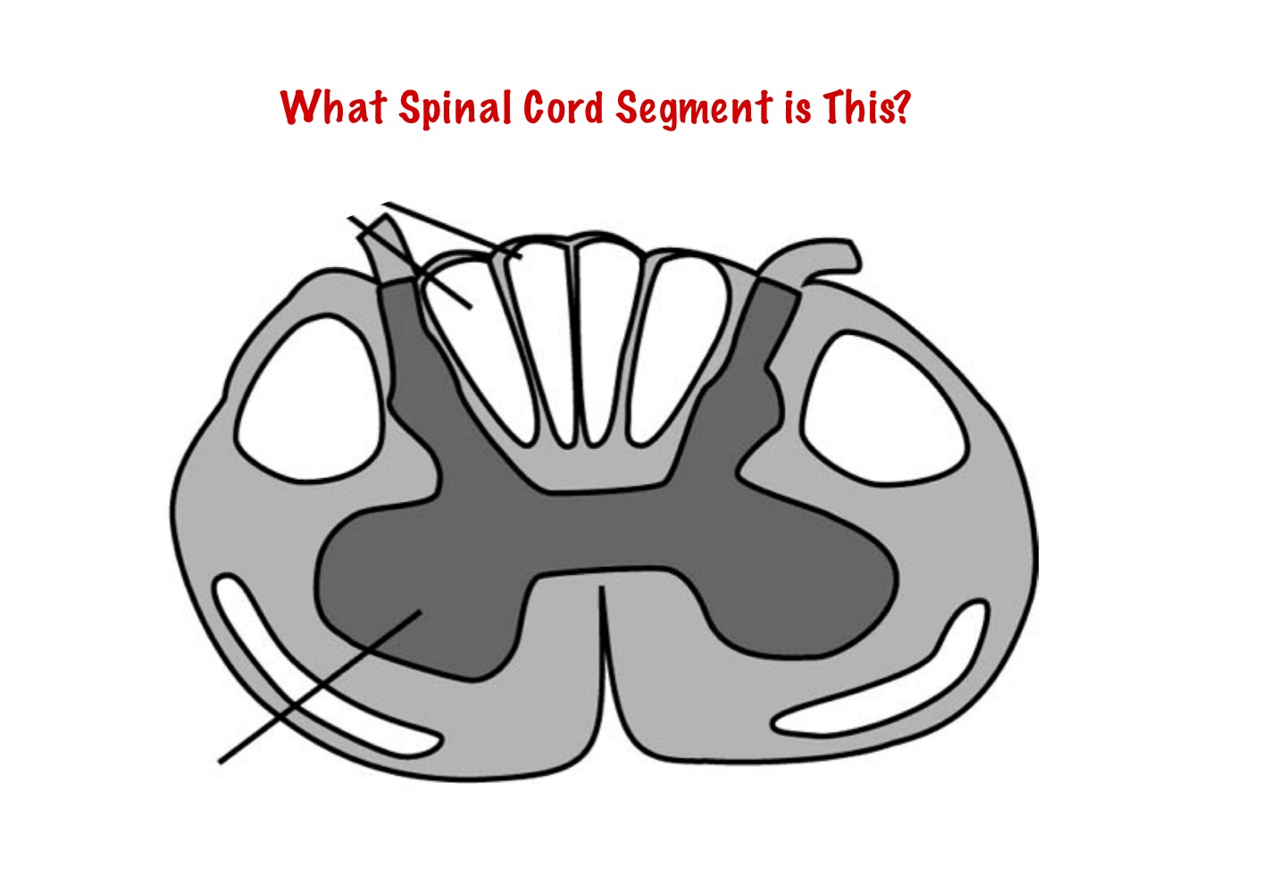
Thoracic
4
New cards
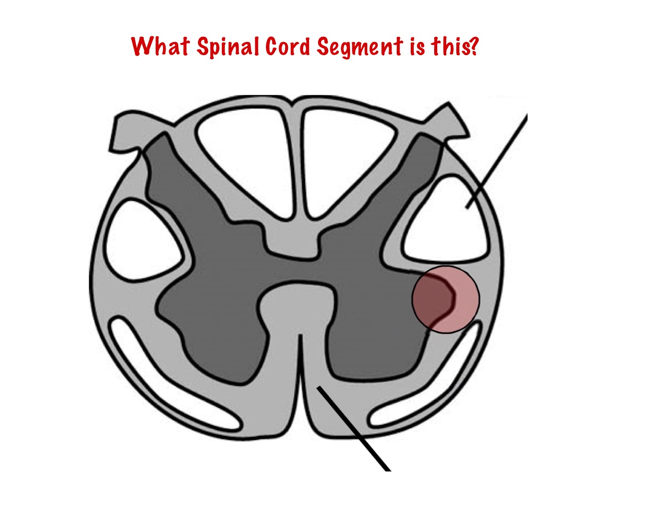
Cervical
5
New cards
Dermatome
6
New cards
Which one is longer/shorter: the spinal cord or the vertebral canal (bones)?
Shorter: Spinal Cord
Longer: Vertebral Canal (bones)
Longer: Vertebral Canal (bones)
7
New cards
What is the Lumbar Cistern?
The Lumbar Cistern is the end of the spinal cord at the vertebral level L1\~L2 to the vertebral level S2 filled with collections of dorsal and ventral roots.
8
New cards
What is the Cauda Equina?
Cauda equina is the collection of dorsal and ventral roots that fill the lumbar cistern at the end of the spinal cord.
9
New cards
What is the significance of the Lumbar Cistern?
It allows for safe sampling of the cerebrospinal fluid
10
New cards
What is the Filum Terminale?
The Filum Terminale is a structure that anchors the the caudal end of the spinal cord to the end of the dura tube.
11
New cards
What is Lumbar Puncture?
A lumbar puncture is a procedure that involves taking cerebral spinal fluid from the spine in the lower back using a hollow needle for diagnostic purposes or anesthesia.
12
New cards
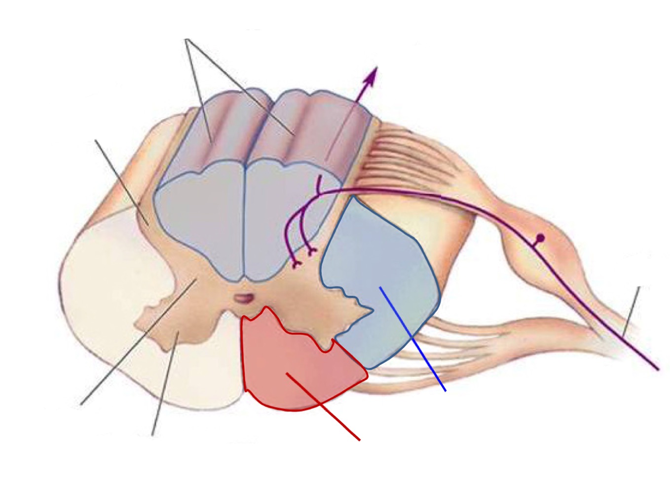
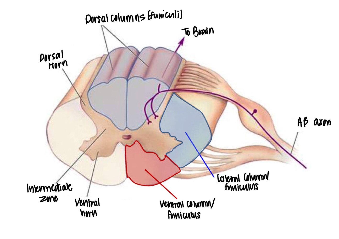
What is the structure of the gray matter in the spinal cord?
Gray matter in the spinal cord has an H-shape and is divided into “horns”
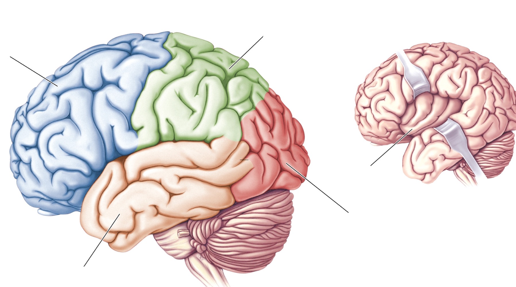
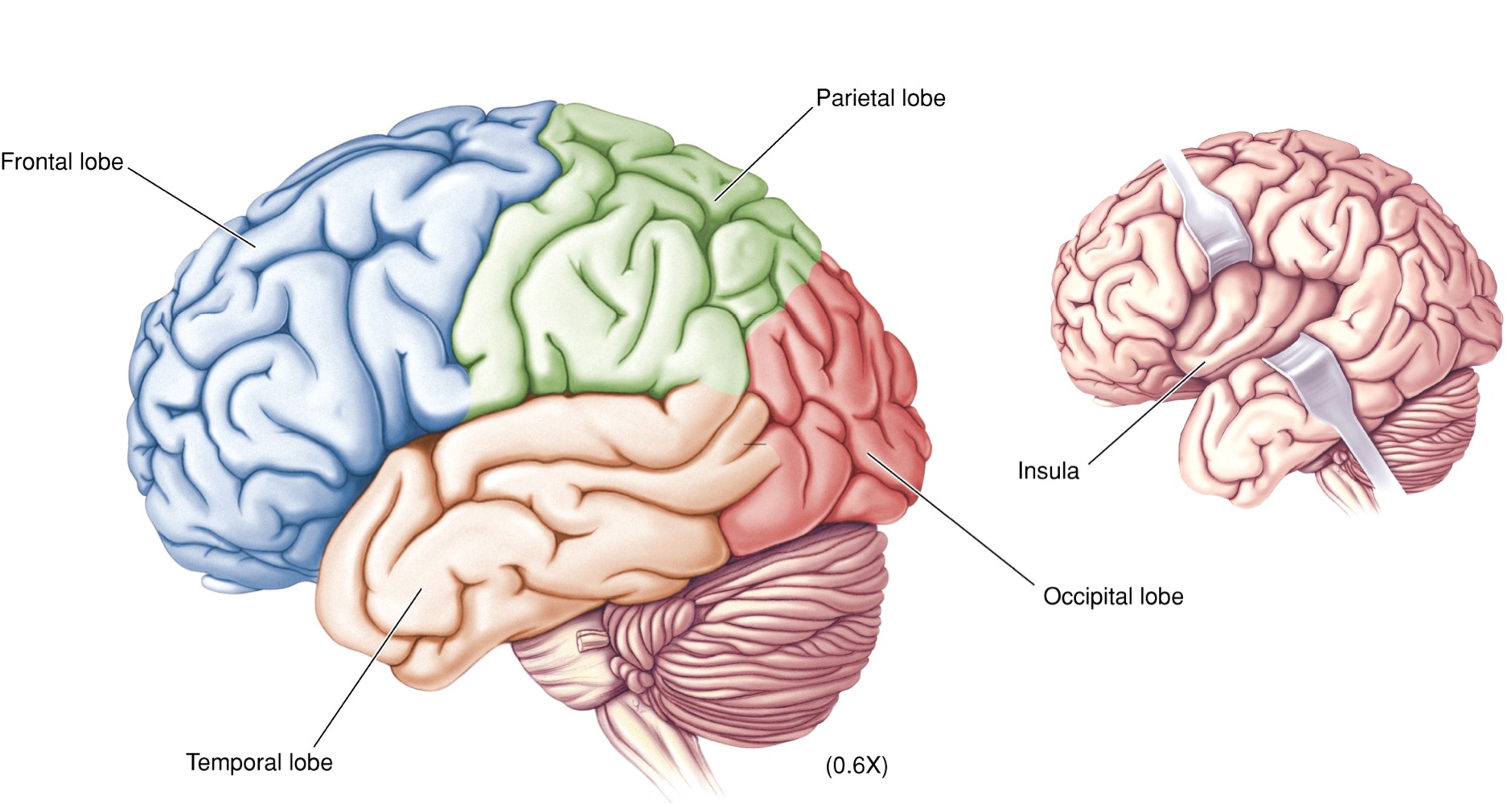
13
New cards
What is located in the gray matter of the spinal cord?
The neuronal cell bodies (soma) and synapses take place in the gray matter of the spinal cord.
14
New cards
What is the function of the white matter in the spinal cord?
The white matter is where myelinated axons (nerve fibers) travel from the spinal cord up to the brain, or from the brain traveling down to the spinal cord.
15
New cards
Where are neuronal cell bodies and synapses located?
Gray matter of the brain and spinal cord
16
New cards
What is the function of the myelinated axons?
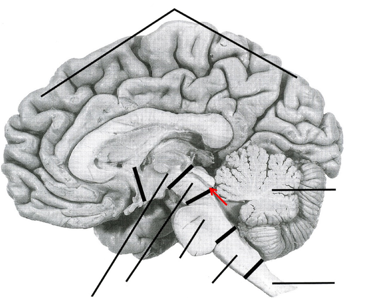
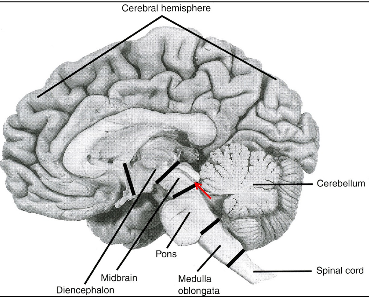
Myelinated axons in the white matter serve to transmit signals between the spinal cord and the brain.
17
New cards
What is the shape of gray matter?
H-shaped and divided into “horns”
18
New cards
What is the function of the ventral (anterior) horn of the gray matter?
Motor Function
19
New cards
What is the function of the dorsal (posterior) horn of the gray matter?
Sensory function
20
New cards
What is the intermediate zone (lateral horn) of gray matter responsible for?
The intermediate zone is responsible for the autonomic system. However, there is no lateral horn in the upper to mid-cervical cord.
21
New cards
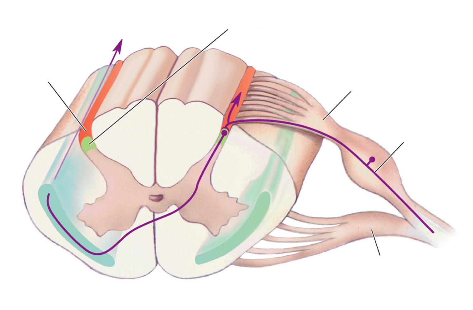
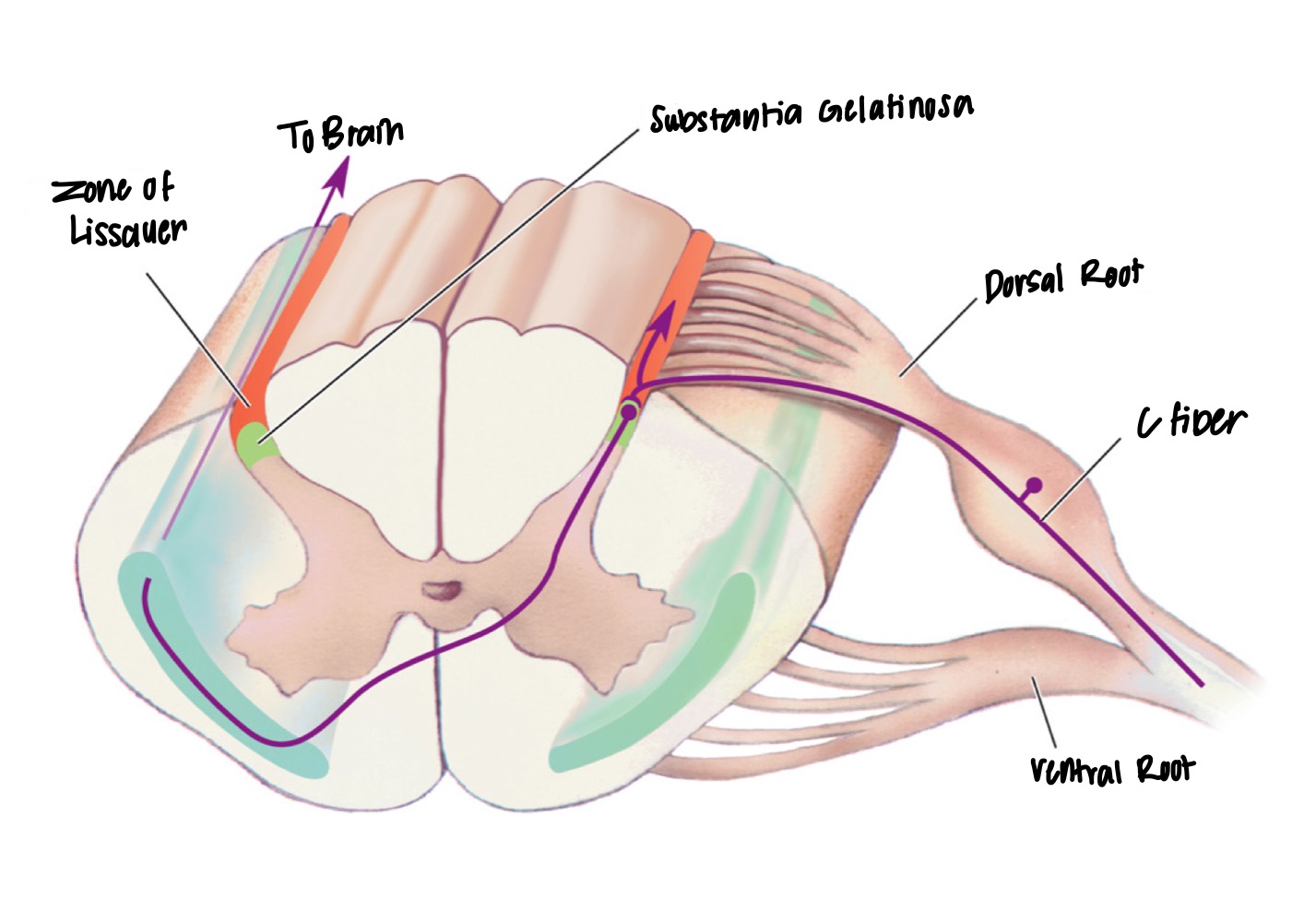
What is the tract/zone of Lissauer’s known as in animals?
Dorsolateral fasciculus.
22
New cards
What is the Lissauer’s Tract?
Group of fibers that originate from the dorsal root ganglia.
23
New cards
What is the function of the substantia gelatinosa?
The substantia gelatinosa contains the first synapses for the sensory information coming from the dorsal roots.
24
New cards
What types of neurons are found in the dorsal horn?
The dorsal horn contains inter neurons and projection neurons that transmit many types of sensory information, including somatic and visceral sensations.
25
New cards
What is the intermediate zone of gray matter responsible for?
The intermediate zone, also known as the lateral horn or intermediolateral cell column, is responsible for autonomic nervous system.
26
New cards
What type of neurons rare found in the intermediate zone in the thoracolumbar column?
Pre-ganglionic sympathetic neurons in T1 to L3.
27
New cards
Which area of the spinal cord houses the sacral parasympathetic nucleus in the autonomic nervous system?
S2 to S4 region of the spinal cord, also known as the craniosacral system.
28
New cards
What type of neurons are found in the sacral parasympathetic nucleus?
Pre-ganglion if parasympathetic neurons that innervate pelvic viscera.
29
New cards
What is the function of the Ventral horn (anterior horn)?
Processes motor information
30
New cards
What are α (alpha) motor neurons?
α (alpha) motor neurons are large motor neurons that innervate the skeletal muscles via the ventral roots. They are also known as lower motor neurons (LMNs).
31
New cards
What is the function of the Small γ (gamma) motor neurons?
Small γ (gamma) motor neurons are neurons that innervate the intrafusal muscle fibers of muscle spindles. They are also called fusimotor neurons.
32
New cards
What are the three funiculi of white matter?
Ventral (anterior) Funiculus, Lateral Funiculus, and Dorsal (posterior) Funiculus.
33
New cards
What is the function of the anterior white commissure?
The anterior white commissure is a bundle of axons that crosses over the midline of the spinal cord and connects the two sides of the cord.
34
New cards
What types of nerve fibers are found in the white matter?
The white matter contains largely ascending and descending myelinated nerve fibers. These ascending and descending fibers are organized into bundles “tracts” which occupy specific regions of the white matter.
35
New cards
What are the three types of nerve fibers in the white matter?
The three types of nerve fibers in the white matter are long ascending fibers projecting to the thalamus, cerebellum, or various brainstem nuclei, long descending fibers projecting from the cerebral cortex or from brainstem nuclei to the spinal cord white matter, and short propriospinal fibers interconnecting various spinal cord levels (fasciculus proprius).
36
New cards
How is the white matter divided?
3 Columns: Posterior (Dorsal), Lateral, and Anterior (Ventral)
37
New cards
What are the major descending tracts found in the spinal cord?
In the lateral funiculus and ventral funiculus.
38
New cards
Where are the ascending tracts found?
Ascending tracts are found in all the funiculi.
39
New cards
What us the propriospinal tract?
Thin layer surrounding the gray matter and is responsible for interconnecting various spinal cord levels.
40
New cards
41
New cards
42
New cards
What are the 2 divisions of the autonomic system?
Sympathetic and Parasympathetic system
43
New cards
What are the functions of the sympathetic system?
* Increased heart rate & blood pressure
* depressed digestive function
* mobilized glucose reserves
* all for “fight or flight” situations
* depressed digestive function
* mobilized glucose reserves
* all for “fight or flight” situations
44
New cards
What are the functions of the parasympathetic system?
* slower heart rate
* fall in pressure
* increased digestive functions
* stop sweating
* all for enhancing energy storage.
* fall in pressure
* increased digestive functions
* stop sweating
* all for enhancing energy storage.
45
New cards
What does the Enteric Nervous System do?
Regulates the digestive system independently of the central nervous system
46
New cards
Where are the pre-ganglionic neurons located?
CNS
47
New cards
Where are the Post-ganglionic neurons located?
Peripheral Ganglia
48
New cards
Where are sympathetic ganglia located?
Near the CNS
49
New cards
Where are the parasympathetic ganglia located?
Near the organs.
50
New cards
What do pre-ganglionic neurons of both systems release?
Acetylcholine
51
New cards
What do postganglionic neurons in the sympathetic system release?
Norepinephrine
52
New cards
What do postganglionic neurons of the parasympathetic system release?
Acetylcholine
53
New cards
What receive only sympathetic innervations?
Sweat glands and limb vasculature
54
New cards
What does the parasympathetic system control?
Pupils and bladder
55
New cards
Where is the enteric division located?
In the lining of the esophagus, stomach, intestines, pancreas, and gallbladder
56
New cards
What is the enteric division composed of?
two complicated nerve networks: myentric plexus and submucous plexus
57
New cards
What is the function of the enteric division?
controls physiological processes involved in transport, digestion of food.
58
New cards
what are the inputs of the enteric division?
From brain via axons of the sympathetic and parasympathetic divisions.
59
New cards
What is the function of the periventricular zone?
connection to the brain stem and spinal cord nuclei
60
New cards
What is the function of the solitary nucleus?
receives and integrates sensory information from all internal organs and coordinates outputs
61
New cards
What is the forebrain called?
Prosencephalon
62
New cards
What does the Telencephalon contain?
cerebral cortex and Basal Ganglia
63
New cards
What is the developmental patterns sequence of the cerebral cortex?
Frontal lobe → parietal lobe → occipital lobe → temporal lobe.
64
New cards
What are the folds called that are present on the cerebral cortex?
The folds present on the cerebral cortex are called cortical gyri.
65
New cards
What separates the cortical gyri on the cerebral cortex?
By deep fissures or shallow sulci.
66
New cards
What are the different fissures (sulci) present in humans on the cerebral cortex?
The central fissure/sulcus (of Rolando), Sylvian fissure (Lateral fissure/sulcus), longitudinal fissure, transverse cerebral fissure, and parieto-occipital fissure.
67
New cards
Label
68
New cards
What is the insular lobe and where is it located?
It is located deep within the temporal lobe and is only present in humans. It forms when growth continues after forming the temporal lobe, and it rolls in upon itself.
69
New cards
Have there been changes in the structure of the cortex among different species?
No, there have not been changes in the structure of the cortex among different species. Rather, the amount of cortex present has changed.
70
New cards
What are the different types of areas in the cortex, according to Leah Krubitzer?
Primary sensory areas, secondary sensory areas, and motor areas in the cortex.
71
New cards
According to Jon Kaas, what has expanded in the cortex?
the secondary sensory areas have expanded in the cortex.
72
New cards
What is the basal ganglia and where is it located?
The basal ganglia, also known as the corpus striatum or striated body, is located deep to the cerebral cortex and is part of the telencephalon.
73
New cards
What are the two parts of the diencephalon?
thalamus (dorsal) and hypothalamus (ventral).
74
New cards
What is the pituitary body, where is it located, and how is it connected to the hypothalamus?
The pituitary body, or gland, is located ventrally from the hypothalamus, and it is hanging through the pituitary stalk.
75
New cards
What do the 2 Foramina of Monroe connect to?
The 2 Foramina of Monroe connect the third ventricle to both the right and left hemispheres.
76
New cards
How do the lateral ventricles follow the growth pattern of the cerebral cortex?
The lateral ventricles follow the growth pattern of the surrounding cerebral cortex, having a frontal horn, a body, occipital and temporal horns
77
New cards
What produces cerebrospinal fluid, and where are they located?
Choroid plexus lining in the lateral, third, and fourth ventricles produce cerebrospinal fluid.
78
New cards
What is the midbrain derived from, and what are its two parts?
he midbrain is derived from the embryonic mesencephalon and is separated into dorsal-tectum and ventral-tegmentum.
79
New cards
What is the cerebral aqueduct, and where is it located?
The cerebral aqueduct, also known as the aqueduct of Sylvius or mesencephalic aqueduct, is a tube-like structure that goes through the midbrain. It connects the third ventricle (above) and the fourth ventricle (below).
80
New cards
Label
81
New cards
What are the substantia nigra?
The substantia nigra are heavily pigmented nuclei lying on top of the cerebral peduncles.
82
New cards
What is the main character of Parkinson’s disease?
Degeneration of the substantia nigra.
83
New cards
What are red nuclei?
Red nuclei are two large round nuclei adjacent to the midline in the midbrain.
84
New cards
What is the hindbrain or rhombencephalon composed of?
The hindbrain or rhombencephalon is composed of three major structures: medulla oblongata, pons, and cerebellum.
85
New cards
What is the pons?
The pons is a large ventral bulge lying just rostral to the medulla. It contains many pontine nuclei, pontine reticular formation, and two major fiber bundles.
86
New cards
What are pyramidal tracts?
Pyramidal tracts are two large fiber bundles that are the major motor pathways, taking origin from the cerebral cortex and terminating in the ventral horn of the spinal cord and certain cranial nerves.
87
New cards
What is the cerebellum?
The cerebellum is the dorsal part of the metencephalon and overlies the fourth ventricle. It has a midline vermis and two large hemispheres, and is not part of the brain stem.
88
New cards
What is the somatic sensory system responsible for?
The somatic sensory system is responsible for enabling the body to feel, ache, and chill and is responsible for touch and pain.
89
New cards
What do receptors encode in the somatic sensory system?
Receptors in the somatic sensory system encode the nature (modality), location, intensity, and duration of stimuli.
90
New cards
What are sensory modalities in the somatic sensory system based on?
The selective sensitivity of its sensory receptors and are different forms of stimuli that evoke different types of sensations.
91
New cards
What are the current classifications of receptors in the somatic sensory system based on?
The current classifications of receptors in the somatic sensory system are based on the type of stimulus to which they are most sensitive, which is called the adequate stimulus.
92
New cards
What are the different types of receptors in the somatic sensory system?
The different types of receptors in the somatic sensory system include chemoreceptors, photoreceptors, thermoreceptors, mechanoreceptors (such as cutaneous receptors for touch, receptors monitoring muscle length and tension, and auditory and vestibular receptors), and nociceptors (pain receptors).
93
New cards
What do visceral structures contain in terms of receptive endings?
Visceral structures contain a variety of receptive endings, with most visceral receptors being supplied by thinly myelinated and unmyelinated fibers that terminate as free nerve endings.
94
New cards
What are the different types of visceral receptors?
1. Mechanoreceptors in the wall of hollow organs (such as the endings in the aortic arch and carotid sinus),
2. Chemoreceptors (such as those in the carotid body to detect blood pH or gases), and nociceptors.
95
New cards
What are sensory receptors and what are the two types?
Sensory receptors are specialized structures that sense particular types of stimuli. There are two types of sensory receptors:
1. Sensory neurons with modified nerve endings
2. Specialized receptor cells that transduce a stimulus.
1. Sensory neurons with modified nerve endings
2. Specialized receptor cells that transduce a stimulus.
96
New cards
What are examples of sensory neurons with modified nerve endings?
Nociceptors, touch receptors, temperature receptors, olfactory receptors, and photoreceptors.
97
New cards
What are examples of specialized receptor cells that transduce a stimulus?
Taste receptors (chemoreceptors), auditory and vestibular hair cells (mechanoreceptors)
98
New cards
Do specialized receptor cells release neurotransmitters onto a sensory neuron?
Yes, specialized receptor cells release neurotransmitters onto a sensory neuron which conveys information to the central nervous system (CNS).
99
New cards
What is a receptive field?
A receptive field is the region of the sensory surface where an appropriate stimulus alters the firing rate of a sensory neuron.
100
New cards
What are some examples of receptors with receptive fields?
Cutaneous receptors have a receptive field on an area of skin where their receptive ending resides, and retinal photoreceptors have a receptive field on some small location in the outside world whose image falls on the particular spot on the retina where that photoreceptor is located.