Neurology of Hearing and Balance
1/55
There's no tags or description
Looks like no tags are added yet.
Name | Mastery | Learn | Test | Matching | Spaced |
|---|
No study sessions yet.
56 Terms
What is another name for hearing
audition
What happens during hearing
A process where acoustic energy waves are changed into neural impluses that are interpreted by the brain
What are the two main divisions of the ear
peripheral auditory system
central auditory system
What is part of the peripheral auditory system?
outer ear
middle ear
inner ear
cranial nerve 8
What is part of the central auditory system?
brainstem
brain
definition of sound
audible variations in air pressure
dependent on the motion of air molecules associated with sound
propagation of sound
The Nature of Sound
pitch = frequency
loudness = amplitude = intensity
The Outer Ear
The canal lined by skin containing hair follicles, sebaceous glands, and ceruminous glands that produce wax. earwax. The cerumen lubricates the skin and coats hairs near the opening to impede the entry of foreign particles into the ear. Excessive accumulation of cerumen can plug the meatus, however, and result in conductive hearing loss
Tympanic Membrane - ear drum
boundary between outer and middle ears
vibrates when sound waves entering the external auditory meatus hit it
a framework of connective tissue fibers covered by skin on the external ear side and mucous membrance on the side of the middle ear cavity
perforation may cause transient or permanent hearing impairment
What are the three ossicles
help with the transduction of vibratory energy into mechanical energy
malleus
incus
stapes
malleus
hammer
attached to the tympanic membrane
incus
anvil
connects the malleus to the stapes
stapes
stirrup
fits into the oval window
What is the structure of the attenuation reflex
tensor tympani inserts on the malleus, stapedius inserts on the stapes
What is the function of the attenuation reflex
protects the innear ear (cochlea) from damaging vibrations
masks background noise in noisy environments
decreases sensitivity to one’s own voice
what is the purpose of the eustachian tube?
equalizes pressure in middle ear to atmospheric pressure
Ear Infections
more common in childrne
their eustahcian tubes are shorter, narrower, more horizontal than adults, makes movement of air and fluid difficult.
Bacteria trapped in the eustachian tube may produce an ear infection that pushes on the eardrum cuasing it to become red, swollen, and sore
Inner Ear: Anatomy
the rocking of the stapes in the oval window creates waves in the cochlear fluid
this is another energy change: mechanical energy of ossicular movement that has been changed into hydraulic energy
these waves disrupt the hair cells in the organ of Corti causing a third energy change: hydraulic energy changed to electrochemical energy
Fluid Movement in the Cochlea
Pressure at oval window, pushes perilymph into scala vestibuli, which then travels around to the scala tympani and causes the round window membrane to bulge out
The Basilar Membrane
At the apex of the cochlea, the scala media is closed off, and the scala tympani becomes continuous with the scala vestibuli at a hole in the membranes called the Helicotrema
The Response of Basilar Membrane to Sound
Structural properties: Wider at apex, stiffness decreases from base to apex. Perilymph movement bends basilar membrane near base, wave moves towards apex
Why the cochlea is spiral-shaped
It increases the strength of vibrations produced by sound waves, especially at low pitch.
Bending of Stereocilia
At rest, the hair cells are held between the reticular lamina and the basilar membrane, and the tips of the outer hair cell stereocilia are attached to the tectorial membrane.
When sound causes the basilar membrane to deflect upward, the reticular lamina moves up and inward toward the modiolus, causing the stereocilia to bend outward.
Depolarization of a Hair Cell
Ion channels on stereocilia tips are opened when the tip links joining the stereocilia are stretched.
The entry of K+ (potassium) depolarizes the hair cell, which opens voltage-gated calcium channels. Incoming Ca2+ leads to the release of neurotransmitter from synaptic vesicles, which then diffuses to the postsynaptic neurite from the spiral ganglion.
Hair Cells: Scanning Electron
(a)Hair cells and their stereocilia.
A higher-resolution view of the stereocilia on an outer hair cell. The stereocilia are approximately 5 µm in length.
Innervation of Hair Cells
One spiral ganglion fiber receives input from only 1 inner hair cell; each inner hair cell feeds about 10 spiral ganglion neurites.
One spiral ganglion fiber synapses with many outer hair cells.
Therefore, vast majority of information leaving cochlea comes from INNER hair cells.
Cochlear Amplifier: Outer Hair Cells Amplify Movement of Basilar Membrane
Outer hair cells amplify the response of the basilar membrane à stereocilia on inner hair cell bend more à increased transduction in inner hair cells à greater response in auditory nerve.
Without cochlear amplifier:
100 fold smaller peak movement of basilar membrane
Excessive exposure to antibiotics (kanamycin) damages outer hair cells cochlear amplifier à deafness
Cochlear Efferent Nerves
About 1000 efferent fibers project from brain stem to cochlea
Synapse on inner and outer hair cells à changes the shape of the outer hair cell à affects response of inner hair cell.
Thus descending input from brain to cochlea can regulate auditory sensitivity
Otoacoustic Emissions
Ears emit sounds present click à echo that can be recorded w/ mic in auditory canal. Normally too faint for us to hear.
Occur because the sensitivity of the cochlear amplifier very high
If spontaneous otoacoustic emissions loud enough:
can cause a form of tinnitus
usually the result of cochlear damage from exposure to extreme loud sound
Can be used to test function of ears: play series of sounds, record evoked echos.
especially useful for those that cannot voluntarily respond – e.g., newborn babies
Overview of Auditory Pathway
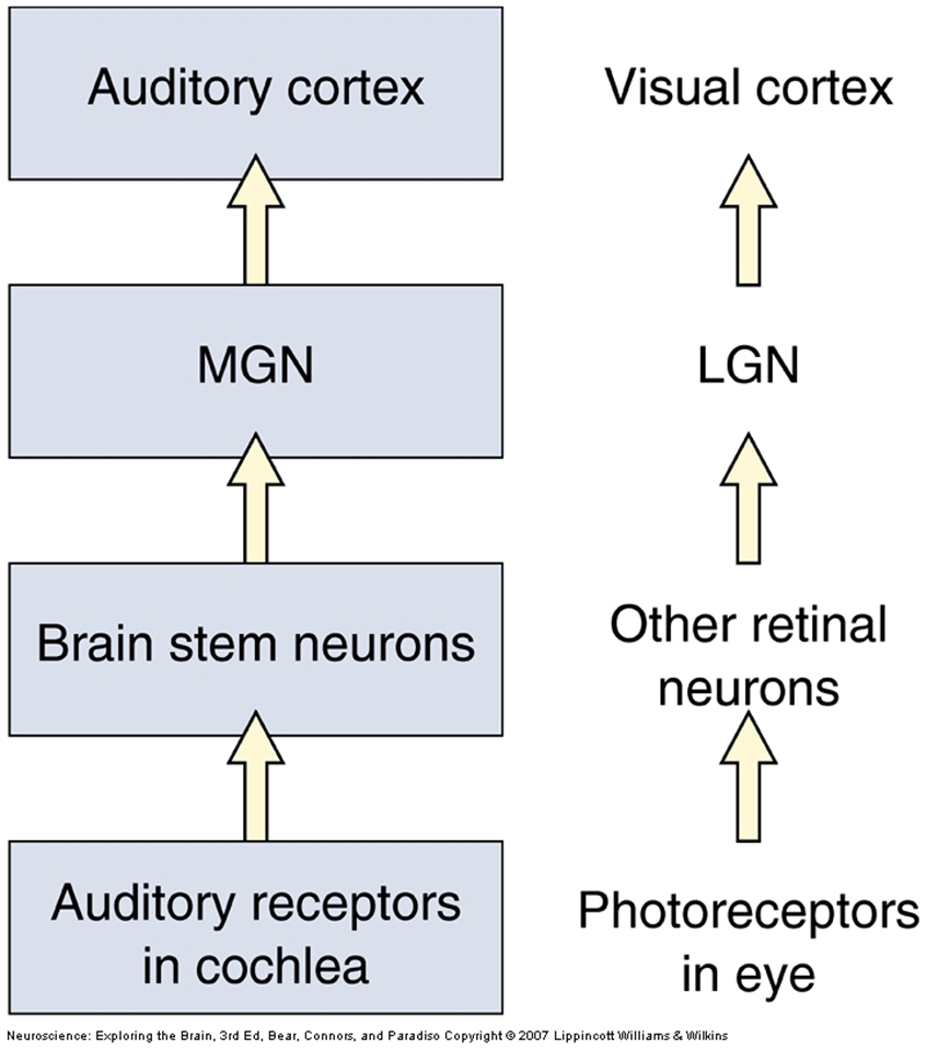
Pathway is complex: many synapses from medullary cochlear nuclei to other brainstem nuclei
Important Pathway
1 a. Ipsilateral cochlea to ventral and dorsal cochlear nuclei in medulla
1 b. Bilateral to Superior Olivary Nuclei Medulla
2. Bilateral thru lateral lemniscus (bundle of fibers) to Inferior Colliculus in Midbrain
3. Bilateral to Medial Geniculate Nucleus Thalamus
4. Bilateral to Primary Auditory Cortex
Majority of ascending fibers cross midline
All ascending pathways converge onto Inferior Colliculus
Important Points
1. Other projections and brainstem nuclei contribute.
e.g., Inferior Colliculus to MGN & also to Superior Colliculus (integration of auditory & visual info)
2. Auditory Pathways have extensive feedback.
e.g., brain stem neurons send axons to outer hair cells
e.g., auditory cortex sends axons to MGN & Inferior Colliculus
3. Each Cochlear Nucleus receives input from only Ipsilateral Ear;
All other auditory nuclei in brainstem receive input from Both ears
à only way brainstem damage can cause deafness in one ear is if cochlear nucleus or auditory nerve damaged on that side
Tonotopic Maps
On basilar membrane and cochlear nucleus. From base to apex, the basilar membrane resonates with increasingly lower frequencies. This tonotopy preserved in auditory nerve and cochlear nucleus. In the cochlear nucleus, bands of cells with similar characteristic frequencies which increase progressively from anterior to posterior.
Sound Localization
Sound waves coming from the right side will reach the right ear first, and there will be a large interaural delay before the sound propagates to the left ear.
If the sound comes from straight ahead, there is no interaural delay. Delays for three different sound directions are shown.
Sound Localization: Ineraural Intensity
With high-frequency sound, the head will cast a sound shadow to the left when sound waves come from the right. Lower-intensity sound in the left ear is a cue that the sound came from the right.
If the sound comes from straight ahead, a sound shadow is cast behind the head but the sound reaches the two ears with the same intensity.
Sound coming from an oblique angle will partially shadow the left ear.
Vertical Sound Localization
based on reflections from the pinna
Tonotopic Maps Pt. 2
Primary auditory cortex (purple) and secondary auditory areas (yellow) on the superior temporal lobe.
Tonotopic organization within primary auditory cortex. The numbers are characteristic frequencies.
Primary Auditory Cortex
Heschl’s gyri
Maintenance of tonotopic representation
Discrimination of timing patterns of auditory stimuli
Essential for human speech perception
Secondary Auditory Area
Superior temporal gyrus
Processing of timing patterns
Recognition of spatial attributes typical of human speech
Acoustic aphasia- impaired perception & discrimination of human speech sounds
Association Cortex of Wernicke
Comprehension of spoken language and generalization of meaning
Hearing Disorders: Conductive
•Interrupted sound transmission to cochlea
•Middle ear pathologies
Otitis media (inflammation of middle ear fluid) and ottosclerosis (impeded stapes movement)
•Symptoms
Fluctuating hearing loss, good word-speech recognition, mostly softly spoken speech, impaired auditory reflex, and air-bone gap
Hearing Disorders: Sensorineural
Dysfunction of hair cells and/or auditory nerve fibers
•Commonly implicated with
Ménière's disease- progressive & fluctuating hearing loss with vertigo & tinnitus
Presbycusis- old age induced hearing loss of high frequencies
Acoustic neuroma/vestibular schwannoma- hearing impairment and disequilibrium, and ataxic symptoms in later stages
•Symptoms
Difficulty in understanding speech, reduced self-monitoring in speech recruitment
Hearing Disorders: Mixed
•A combination of conductive and sensorineural hearing loss.
•There may be a problem in the outer or middle ear and in the inner ear or auditory nerve.
•
•It can happen after a head injury, long-term infection, or because of a disorder that runs in your family.
Hearing Disorders: Central
•Can result from damage to lower brainstem upper brainstem or primary auditory cortex
•
•Clinical Characteristics
•Near-normal sensitivity to stimuli
•Impaired processing of linguistic signals
Cortical Involvement: Unilateral Cortical Lesion
normal hearing thresholds with imparied perception and discrimination of speech
Cortical Involvement: Bilateral Cortical Lesion
profound hearing impairment
Cortical Involvement: Primary and Secondary Auditory Lesion
Acoustic aphasia
•Impaired discrimination of speech sounds & phonemes- skills necessary for learning phonemes and understanding language
Cortical Invovlement: Language association cortex lesion
Wernicke aphasia
•Impaired comprehension of spoken and written language
•Word finding deficit & asemantic verbal output
Cortical Invovlement: Right Temporal lesion
Impaired processing of environmental sounds, non-verbal memory, & musical properties
Balance
Also known as vestibular system
Our spatial orientation system that helps us sit upright, walk, and perform other functions in our spatial environment
The Vestibular Labyrinth
Locations of the otolith organs (utricle and saccule) and semicircular canals.
A vestibular labyrinth resides on each side of the head, with the semicircular canals arranged in parallel planes
Macular Hair Cells Responding To Tilt
When the utricular macula is level (the head is straight), the cilia from the hair cells also stand straight.
When the head and macula are tilted, gravity pulls the otoliths, which deform the gelatinous cap, and the cilia bend.
Macular Orientation
The macula in the utricle is horizontal.
The macula in the saccule is vertical. The arrows on each macula show how the hair cells are polarized. Bending the hairs in the direction of the arrow depolarizes them.
Essentially, these otolithic organs sense how quickly you are accelerating forward or backward, left or right, or up or down
What do semicircular canals do?
Sense rotation of the head
Ampula of Semicircular Canals
The cilia of hair cells penetrate into the gelatinous cupula, which is bathed in the endolymph that fills the canals.
When the canal rotates leftward, the endolymphlags behind and applies force to the cupula, bending the cilia within it.
Activation of Semicircular Canals
Head rotation causes the excitation of hair cells in one horizontal semicircular canal and the inhibition of hair cells in the other. (Graphs) Long-lasting head rotation leads to adaptation of the firing in vestibular axons. When rotation is stopped, the vestibular axons from each side begin firing again, but with opposite patterns of excitation and inhibition
Vestibular Pathlogies
•Vestibular schwannoma – slow-growing, unilateral, benign tumor on vestibular branch of cranial nerve VIII
main symptoms are hearing loss and tinnitus as well as vertigo and balance issues
•Labyrinthitis – infection of the inner ear
leading to vertigo, nausea, and vomiting
•Ménière's disease – unknown cause likely involves both genetic and environmental factors
episodes of feeling like the world is spinning (vertigo), ringing in the ears (tinnitus), hearing loss, and a fullness in the ear
•Benign paroxysmal positional vertigo (BPPV) - probably caused when pieces that have broken off otoliths have slipped into one of the semicircular canals
dizziness, vertigo and nystagmus
•Motion Sickness - the vestibular system reports no movement but the visual system reports movement
nausea