SNAB TOPIC 6- immunity, infection and forensics
1/24
There's no tags or description
Looks like no tags are added yet.
Name | Mastery | Learn | Test | Matching | Spaced |
|---|
No study sessions yet.
25 Terms
6.1 Understand how to determine the time of death of a mammal by examining the extent of decomposition, stage of succession, forensic entomology, body temperature and degree of muscle contraction.
Extent of decomposition
Pattern/stage of decay can be examined-
stage 1) Is there discolouring? (few hrs-days)
stage 2) Is there bloating? Is the skin falling off? (few days to weeks)
stage 3) Is there liquefication of tissues? (several weeks)
stage 4) Are there still any tissues left? (few months to few years)
These can all be asked successively to determine the time of death
This all happens because of digestive enzymes that are released, both within the cells and the microorganisms on/in the body
6.1 (PART B) Understand how to determine the time of death of a mammal by examining the stage of succession and forensic entomology
Forensic entomology
Depending on the species present, and the life cycle stage it is in, a time of death can be estimated
Stage of succession:
•Coloniser species-flies, maggots (eggs take about 24 hours to hatch)
•Secondary succession, e.g. organisms that feed on the coloniser species/decomposing body fat
•Further species arrive to break down the corpse as it reaches further stages of decomposition
etc.
Indicator species for each succession stage, and the life cycle stage tell us how long ago time of death was.
6.1 (PART C) Understand how to determine the time of death of a mammal by examining the body temperature
Respiration/metabolic processes produce heat.
Once a person dies, no more heat is produced
ALGOR MORTIS occurs- the process of cooling.
The body temperature decreases around the same rate per hour, which allows the time of death to be estimated if the corpse's core temp. is measured and the recent local temperature records are used
conditions affecting the temperature change rate
•Air temperature
•Ambient temperature
•Weather/precipitation
•Humidity
•Presence of clothing
•Surface area:volume ratio.
6.1 (PART D) Understand how to determine the time of death of a mammal by examining the degree of muscle contraction.
Degree of muscle contraction
After death, muscles stiffen- rigor mortis. Rigor mortis lasts between the 2nd/4th and 36th hour after death (so it is a limited indicator but shows recent death)
This is because of calcium ions building up in the muscle cells, as ATP is used up.
[topic 7 link] ATP is no longer there to break the bridges between the myosin head and actin filament
6.2 Know the role of micro-organisms in the decomposition of organic matter and the recycling of carbon.
Bacteria and fungi release digestive enzymes.
These decompose dead organic matter into molecules that are used by the microorganisms.
They are used for respiration too, and CO2/Methane are both released, recycling carbon into the atmosphere
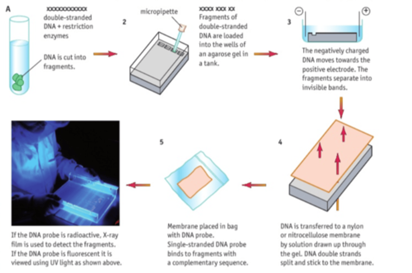
6.3 Know how DNA profiling is used for identification and determining genetic relationships between organisms (plants and animals). [gel electrophoresis]
This method separates and visualises a DNA sample, which can be compared to a known DNA profile.
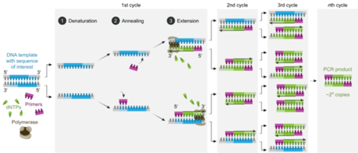
6.4 Know how DNA can be amplified using the polymerase chain reaction (PCR).
Primers, DNA polymerase, free nucleotides, buffer solution are all used
Denaturation (95°C) - breaking apart DNA
Annealing- (55°C)- primers combining to the ends of each strand
Elongation -(72°C)- DNA polymerase produces a new double stranded DNA molecule.
72°C is the optimum temp. for this type of DNA polymerase.
CORE PRACTICAL 14:
Use gel electrophoresis to separate DNA fragments of different length.
Equipment
•Restriction enzymes
•Agar gel
•Gel tank
•Electrical supply + electrodes
•Micropipettes
•DNA sample
•Loading dye
•UV light camera
Buffer solution
•Beaker
Method
Mix DNA with restriction enzymes and loading dye.
Prepare agar gel in tank, with wells
Fill tank with buffer solution
Use micropipette to load the restriction ladder in first well
Connect tank to electrical supply. Turn on and leave til dye has moved to opposite end of tank
Turn off, remove gel carefully and view under UV light.
Compare with known DNA profile.
Explanation/conclusion
DNA has an overall negative charge, which means it moves to the positive electrode
Different sized fragments travel different distances due to varying STRs within the introns (non coding regions of the DNA).
This creates a unique DNA fingerprint.

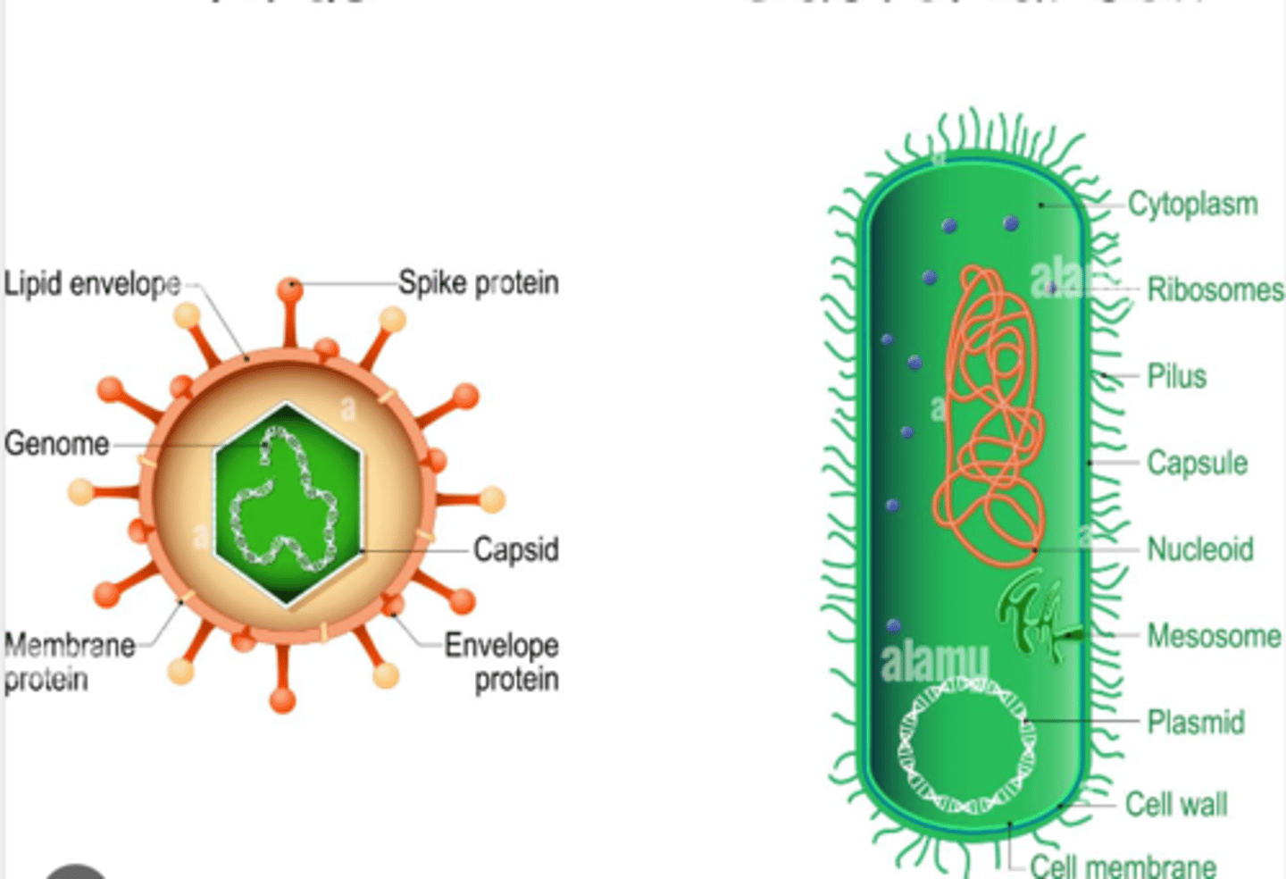
6.5 Be able to compare the structure of bacteria and viruses.
Virus
Lipid envelope ---- allows viruses to easily enter a cell by fusing of membranes
Spike protein ---- helps viral entry, binds to cell receptors
Protein capsid ---- protects genetic code
Nucleic acids (DNA or RNA)
NON LIVING
Bacteria
Cell wall
Cell membrane
Mesosome ----contains enzymes for cellular respiration
Plasmid ---- contains genetic code for antibiotic resistance
Pilus/Pili ----attach to other bacterial cells for exchange of plasmids
Cytoplasm
Ribosomes (70s)
Slime capsule ----- protects cell from physical/chemical attacks, helps the cell retain moisture and adhere to surfaces.
Circular nucleic acids (DNA)
Does not have histone proteins
DNA is within the cytoplasm, no membrane surrounding it
PROKARYOTES
6.6 Understand how Mycobacterium tuberculosis (TB) infects human cells, causing a sequence of
symptoms that may result in death.
TB infects phagocytes in the lungs
Phagocytes that engulf TB bacteria are sealed in tubercles, in the lungs
TB bacteria lie dormant, as they are covered with a thick waxy coat
When immune system is weakened, the bacteria can become active again and start slowly destroying lung tissue
Symptoms
Breathing problems, coughing, weight loss, fever, can be fatal
6.6 Understand how Human Immunodeficiency Virus (HIV) infects human cells, causing a sequence of symptoms that may result in death.
HIV infects and destroys T-helper cells. They bind to their CD4 receptor and the capsid enters the cells after membranes fuse.
Viral RNA is used as a template by reverse transcriptase enzyme, to produce complementary DNA which is turned into a double stranded DNA molecule and inserted into the DNA of the T cell by the integrase enzyme. T cell now has the genes to produce viral proteins to build new virus particles, which move out of the T cell and infect other cells.
Reduction in T-helper cells leads to AIDS:
The first symptoms of HIV are flu-like: fevers, tiredness, headaches
HIV antibodies appear in the blood, making a person HIV positive.
Symptoms disappear until the immune system becomes weakened again, leading to AIDS.
Symptoms
Weight loss, diarrhoea, dementia, cancers and opportunistic infections that can lead to death.
structure of hiv
Protein capsid
Glycoproteins
Two strands of RNA
Spherical shape
GP120 antigen
6.7 Understand the non-specific responses of the body to infection, including inflammation, lysozyme action, interferon, and phagocytosis.
Non specific response includes
Inflammation caused by histamines: vasodilation, leaky capillaries. More blood flow, more plasma and white blood cells leaking out into infected tissue
Fever- higher temperature decreases speed of pathogen reproduction, increases rate of specific immune response.
Lysozyme action- found in tears/mucus, damages bacterial cell walls
Interferon - helps reduce viral replication/cancerous cell replication. They are released by T cells, Natural killer cells and Macrophages.
They also alert other T killer cells of the presence of pathogens/cancer
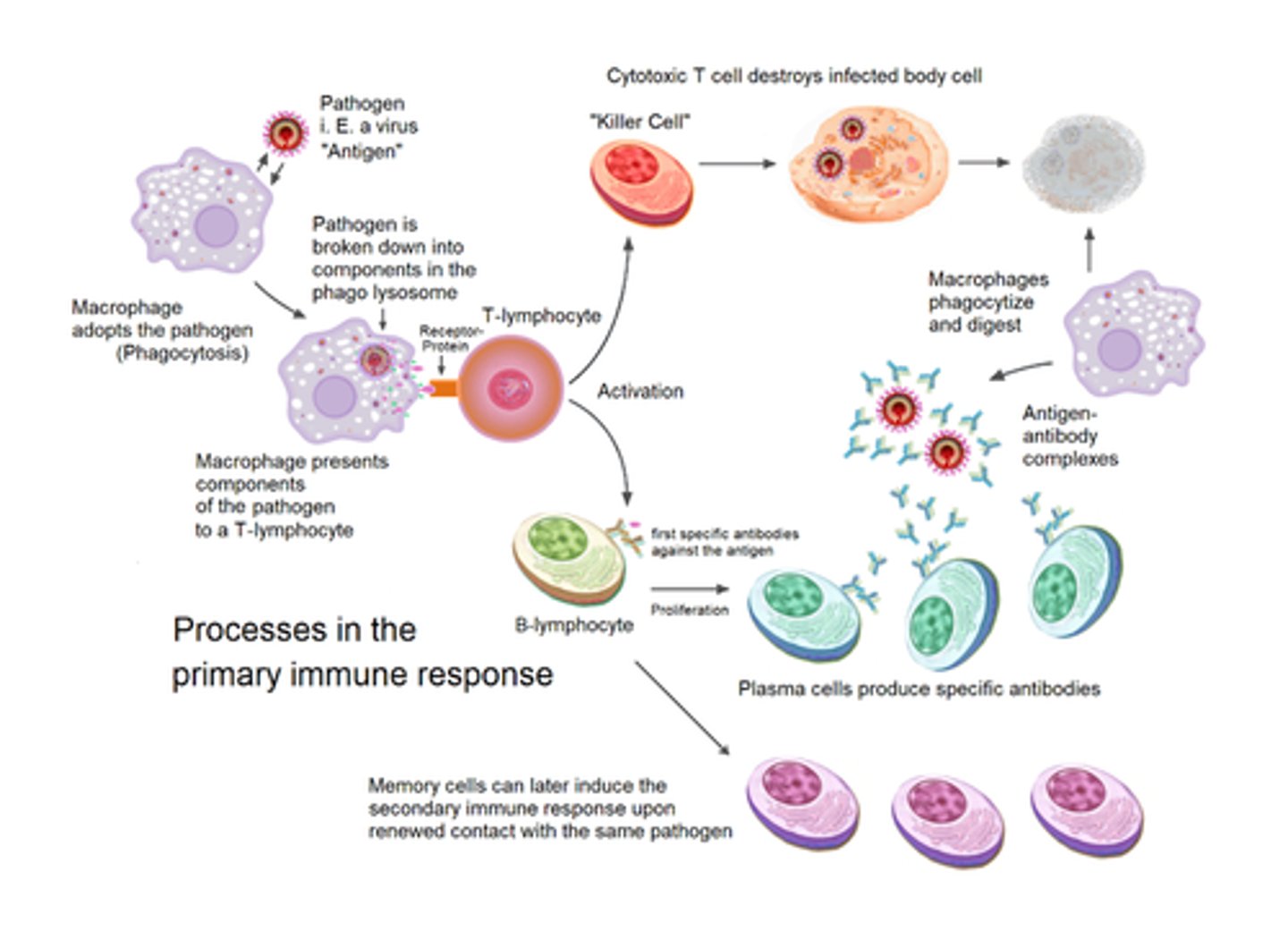
Phagocytosis- white blood cells engulfing pathogens and destroying them in a phagocytic vacuole with a lysosome. They become APCs, presenting an antigen-MHC complex on the surface. These bind to T cells.
Two types of phagocytes : macrophages, and neutrophils.
Neutrophils are the first responders and can easily squeeze out of capillaries.
Macrophages respond to cytokines from other T-cells.
[Natural killer cells are also part of the non specific cellular response]
6.8 Understand the roles of antigens and antibodies in the body's immune response including the involvement of plasma cells, macrophages and antigen-presenting cells.
APCs
antigen presenting cells that have processed and produced antigens on its surface
T cells, B cells, infected cells and macrophages can all be APCs
The antigen is presented with an MHC molecule on the cell surface
Macrophages
Macrophages circulate the blood
They move to sites of inflammation
Macrophages engulf pathogens/antigens. The pathogen is digested in a phagocytic vacuole when a lysosome fuses
the antigens are not digested
They become an APC
They may also engulf cell debris
Plasma cells
Produce antibodies.
Differentiated from B effector cells
Antibodies help by
Binding to antigens for:
clumping microbes together, making phagocytosis easier (agglutinisation)
lysing/bursting bacterial cells
marking them for phagocytosis
making soluble toxins insoluble
neutralising harmful toxins
Antibodies are Y shaped, heavy/light polypeptide chained with a hinge region (disulfide bonds) and a variable region.
6.9 Understand the roles of B/T cells in the body's specific immune response.
Specific immune response
Antigen specific, responds to one type of pathogen only.
This response relies on lymphocytes produced in the bone marrow.
cellular immunity means being able to respond to cancerous/infected cells
humoral immunity means circulating antibodies in the blood/lymph in response to toxins/pathogens
T cells= cellular immunity (but t-helper involved in humoral)
Mature in the Thymus.
T-Lymphocytes only respond to antigens that are attached to a body cell.
Inactive T cells:
•have receptors
•these bind to a specific antigen, causing it to be activated, and differentiate into T killer or T helper cells
T helper cell
1) when it binds to a complementary cell, it rapidly divides
Releases cytokines:
-->involved in activation of B cells
--->stimulates clonal expansion of T killer cells
--->makes macrophage APCs more efficient
It also helps mark pathogens for phagocytosis
T killer:
1) binds to infected cells/cancerous cells' antigens
2) once it binds, it instructs the cell to self-destruct
3) It is stimulated by complementary T helper cells
4) it multiplies rapidly to form clones:
active T killer cells or T memory cells
T memory:
•remains in the body to ensure faster secondary response if antigen is encountered again
B cells= humoral immunity
•mature in the Bone marrow
•involved in helping circulating antibodies in blood/lymph
•responds to pathogens/toxins
•binds to antigen, engulfs the antigen, becomes an APC
Differentiates into:
B Effector cells: differentiates further into plasma cells that produce antibodies
B Memory cells: remains in the body for a faster secondary response.
6.10 Understand how one gene can give rise to more than one protein through posttranscriptional changes to messenger RNA (mRNA).
One gene can give rise to more than one protein through posttranscriptional changes to messenger RNA (mRNA).
Introns (non coding DNA) are present on a gene, separated by exons (coding DNA).
During transcription, both introns and exons are copied
During post transcriptional modifications through splicing, introns are removed, leaving only the exons.
Different combinations of exons can be joined, to translate for different proteins, all from one mRNA
6.11 i) Know the major routes pathogens may take when entering the body.
Major routes pathogens take when entering the body
Cut skin
Digestive system (contaminated food)
Mucosal surfaces (genitals, nose or mouth)
Through respiratory system
6.11 ii) Understand the role of barriers in protecting the body from infection, including skin, stomach acid, and gut and skin flora.
Barriers protecting body from infection
Skin-dry, watertight barrier. Acidic pH preventing most bacteria from growing.
Skin flora-microorganisms secrete fatty acids that inhibit colonisation by other microbes
Stomach acid- most pathogens are killed by the acid
The gut-many microorganisms present, can outcompete pathogens for nutrition and space.
6.11 ii (extended info on gut/skin flora)
Gut/skin flora:
•they secrete chemicals/lactic acid that help destroy pathogens
•they outcompete pathogens as they are better adapted to the body's conditions
6.12 Understand how individuals may develop immunity (natural, artificial, active, passive).
Natural immunity
When individual is exposed to pathogen, and memory cells remain in body for a secondary specific immune response
Artificial immunity
Vaccination
Active immunity
When the body itself makes antibodies after encountering antigen
Passive immunity
When antibodies made by another organism enters the immune system of antoher
6.13 Understand how the theory of an 'evolutionary race' between pathogens and their hosts is supported by the evasion mechanisms shown by pathogens.
Evolutionary race: immune system evolution vs pathogen evading immune system evolution
Pathogens have developed some evasion mechanisms:
e.g.
•HIV killing host T cells to reduce effective immune cells of body
•TB virus, when dormant, preventing the lysosome from fusing with the phagocytic vacuole
•New strains of viruses appearing due to mutation means a different specific primary response has to occur
6.13 (PART B) Understand how the theory of an 'evolutionary race' between pathogens and their hosts is supported by the evasion mechanisms shown by pathogens
Pathogens can evolve faster than organisms such as ourselves because they replicate much faster
Another mechanism that pathogens use to evade immune systems:
•Conjugation: Type of cell-cell contact that allows the transfer of replicated Plasmid DNA between two bacteria
6.14 Understand the difference between bacteriostatic and bactericidal antibiotics.
Bactericidal:
Directly kills bacteria by preventing bacterial cell wall formation. This leads to lysis of the cell.
Bacteriostatic
Slows down growth/replication by preventing protein synthesis or DNA replication
CORE PRACTICAL 15:
Investigate the effect of different antibiotics on bacteria.
Equipment
•Petri dish, agar seeded with E.coli
•Sterile forceps & paper discs
•Marker pen
•Mast ring with at least 5 different antibiotics
•Ruler
Method
•Place the mast ring with antibiotics on the petri dish.
•Close dish, seal with tape but leave a gap to allow for aerobic conditions
•Leave cultures to incubate at 25°C for a day.
•Use ruler to measure the radius of the clear zone of inhibition circle for each antibiotic disc.
Control
The control can be a plain paper disc with distilled water, to compare results with
Calculation
Area of zone of inhibition =πr² , r being measured by ruler.
Means can be calculated from repeats.
Conclusion
Disc with biggest ZoI= most effective antibiotic
6.15 Know how an understanding of the contributory causes of hospital acquired infections have led to codes of practice regarding antibiotic prescription and hospital practice that relate to infection prevention and control.
Patients are prone to infection.
Codes of hospital practice
•Wash hands before and after visiting patients
•Disinfect hospital beds/surfaces
•Quarantine patients with hospital acquired infections
Preventing antibiotic resistance
•Preventing prescription of antibiotics for minor infections or for preventing infections
•Prescribing more specific antibiotics for a particular bacterium
•Rotate the antibiotic
•Patient should adhere to treatment plan