Vitreous Humor
1/41
There's no tags or description
Looks like no tags are added yet.
Name | Mastery | Learn | Test | Matching | Spaced |
|---|
No study sessions yet.
42 Terms
Where is the vitreous humor located?
Adjacent to the lens and zonules, pars plana, the retina, and the optic disc. Takes up the posterior 80% of the globe.
What is the composition of the vitreous?
It’s similar to the AH but also contains a high content of collagen (II) and Hyaluronic Acid. Its functional cell is the hyalocyte which are similar in function and lifespan to keratocytes.
What is the function of the collagen and HA in the vitreous?
Provides consistency and transparency
Hyalocyte
Endogenous pluripotent cell of the vitreous humor. Responsible for secretion of the fibrinous component of the VH, phagocytosis of debris, act as macrophages for anti-inflammatory purposes, and have a contractile capability for fibrinous scar production.
Vitreous surfaces
Split into a posterior and an anterior region; made up of fine collagen fibers
Anterior hyaloid surface
More “formed” to keep out the AH but still very thin to promote transparency; anterior relative to the ora serrata
Posterior hyaloid surface
Less formed; limiting membrane helps to keep separation from the retina; posterior relative to the ora serrata
Where does the vitreous attach to the eye?
Vitreous base; Lens (Hyaloideocapsular ligament/Ligament of Weiger); Posterior pole (Peripapillary and Perimacular attachments)
Vitreous base attachment
Thickest collagen fibers here, which run perpendicular to the ILM and the hyaloid surface
Hyaloideocapsular Ligament/Ligament of Wieger
Posterior lens attaches to the anterior vitreous face and the posterior zonules (orbiculoposterior fibers) are involved in attachment
Patellar/Hyaloid Fossa
Space where the lens hangs out
Space of Berger
Potential space underneath the lens
Peripapillary Attachment
Ring of attachment at the optic disc, around the optic nerve.
Perimacular Attachment
Loose attachment at the macular region around large retinal blood vessels
How is the posterior hyaloid surface attached to the retina?
Attachment via collagen fibers that attach the ILM of the retina to the posterior face of the vitreous
Posterior Vitreal Detachment
Posterior vitreal face pops off and pulls away from the retina
Zones of the vitreous
Vitreal cortex (anterior and posterior), Intermediate zone, and central zone
Vitreal Cortex
Outermost zone surrounding the vitreous; made up of a thin layer of collagen fibers and HA; interior to the vitreal surface. High concentration of collagen and HA contributes to structural stability
Anterior cortex
Anterior to the vitreal base and adjacent to the ciliary body, posterior chamber, and the lens.
Posterior cortex
Extends posterior to the vitreal base
Intermediate zone
Core of the vitreous. Fibers are continuous and arise in the vitreal base. Decreased concentration of HA and collagen when compared to the cortex
Vitreal base
Part of the intermediate zone near the ora serrata. The cortex goes around it. It separates the anterior and posterior cortex as well as pars plana from the retina.
Central zone
Region at the center of the vitreous which runs from the posterior middle of the lens to the optic nerve. Contains Cloquet’s canal, the patellar fossa, and the prepapillary attachment.
Cloquet’s Canal
It is a leftover remnant of the Hyaloid artery that supplied nutrients to the eye during embryology.

1
Vitreous base (attachment)

2
Hyaloideocapsular ligament

3
Patellar fossa

4
Vitreal cortex

5
Berger’s space

6
Cloquet’s canal

7
Perimacular attachment/ bursa premacularis
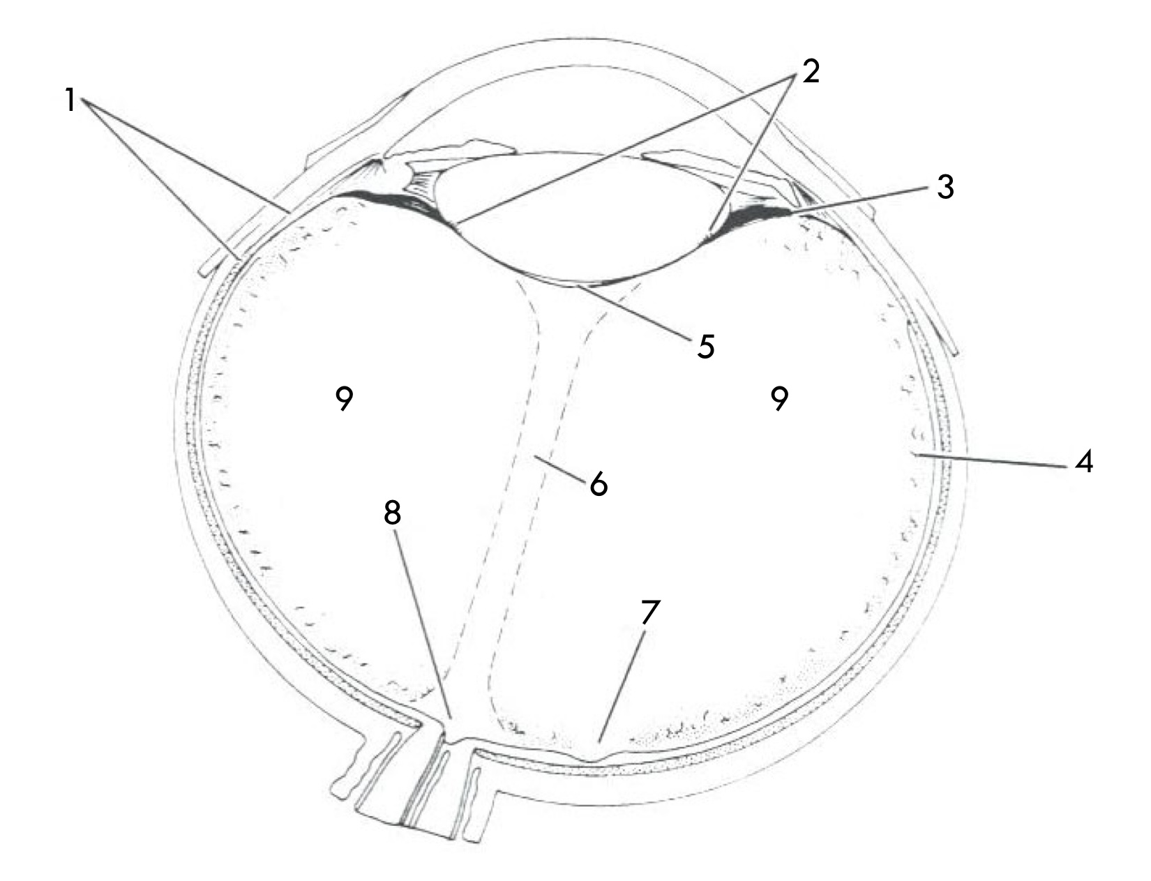
8
Peripapillary attachment/area of Martegiani

9
Intermediate zone/ vitreal core
Vitreous functions
Exchange of metabolites, support the retina, optical transmission, eye growth, barrier
Exchange of metabolites
Exchange with the retina, lens, the choroid, aqueous humor, optic nerve, and the ciliary body
Optical transmission
Transparency d/t Hyaluronic acid and water composition; has a lower refractive index than the lens
Eye growth
VH provides signals for eye growth. Can be normal or abnormal
Barrier function
Keeps the aqueous and the vitreous where they’re suppposed to be
How does the vitreous change over time?
With age, the liquid:gel ratio changes (gets more watery); the vitreous shrinks and may pull away; fibres aggregate into sheets and collapse into bands; in general there is a freeing up of water
Vitreal syneresis
Vitreal shrinking that may result in the vitreous pulling away from its attachments
Persistent remnants of the vitreous humor
Floaters are likely debris, noticed d/t their motion and shadows.
Floaters
Muscae Volitantes