6 - Development of Nervous System
1/15
There's no tags or description
Looks like no tags are added yet.
Name | Mastery | Learn | Test | Matching | Spaced | Call with Kai |
|---|
No analytics yet
Send a link to your students to track their progress
16 Terms
Axis within Adult Nervous System
Most of nervous system
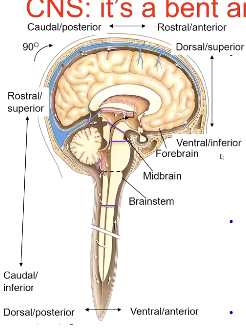
rostral — up (towards head)
caudal — towards tail
ventral (towards front) — anterior
dorsal (towards back) — posterior
ventricle demarcates the rostrocaudal axis — till midbrain
rostrocaudal axis of nervous system “neuraxis” bends approx 90 deg btn midbrain and forebrain
in human forebrain
rostral — sits anterior
caudal — sits posterior
ventral — sits inferior
dorsal — sits superior
remnant of embryonic brain flexure
Trilaminar Emrbyo (18 Days)
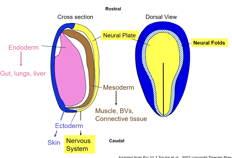
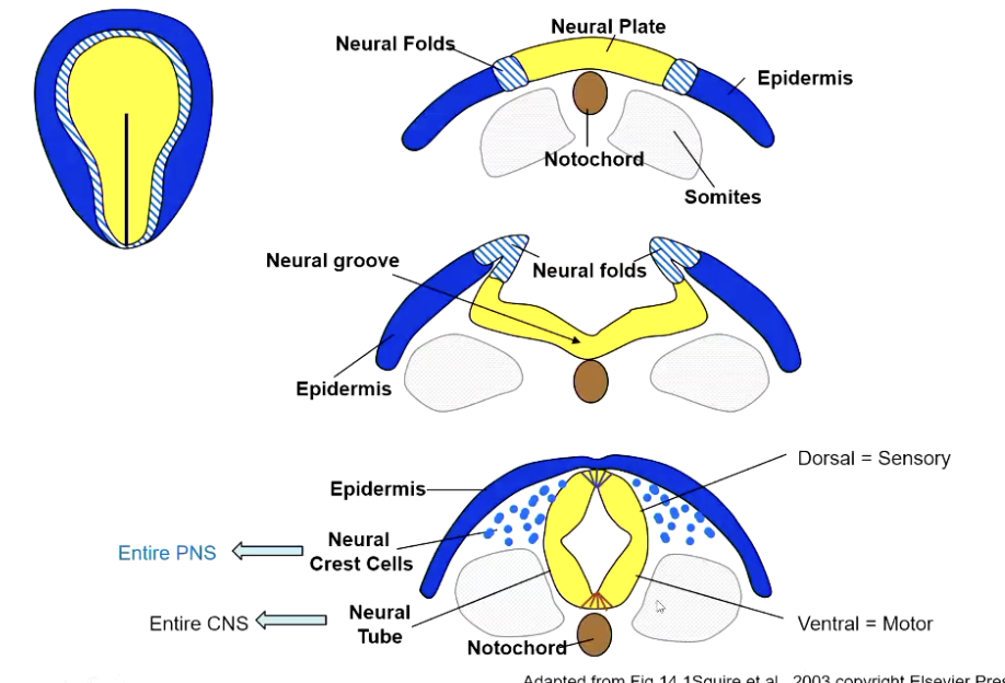
Folding of Neural Tube
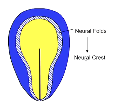
(1→2) neural folds fold downwards and push neural plate away from dorsal surface of embryo and start pushing it down inside
(1→2) simultaneously — midline of neural plate buckles in opposite direction — forms v shape going upwards
(2→3) next stage — neural folds reach midline — migrate inside and form neural crest cells — forms whole of the PNS
(2→3) medial border of epidermis fuse at the midline — one continuous layer of skin — separates NS from outside world
(2→3) lateral borders of neural plate meet in centre and form one continuous tube
(3) neural tube forms whole of CNS
(3) hollow in the centre — goes on to form ventricles
dorsal = sensory
ventral = motor
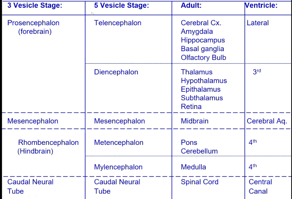
Differentiation of Neural Tube — 3 Vesicle Stage
1) prosencephalon (rostral end) — forms forebrain structures
2) mesencephalon — forms midbrain structures
3) rhombencephalon — forms hindbrain structures
4) caudal neural tube — develops into spinal cord

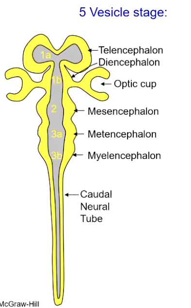
Differentiation of Neural Tube — 5 Vesicle Stage
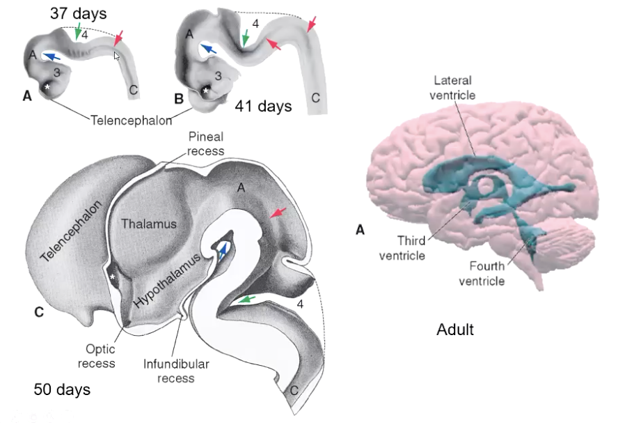
1a) upper subdiv of prosencephalon — telencephalon — forms cortex — hollow forms lateral ventricles
1b) subdiv 2 of prosencephalon — diencephalon — hollow bit forms 3rd ventricle — c shaped structure is optic cup (neural structures associated w eye forms)
2) mesencephalon
3a) upper subdiv of rhombencephalon — metencephalon — forms pons and cerebellum
3b) lower subdiv of rhombencephalon — mylenecephalon — forms medulla
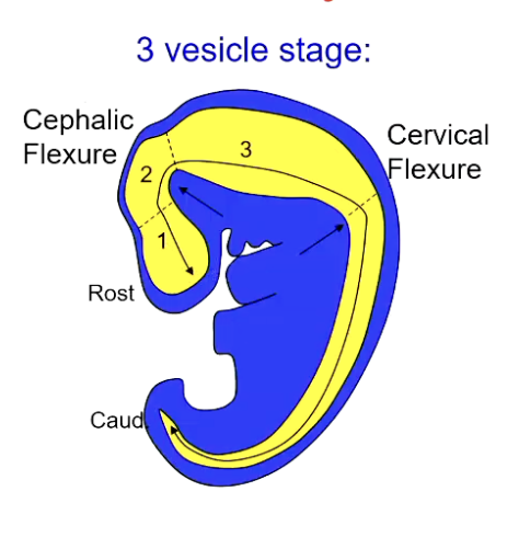
Embryonic Brain Flexures — 3 Vesicle Stage
1) bend between midbrain and forebrain — cephalic flexure
2) bend between spinal cord and brainstem — cervical flexure
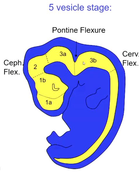
Embryonic Brain Flexures — 5 Vesicle Stage
1) ceph flex — maintained in adult
2) cerv flex
3) pontine flexure — divides upper half of brain stem (metencephalon) from lower vesicle (myelenecephalon) — responsible for open medulla/fourth ventricle
Derivatives of Neural Tube — Summary Sheet
Derivatives of Neural Crest
neural crest cells can migrate throughout the body and give rise to many types of cells e.g.
sensory neurons (DRG)
autonomic ganglia
enteric neurons
schwann cells
Neuroepithelial Stem Cells in Ventricular Zone + Microglia
produce diff CNS cells at diff times which then migrate into position
all neurons and most glia of CNS are generated from NSC
exception — microglia
this lineage migrates in from the yolk sac during early dev and then differentiates in CNS
brains immune cells that play key roles in brain dev, plasticity and health
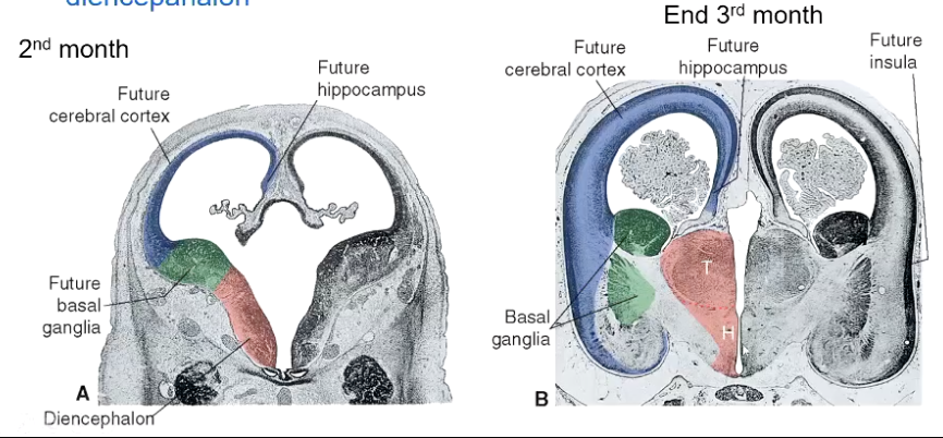
Shaping the Telencephalon — Weeks 6 to 12
basal part of telencephalon thickens to form pre-cursor of basal ganglia
diencephalon thickens to form thalamus and hypothalamus — separated by a sulcus
by end of 3rd month — massive fibre bundles form the internal capsule which connects the telencephalon and diencephalon
axons start to grow in and out of cortex into thalamus — create massive white matter tracts incl internal capsules
Expanding the Cortex
up to 15 million neurons born per hour
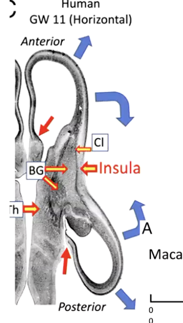
centre of cerebral hemispheres anchored to deep structures below (basal ganglia, region of future insula)
anterior and posterior regions are free to move and fill available space
Formation of Temporal and Frontal Lobes
bones of skull and attachment via insula constrain the shape of the rapidly expanding cortex
frontal lobe expands to fill anterior region
originally caudal regions get pushed ard by skull to lie inferior to insula — forming temporal lobe
results in formation of great C-shaped cortical arc
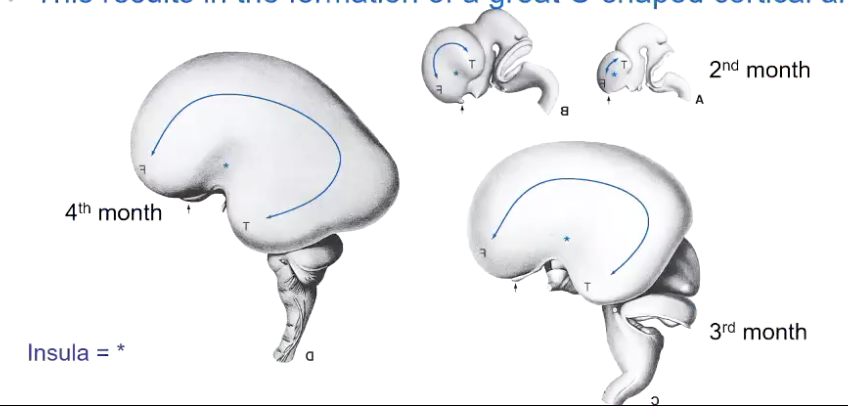
cavity of neural tube is drawn into C shape — forming lateral ventricles in the telencephalon
Brain Growth Continues After Neurogenesis Finishes?
because
neurons grow — somata + dendrites
axons grow
synapses form (and are refined)
glia cells born
axons myelinated
together — causes cortical sheet to grow a little thicker — huge increase surface area
expanding cortical sheet gets pushed into hills (gyri) and valleys (sulci)
enables developing brain to make best use of available volume
compared to adult brain volume
birth — 25%
2 years — 80%
14 years — 100%
Rates of Maturation Differ Greatly Btn Brain Regions
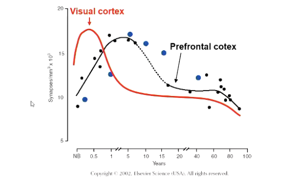
measures synaptic density
overproduction and then synaptic pruning
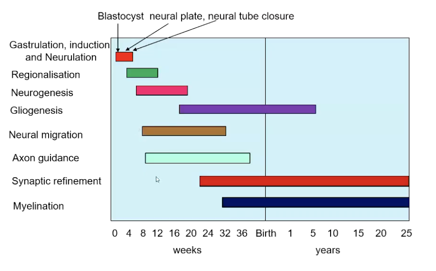
Timeline of Human Brain Development