digestive system, comparative anatomy of the liver and pancreas
1/19
Earn XP
Description and Tags
Describe the topography of the liver and pancreas in the domestic species ✓ Describe the lobulation of the liver and pancreas in the domestic species ✓ Identify the isolated liver and the pancreas of each of the domestic species ✓ Describe the microscopic structure of the associated organs within the digestive system ✓ Describe the type of cells within the associated organs ✓ Describe the function and the ducts of the liver and the pancreas ✓ Describe the blood supply and venous drainage of the liver and the pancreas
Name | Mastery | Learn | Test | Matching | Spaced |
|---|
No study sessions yet.
20 Terms
what are the functions of the liver?
Detoxification and excretion (waste products)
Secretion (Bile)
Storage (lipids, vitamins A and B, glycogen)
Synthesis (Albumin, clotting factors, globulins)
Protein, carbohydrate and lipid metabolism
Immune function (Kupffer cells)
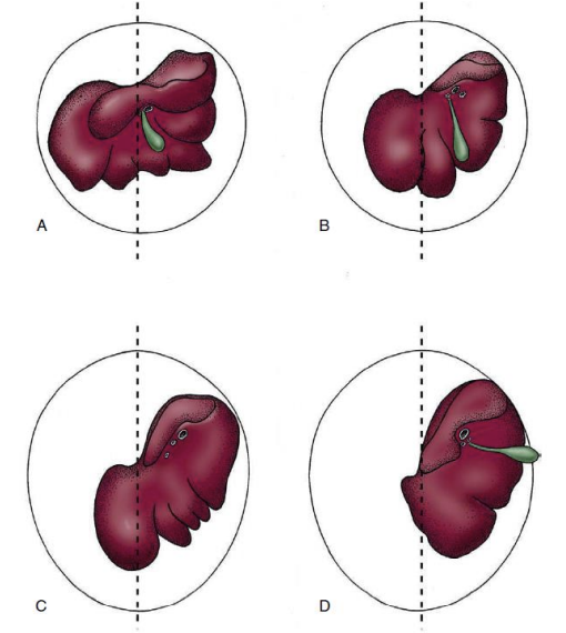
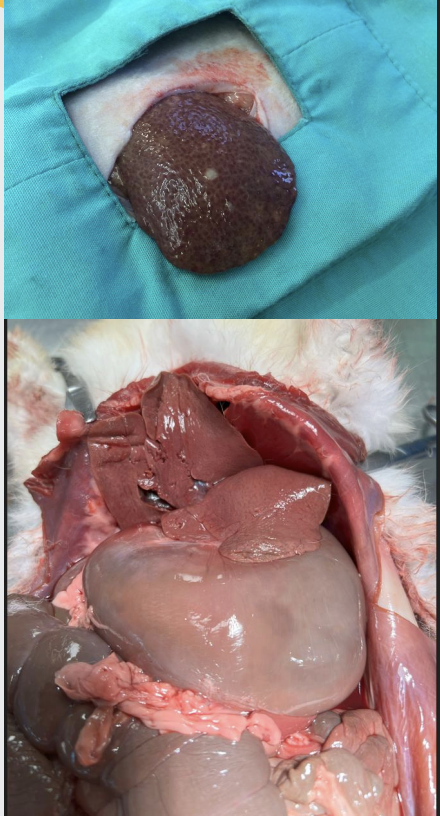
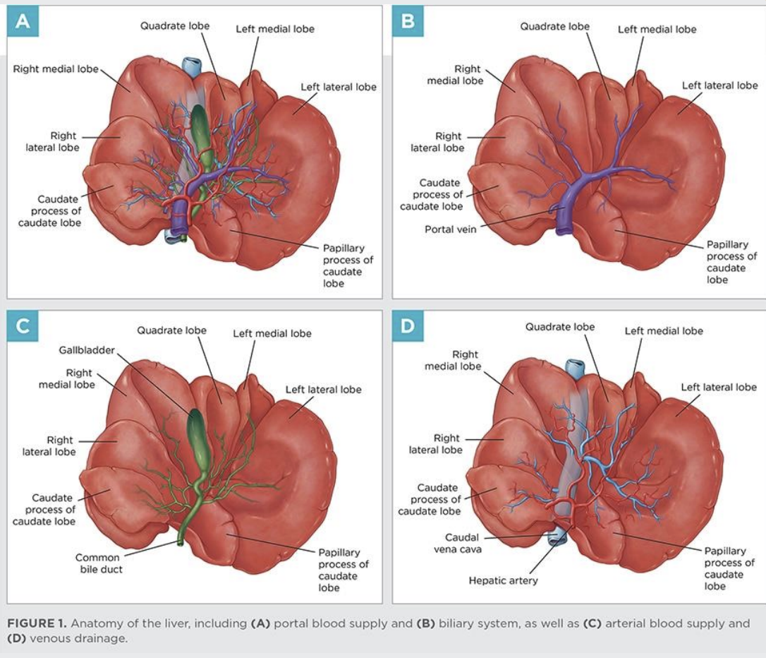
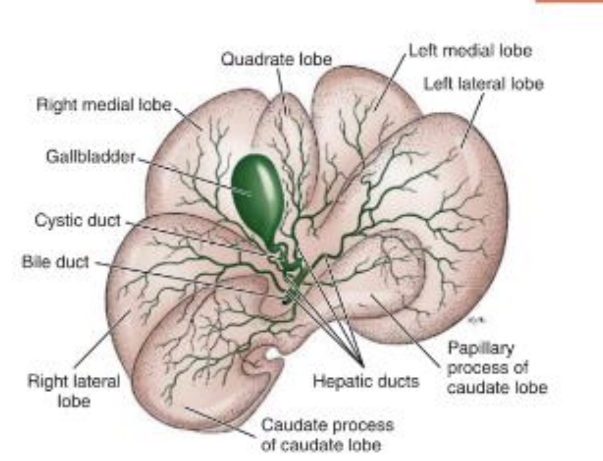
how do we compare the liver in the body?
it is the largest gland in the abdominal cavity, caudal to the diaphragm
bulk lies to the right in all species and in ruminants entirely moved to the right
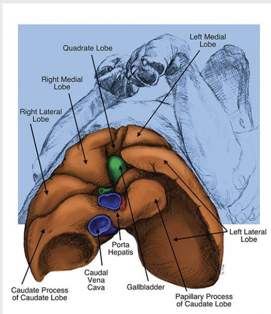
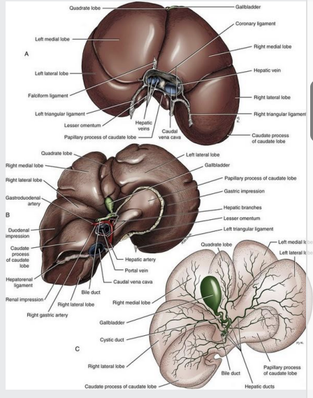
gall bladder between the right medial and quadrate lobe
some species have no gallbladder in the fossa (horses, rats)
A=dog
D=ruminant
whats the surface of the liver like?
strongly convex surface towards the diaphragm (parietal surface or diaphragmatic surface)
concave towards viscera (lies against and impressed by stomach, duodenum, etc) hence visceral surface
the visceral surface is marked by the porta (gate) of the liver
what is the porta of a liver
image shows dog lying on its back (paws on top)
the visceral surface of the liver
bile duct, portal vein, and hepatic vessels enter and leave)
porta is allso known as. the hilar region on the liver
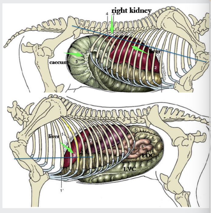
what is the topography of the liver
Located cranial part of the abdomen
Cranial to liver is diaphragm
Caudal to liver is the stomach, intestinal mass, and right kidney
The right kidney makes contact with liver
Adopts to the form of adjacent organs (e.g. renal impression
of the caudate lobe)
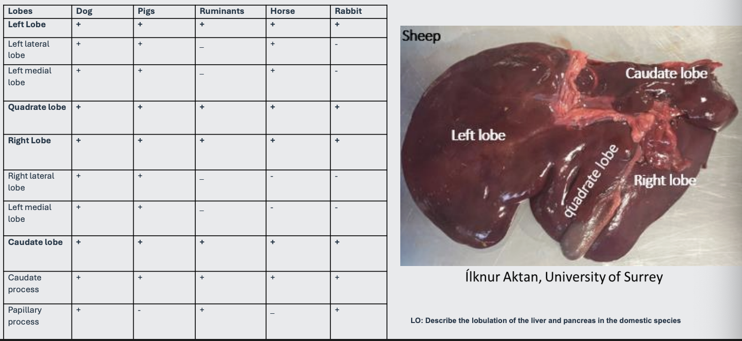
what is the liver lobes
lobes are created by fissures (indentations)
where is the liver located in the dog?
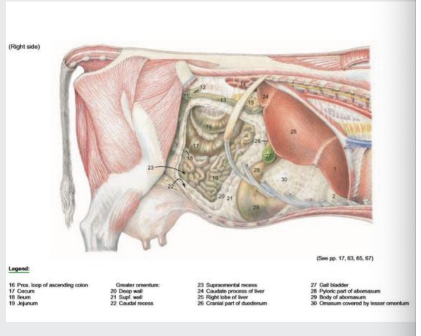
Liver is related on the left to the stomach (spleen); and on the right to the pancreas,
kidney, and duodenum; and ventrally to the greater omentum
Liver reaches the umbilical region on the ventral abdominal wall
Its most caudal part covers the cranial extremity of the right kidney and reaches the 13th thoracic vertebra
Extends slightly beyond the costal arch (lower edge of ribs cartilage bit -where it connects to sternum) ventrally (more so in pups and in congestive heart failure)
Liver biopsy-puncture caudal to the xiphoid process
xyphoid process=tip of sternum - can pulpate - where you puncture to get biopsy of liver
where is the liver located in the horse?
Lies completely within the ribcage
Located obliquely across the diaphragm with the left lobe ventral and right lobe dorsal
Caudal margin reaches the 15th ICS
You can do liver biopsy at either the :
12th ICS (intercostal space- count from caudal end): on a line between the tuber coxae (hip) and point of the shoulder (right-hand side)
8th ICS: at the level of the deltoid tuberosity of the humerus (right-hand side)
liver in rabbit
The liver has five lobes with a deep cleft dividing it into right and left lobes
Caudate lobe has a narrow attachment to the porta region of the liver and could be a site of liver torsion
Gallbladder present
Liver biopsy-Ultrasound-guided biopsy can be taken ventrally from
any lobe
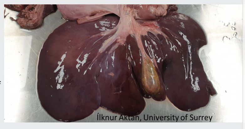
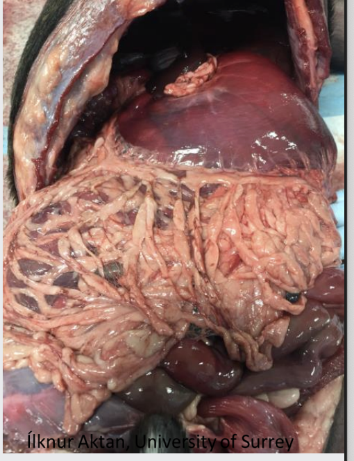
cow liver
Liver reaches dorsally as far as the last rib
The gall bladder also rests 10th ICS
Note the left lobe of the liver here (1), right lobe would be 24/25 (imagine liver rotated forward)
Liver biopsy -10th ICS, ¼ of the length of the rib
what are the peritoneal folds and ligaments
Peritoneal folds serve more to fix organs in position than as channels for blood vessels. These are called ligaments: -supportive function- attach liver firmly
Coronary ligament- surround the caudal vena cava-attaches liver to the central tendon of the diaphragm
Triangular ligaments extend between the dorsal part of the liver on each side of the diaphragm
Right and left triangular and coronary ligaments that pass to the diaphragm from the parietal surface have fibrous cores
Liver is covered by serosa (visceral peritoneum) overlying a thin connective tissue capsule (tunica fibrosa-especially strong in Pigs)
Some peritoneal folds have no supportive function but carry blood Vessels, nerves, and lymphatics
Falciform ligament- between the liver and the diaphragm, and the ventral abdominal wall
Round ligament –a fibrous strand in the free edge of the falciform ligament (umbilical vein remnant)
Hepatoduodenal (liver to duodenum) and Hepatogastric (liver to stomach) ligament- (Lesser omentum)
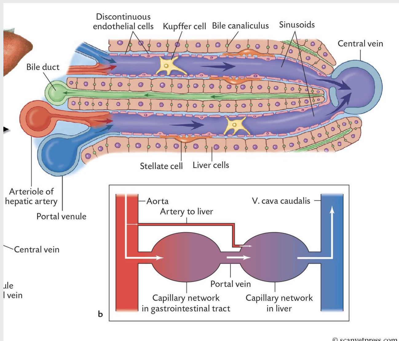
what is the blood supply like in liver?
dual
Hepatic artery
-Fresh oxygenated (nutritional) blood
-Branch of the coeliac artery
Hepatic portal vein
- Large vein carrying nutrient-rich (functional) blood from the unpaired organs (like intestines, pancreas, stomach, spleen, not kidneys cos theres two of them) into the liver
-Supplying 60-70% of the blood flow
then the blood coming from the artery and portal vein mix once theyre in the liver
tell me more about the blood vessels in the dual blood supply of the liver
image shows how the blood from the artery and vein join in one tube in the middle (sinusoid)
The hepatic portal vein receives the venous blood form all unpaired abdominal organs (stomach, pancreas, spleen and intestines)
Blood from the portal vein and hepatic artery branches mixes within the hepatic sinusoids before collecting in central veins
These central veins merge and eventually form hepatic veins that drain into the abdominal portion of the caudal vena cava
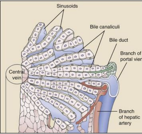
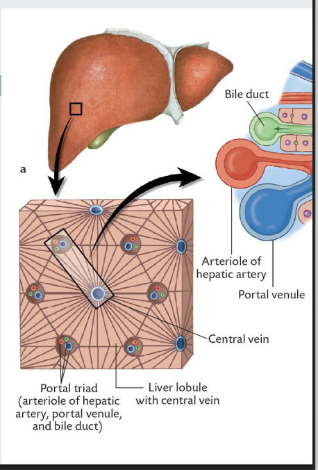
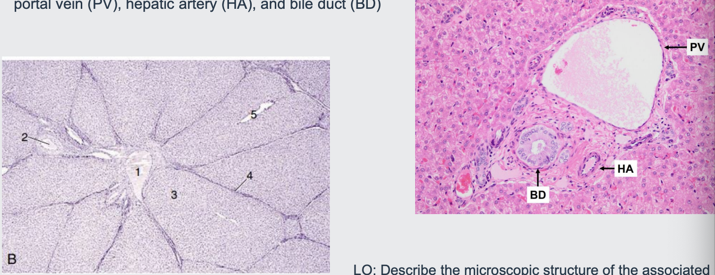
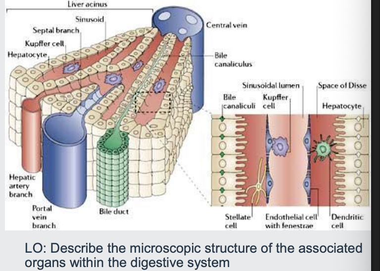
what is the hepatic lobule?
The basic structure of the liver is the hepatic lobule-hexagon shaped
At each corner - an arteriole (from hepatic a.); a venule (from portal vein) and a small bile duct form the bile duct
These three structures are commonly referred to as portal triad
Each portal triad may provide blood to the several adjacent hepatic lobules
surrounded by hepatocytes
lets break down this portal triad
sinusoids are very leaky to soak the hepatocytes
kupffer cells catch harmful like pathogens or tumours
Arteriole and venule empty their blood into the spongy liver lobule
Blood capillary beds that run between portal triad and central vein
Capillary beds are lined by fenestrated, leaky endothelium known as liver sinusoids
Blood flowing into the lobule is drained out via the central vein
central vein then eventually goes to the cranial vena cava - its a loop in terms of circulation
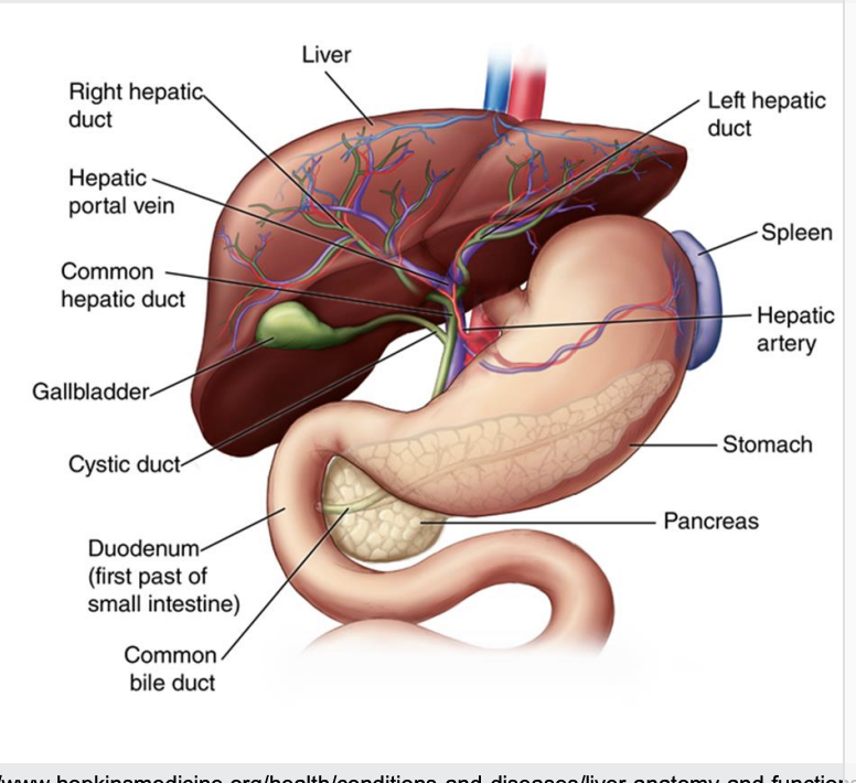
what is the bile and bile duct
whilst theres a portal triad we also have the bile duct next to it
Bile is produced by the sheets of hepatocytes
Discharged into bile canaliculi (microscopic channels)
Canaliculi unite to form the interlobular ducts -lobular ducts
Hepatic ducts joins with cystic duct (the one that attaches to gall bladder-think of it as like holding the cyst) and the bile duct (its the one that enters the small intestine)
hepatic ducts are slightly larger than bile duct
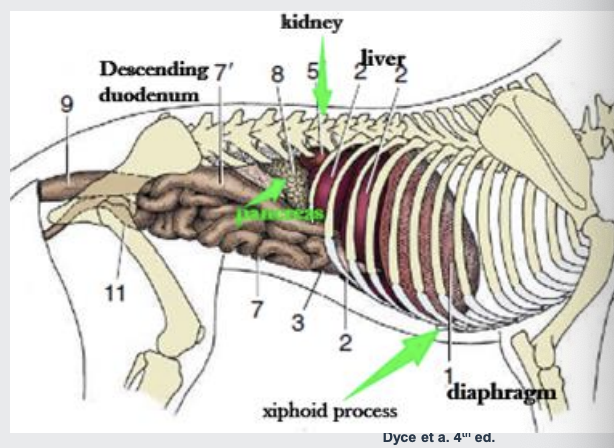
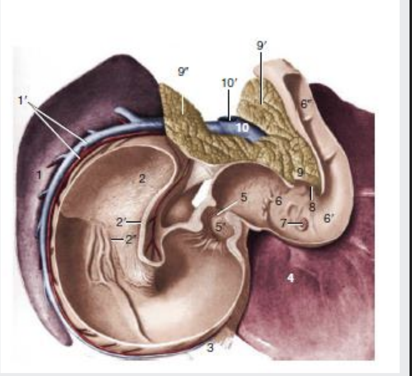
what is the pancreas
it is located in the dorsal part of the abdominal cavity, often abuts the stomach and liver
Small lobulated gland with two lobes joined by the body which is in contact with the pylorus
Right lobe (7) is within mesoduodenum next to DD
Left lobe (5) lies in the deep leaf of the greater omentum
Coeliac and cranial mesenteric a. branches supply blood
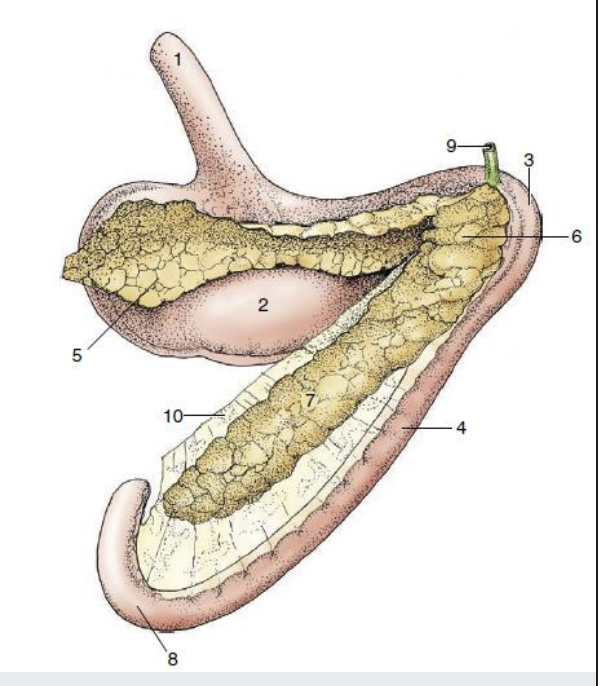
what are the pancreatic ducts
Pancreatic duct (greater) opens into the duodenum together or just beside the bile duct
Accessory pancreatic duct (lesser) opens on the opposite
In some species only one duct commonly survives (sheep, pig)
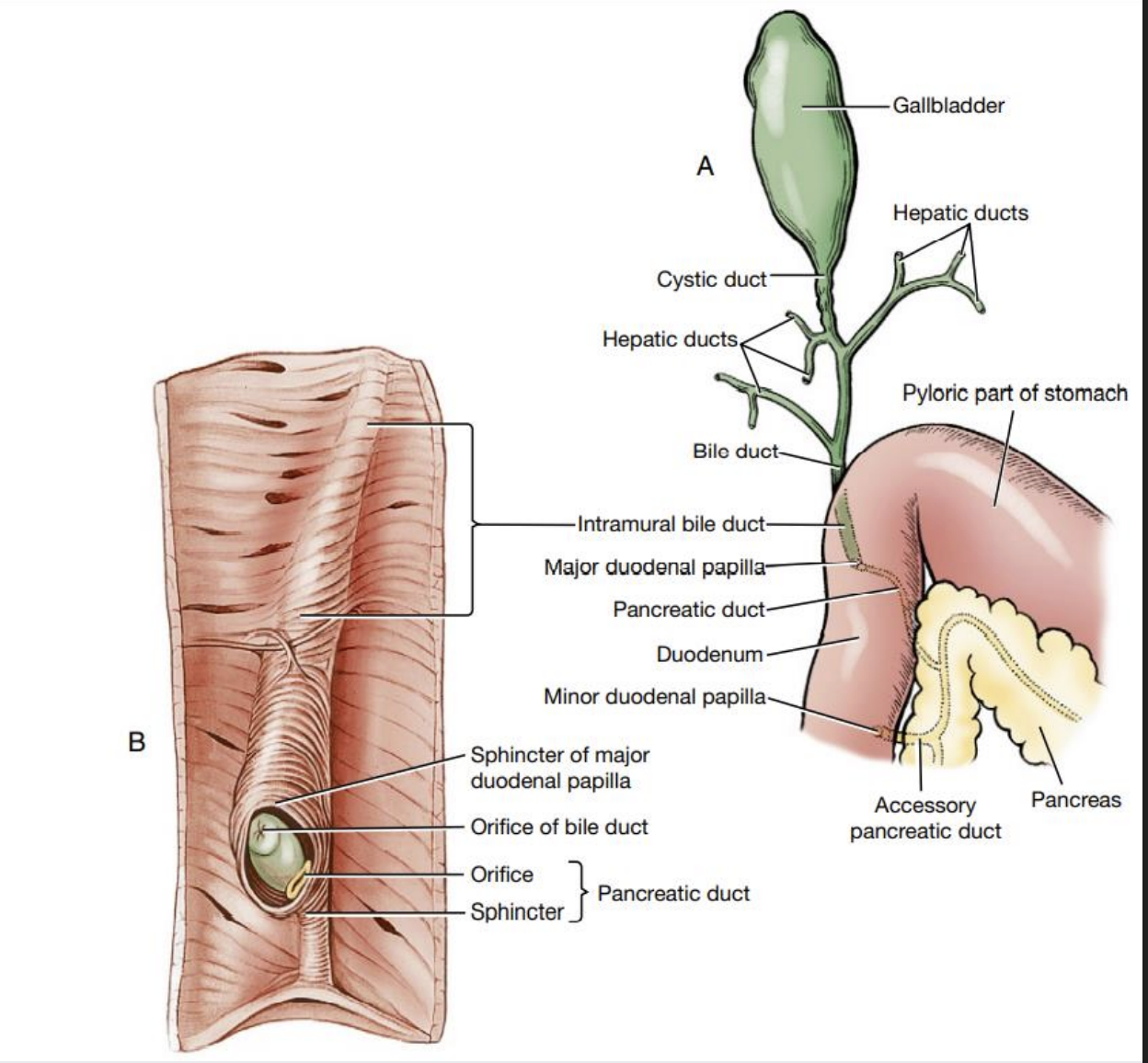
what do the pancreatic and bile ducts compare in each species
Horse- Bile and pancreatic ducts open at the first bend of the duodenal sigmoid flexure
Dog-common bile duct enters the duodenum at the major duodenal papilla adjacent to, the pancreatic duct
Cat-common bile duct and the pancreatic duct conjoin just before they enter into the major duodenal papilla
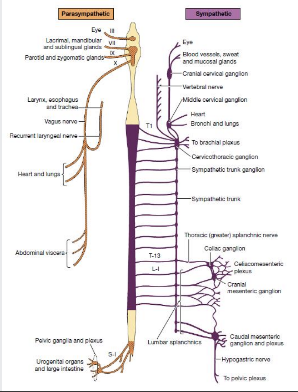
how is the liver innervated?
Liver is innervated both sympathetic and parasympathetic nerves
It receives fibers from the vagal trunk and sympathetic axons from celiac plexus/ganglion
Vagal axons reach the abdomen by passing through the diaphragm with the oesophagus