pancreas pt.1
1/26
There's no tags or description
Looks like no tags are added yet.
Name | Mastery | Learn | Test | Matching | Spaced | Call with Kai |
|---|
No analytics yet
Send a link to your students to track their progress
27 Terms
Normal anatomy of pancreas
Dumbbell or sausage shaped
Non-capsulated
Lies transversely in the retroperitoneal cavity
Extends from C-loop of duodenal to the splenic hilum*
Pancreas echogenicity to the liver
Isoechoic or hyperechoic to the liver
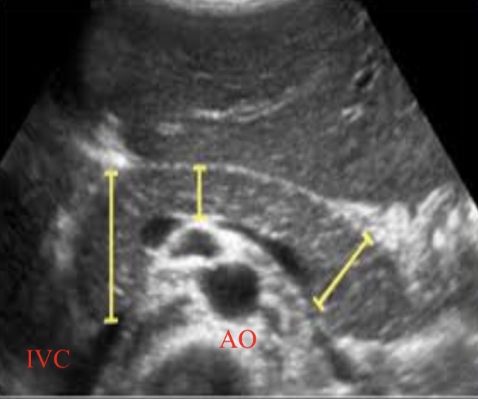
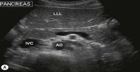
Posterior vascular landmarks for pancreas
IVC and aorta
Parts of the pancreas
Head, neck, body, uncinate process
Size of head, neck, tail
Head<3cm
Neck<2.5cm
Body<2.5cm
Tail<2.0cm
Hyperechoic to hypoechoic
KIDS LOVE SODA POP
Renal sinus, pancreas, spleen/liver, renal cortex
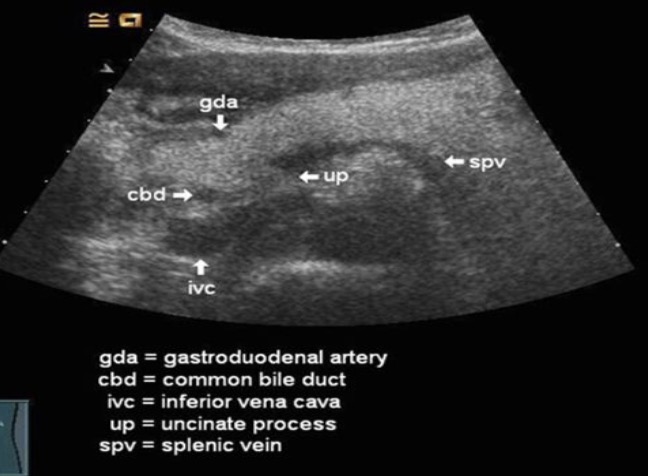
CBD in relation to pancreas
Seen at the posterior lateral margin of the pancreatic head

GDA in relation to pancreas
Can be seen at the anterior lateral margin
probe used for pancreas
2.5MHz curved linear probe
5MHz curved linear probe for very thin or young patients
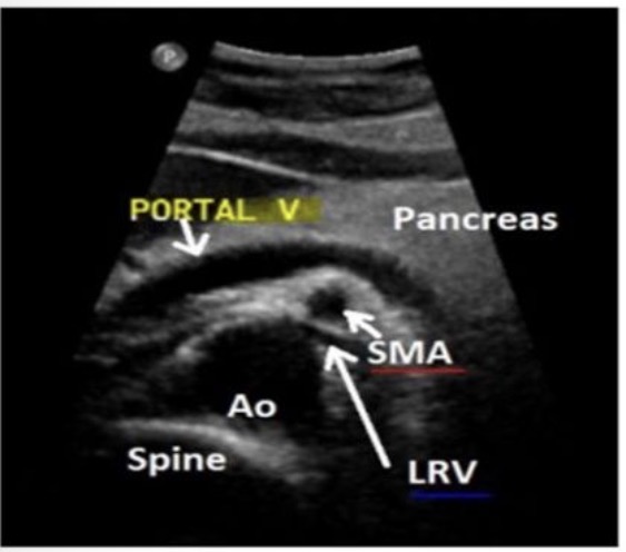
PV location in relation to the pancreas
PV is shown posterior to the head of the pancreas

SMA in relation to the pancreas
SMA is sandwiched between the aorta and splenic vein

left renal vein location in relation to the pancreas
LRV is anterior to the aorta and posterior to the SMA
superior border of the pancreas
the celiac axis
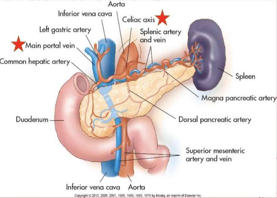
Posterior border of the pancreas
The portal venous system
The head of the pancreas lies
“in the lap” of the C-loop of the duodenum

The SMV in relation to the ucinate process and neck and body of the pancreas
The SMV crosses anterior to the ucinate process and posterior to the neck and body of the pancreas
The uncinate process
The small, curved tip at the end of the head of the pancreas
Most inferior portion of the pancreas
The head
Tongue-like extension of the pancreatic neck
The uncinate process
The portal vein is formed
posterior to the neck by the junction of the SMV and splenic veins
The pancreatic head and body is often included as
part of the body
The largest portion of the pancreas
the body
The portal splenic confluence lies posterior to
The neck of the pancreas
The splenic artery and vein form the
Posterior and superior border of the tail
The posterior wall of the stomach in relation to the tail of the pancreas
The stomach overlies the anterior borded of the tail of the pancreas
To better visualize the pancreas (specifically the pancreatic tail)
Lay the patient LLD, supine, or RLD
drink water to fill the stomach
Main pancreatic ducts
Duct of wirsung (connects directly to 2nd portion of duodenum
Duct of santorini (secondary duct that drains the upper anterior head)