Grade 11 Lab Assessment
1/98
There's no tags or description
Looks like no tags are added yet.
Name | Mastery | Learn | Test | Matching | Spaced | Call with Kai |
|---|
No analytics yet
Send a link to your students to track their progress
99 Terms
What is osmosis?
movement of water across a semipermeable membrane toward areas of high solute concentration
What is a hypertonic solution?
solution with a high solute concentration
Cells in a hypertonic solution will __
shrivel
in a hypertonic solution, water will __
move out into solution
What is a hypotonic solution?
solution with a low solute concentration
In a hypotonic solution water will__
move into the cell
Cells in a hypotonic solution will __
swell
What is an isotonic solution?
solution where solute/solvent concentration is equal
Proximal
close to the core
Distal
away from the core
dorsal
back view
Ventral
front view
Anterior
closer to the head/top
Posterior
closer to tail/bottom
What does iodine test for?
starch
if starch is present, what colour will the iodine turn?
purple/blue
if no starch is present, what colour will the iodine turn?
brown
What does Benedict's solution test for?
simple sugars or glucose using benedicts solution
if low amounts of reducing sugars are present, what colour will the solution be?
green
if medium amounts of reducing sugars are present, what colour will the solution be?
yellow
if high amounts of reducing sugars are present, what colour will the solution be?
orange/red
if no reducing sugars are present, what colour will the solution be?
blue/purple
What does Biuret test for?
proteins and amino acids
if protein is present, what colour will the solution be?
purple
if no protein is present, what colour will the solution be?
blue
An unknown solution turns purple with biuret's reagent and light blue with Benedict's Test. What would you expect this solution to contain?
protein
A solution changes to dark orange with Benedict's Test and purple with Biuret's reagent. What can you conclude about the sample?
contains sugars and protein
blood cells are covered in __
antigens
What are antigens?
Proteins on cell surface for cell recognition
What are antibodies?
Substances created in the body to attack foreign antigens.
A,B and Rh antigens determine__
blood type
If clumping (or agglutination) occurs, the _ is present as the antibody recognized and attacked it.
antigen
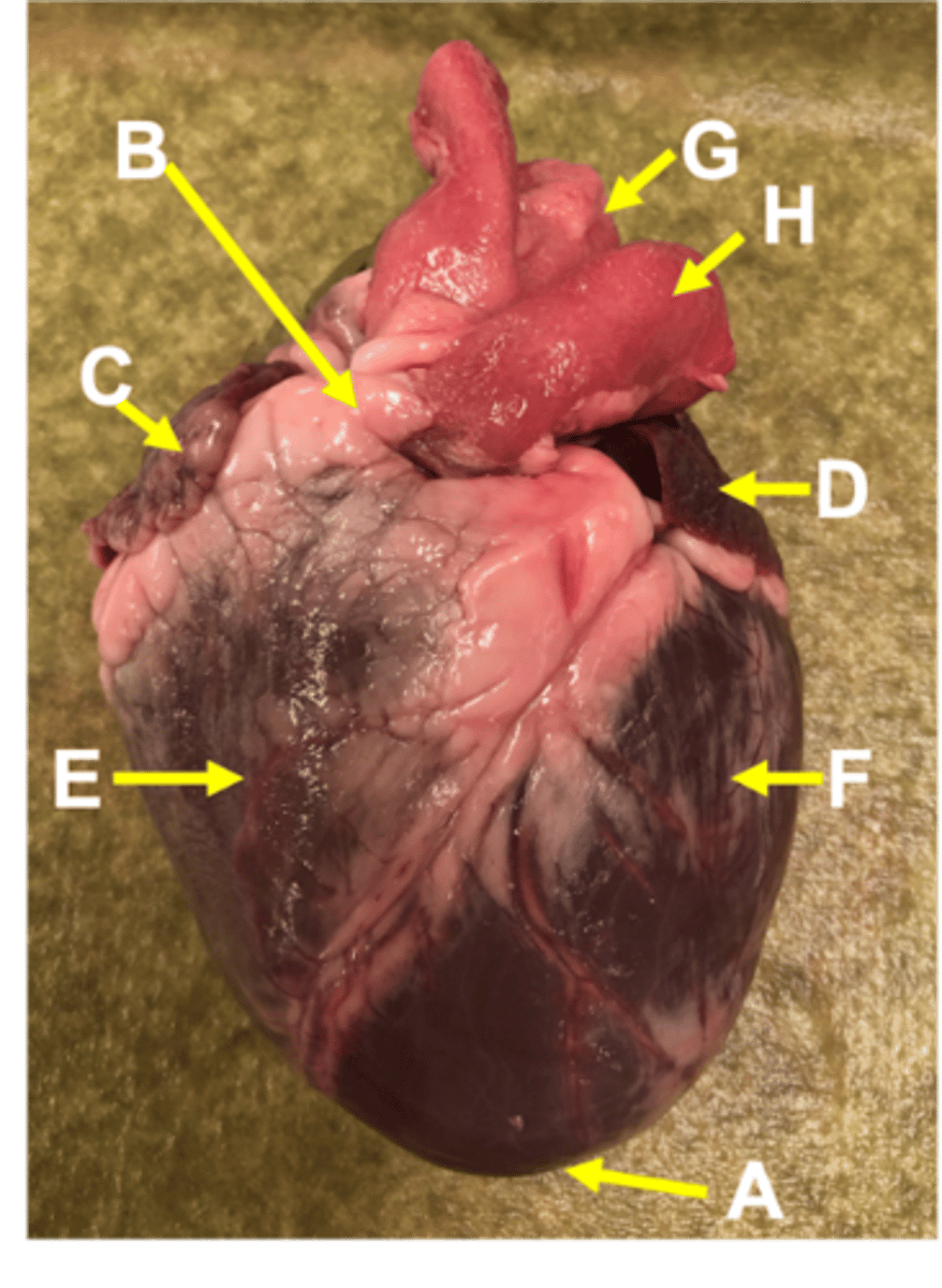
What view are you observing this heart in?
ventral

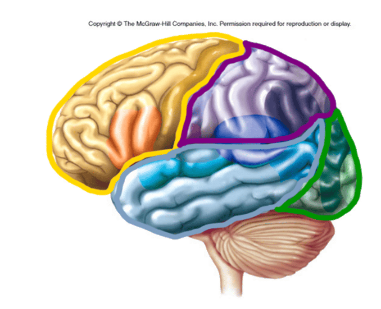
Which lobe is in yellow?
frontal

which lobe is in purple?
Parietal
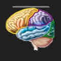
which love is in blue?
temporal

which lobe is green
occipital
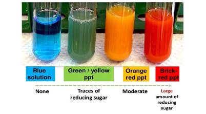
which test tube contains glucose
The test tube that turns orange contains glucose.
what does onion lab test show
Osmosis in plant cells using different concentrations of salt solutions, to draw water into or out of the cell
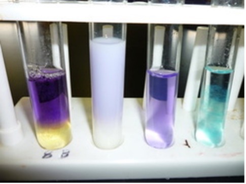
which sample contains proteins
turns purple contains proteins.
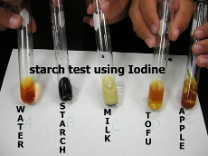
Which sample is positive for starch?
purple or black one
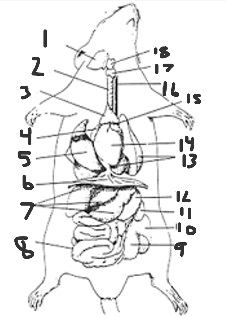
name them all
Submandibular salivary gland
trachea
thymus gland
right atrium
right lung
diaphragm
liver
small intestine
cecum
large intestine
spleen
stomach
left lung
ventricles
left atrium
esophagus
thyroid gland
larynx
treacha
resp sys
thymus gland
immune/lympatic system
right atrium
cardio sys
right lung
resp sys
disphragm
resp system
liver
dig sys
small intestine
digest sys
cecum
diges sys
large intes
digestive sys
spleen
Digestive sys
stomach
digestive system
left lung
resp sys
ventricles
cardio sys
esophagus
digestive
thyroid gland
endocrine sys
larynx
resp sys
which lobe is blue
temporal
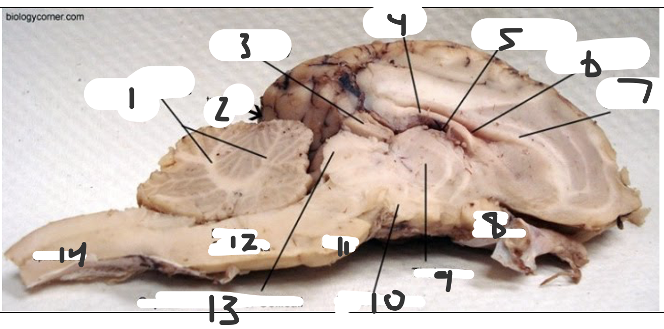
name each
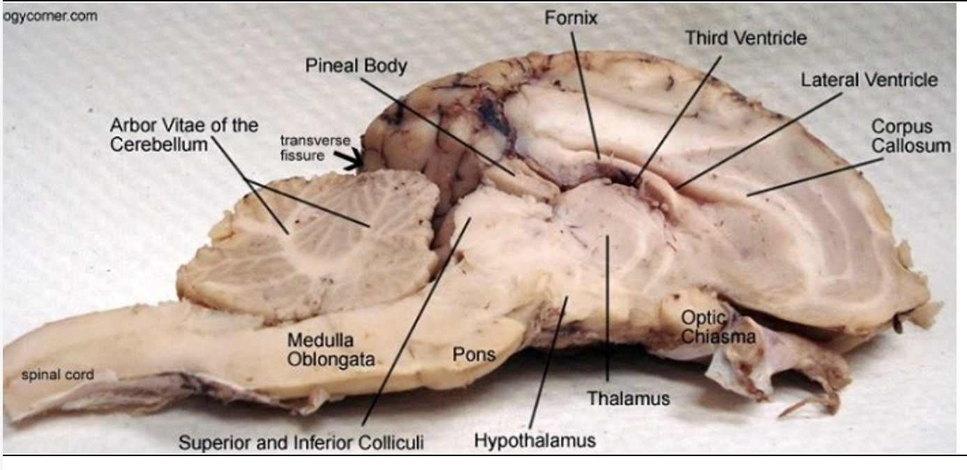
Pineal body
Midbrain (part of the diencephalon)
Fornix
Part of limbic system, connects hippocampus with hypothalamus
Third ventricle
diencephalon
Lateral ventricle
in each cerebral hemisphere
Corpus callosum
Connects the two cerebral hemispheres
Optic chiasma
diencephalon
Thalamus
Part of the diencephalon
Hypothalamus
diencephalon
Pons
brainstem
Superior and inferior colliculi
midbrain
Medulla oblongata
brainstem
Spinal cord
the brainstem down the vertebral column
Arbor vitae of the cerebellum
Cerebellum
Transverse fissure
Separates the cerebrum from the cerebellum
what blood typing results mean
Clumping means that antigen is present. I.e. crumps with anti A and Anti Rh meant A+
how many chambers in heart
4
Right Atrium
Receives deoxygenated blood from the body via the superior and inferior vena cavae.
Right Ventricle
Receives deoxygenated blood from the right atrium and pumps it to the lungs through the pulmonary artery.
Left Atrium
Receives oxygenated blood from the lungs via the pulmonary veins.
Left Ventricle
Receives oxygenated blood from the left atrium and pumps it to the body through the aorta.
Superior and Inferior Vena Cavae
Large veins that carry deoxygenated blood from the body to the right atrium.
Pulmonary Arteries
Carry deoxygenated blood from the right ventricle to the lungs.
Pulmonary Veins
Carry oxygenated blood from the lungs to the left atrium.
Aorta
The largest artery in the body, it carries oxygenated blood from the left ventricle to the rest of the body.
Deoxygenated Blood
Flows through the right side of the heart (right atrium and right ventricle) and is pumped to the lungs to become oxygenated.
Oxygenated Blood
Flows through the left side of the heart (left atrium and left ventricle) and is pumped to the rest of the body to deliver oxygen to tissues and organs.
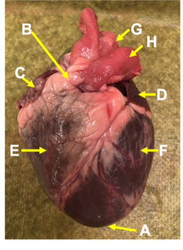
label each
a. apex
b. base
c.right atrium
d.left atrium
e.right ventricle
f.let ventricle
g. aorta
h.pulmonary trunk
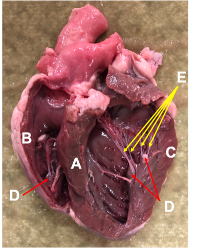
label each
a.interventricular septum
b.right ventricle
c.left ventricle
d.papillary muscles
e.chordae tendineae (heart strings)
segmental artery
Blood supplied to specific segments of the kidney.
renal medulla
Urine concentrated and transported through the collecting ducts.
fat in renal sinus
Adipose tissue.
renal column
Contains blood vessels, nerves, and renal tubules.
renal pyramid
Urine formed in the nephrons
arcuate vein
Collected blood from interlobular veins.
minor calyx
Urine from the renal papillae
renal papilla
Urine from the collecting ducts.
interlobar vein in renal column
Collected blood from the arcuate veins.
renal capsule
doesnt collect anything
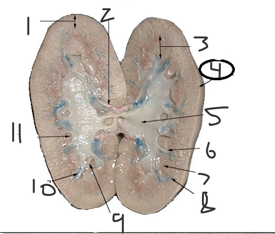
label
1 renal cortex
2 segmental artery
3 renal medulla
4 renal capsule
5 fat in renal sinus
6 renal column
7 renal pyramid
8 arcuate vein
9 minor calyx
10 interlobar vein in renal column
11 renal papilla