Neuroscience 2
1/64
There's no tags or description
Looks like no tags are added yet.
Name | Mastery | Learn | Test | Matching | Spaced | Call with Kai |
|---|
No analytics yet
Send a link to your students to track their progress
65 Terms
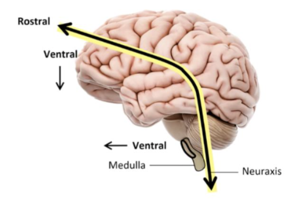
Neuraxis
Dorsal - top of neuraxis
Ventral - bottom of neuraxis
Rostral - front
Caudal - back
Phineas Gage case
A historic brain injury showing that damage to the frontal lobe can dramatically alter personality and behavior.
Advantage of lesion studies
Reveal relationships between brain structure and function.
Limitation of lesion studies
Brain regions are highly interconnected so lesions often affect multiple behaviors.
Penfield’s stimulation studies
Electrical stimulation during surgery showed specific brain regions are responsible for specific functions.
Single cell recording
A method using a microelectrode inserted into nervous tissue to record activity from a single neuron during a task.
Computed tomography (CT)
Structural imaging technique using X-ray slices to produce images of the brain. Useful for diagnosing brain injuries
CT scan limitation
Relatively low resolution compared to MRI.
Magnetic resonance imaging (MRI)
Structural neuroimaging technique using magnetic fields to create high-resolution brain images.
MRI limitation
Time-consuming and expensive.
Positron emission tomography (PET)
Functional imaging using radioactive tracers to measure brain metabolic activity. The more active the area of the brain, the more metabolic resources will be used.
PET scan limitation
Invasive due to radioactive tracer injection.
Functional magnetic resonance imaging (fMRI)
Measures the use of oxygen throughout the brain and operates assuming that more active areas of the brain require more metabolic resources.
fMRI limitation
Oxygen use in the brain spikes a few seconds later after activity
Not the best method if researcher wants to capture the precise timing of brain activation and function
Electroencephalogram (EEG)
Electrical activity of the brain can be recorded through the scalp by wearing a cap of sensitive electrodes.
Brain regions
Forebrain, midbrain, hindbrain
Hindbrain
Brain region involved in basic life functions and movement coordination.
Hindbrain Structures include:
Reticular formation, cerebellum, medulla, pons
Reticular formation
Regulates arousal and motivation, circadian rhythms, posture and balance
Cerebellum
Facilitates coordinated movement.
Medulla
Controls autonomic functions such as breathing
Pons
Involved in movement, auditory perception, emotional processing
Midbrain
Small brain region involved in sensory processing and motor control.
Tectum
Midbrain structure involved in visual (superior colliculus) and auditory processing (inferior colliculus)
Tegmentum
Midbrain structure that contains red nucleus (motor control) and substantia nigra (reward center, dopamine)
Forebrain structures:
Hypothalamus, pituitary gland, amygdala, hippocampus, thalamus, cortex
Hypothalamus
Stress response, energy metabolism, four F’s (fight, flight, feeding, reproduction)
Pituitary gland
Master endocrine gland under hypothalamic control.
Anterior pituitary
Receives signals from the brain via the hypothalamus and releases stimulating hormones to other important endocrine glands
Posterior pituitary
An extension of the hypothalamus that releases two hormones called oxytocin and vasopressin
Thalamus
Relay station for all senses except smell.
Amygdala
Receives sensory information and decodes emotions. Receives stimuli that may be threatening, associated with PTSD.
Hippocampus
Short term memory, navigation, one of the few regions in the brain where neurogenesis continues throughout adulthood
What condition do we see accumulated damage to hippocampus?
Neurodeneration in Alzheimer’s where it leads to amnesia
Cerebral cortex
Thin outer layer responsible for controlling information processing and cognition
Limbic system Structures
PHAT-H
Gyri
Cortical ridges that increase surface area linked to specific mental functions
Sulci
Cortical grooves that separate functional regions like longitudinal fissure for left and right hemisphere
Frontal lobe
Processes complex decision making and “higher-order thinking”
Responsible for complex functions such as language, inhibition (restraint), manipulation of items in short-term memory.
Occipital lobe
Exclusively processes vision
Handles most basic visual input and some complex input
Functional blindness
condition where an individual can physically see but is not able to process what is happening around them, therefore “blind”
Temporal lobe
Processes form and identity of visual stimuli
Location of primary auditory cortex where auditory processing begins
What can damage in the temporal lobe result in?
Deficits in memory, auditory processing, production of speech
Parietal lobe
Processes touch, involved in complex visual and spatial functions
What can damage to parietal lobe result in?
Deficits in orientation, attention, coordination of targeted movements
Brain lateralization
Specialization of functions to one hemisphere of the brain.
Broca’s Area
Left frontal lobe responsible for motor production of speech
Expressive aphasia
Damage to Broca’s area where you cannot properly articulate your words
Wernicke’s Area
Left temporal gyrus responsible for language comprehension
Receptive Aphasia
Damage to Wernicke’s Area where you struggle to understand language and often speak meaningless sentences, words are pronounced fluently and phrases seem to have normal rhythm and grammar.
Corpus Callosum
Thick bundle of axons which allows for communication of the two hemispheres of the brain
Split brain syndrome
Where corpus callosum is severed leading to two independently operating hemispheres
What happens when objects are presented to different visual fields in split-brain patients?
Objects in the left visual field are processed by the right hemisphere, which cannot verbally identify them but can recognize them through touch due to spatial and visual specialization, while objects in the right visual field are processed by the left hemisphere, which can name them but cannot identify them well through touch.
What do split-brain object recognition experiments demonstrate?
They show brain lateralization, where the right hemisphere specializes in spatial awareness and visualization and the left hemisphere specializes in language and naming.
Theory of Mind
The ability to attribute mental states (beliefs, intents, desires, emotions, knowledge) to oneself and to others
Mirror and Motor Neurons
Mirror neurons which fire after watching someone perform an action and then you perform it
Treatment for Phantom Limb Pain
Patients with phantom limb look at their non-amputated limb in the mirror and clench and unclench it over and over again and the pain subsides because for humans, “seeing is believing”
Capgras Delusion (what is it and cause)
Belief that an identical person has replaced a person close to him/her caused by damage to the visual processing stage involved with assessing the emotional significance of faces.
Hemi-Spatial Neglect
Ignoring half the world due to damage to right parietal lobe
Ablation Studies
Studies that remove part of the brain in order to study the results.
Declarative Memory
Also known as explicit memory; refers to memories that can be consciously recalled such as facts and knowledge.
Eloquent Cortex
Areas where damage would lead to paralysis, loss of language ability, or loss of sensory processing.
Metencephalon
Subdivision of the brain consisting of the cerebellum and the pons.
Myelencephalon
Subdivision of the brain consisting of the medulla oblongata.
Phrenology
Determining character and mental abilities based on the shape and size of the cranium.