PSYC 102 Final Exam (non-cumulative)
1/76
Earn XP
Description and Tags
covers LE 14-17 (refer to notes for covered slides), MB study guide problems, MB overview/test review
Name | Mastery | Learn | Test | Matching | Spaced | Call with Kai |
|---|
No analytics yet
Send a link to your students to track their progress
77 Terms
def. somatosensory system
what are the 3 parts of somatosensation (1 of them has 2) & def. them
part of the sensory system related to conscious perception of touch, pressure, pain, temperature, position, movement, and vibration which comes from the skin, muscles, joints, and fascia/connective tissues
__
cutaneous senses - sensation due to stimulation of the skin
tactile/touch - perception of touch & pressure
nociception - pain from stimulation of the skin
proprioception - ability to sense position of the body and limbs
kinesthesis - ability to sense movement of body and limbs
for cutaneous system (← part of somatosensory system related to detecting sensations from the skin)
what is the heaviest organ in the body?
state the 2 layers of it & def. (2 each)
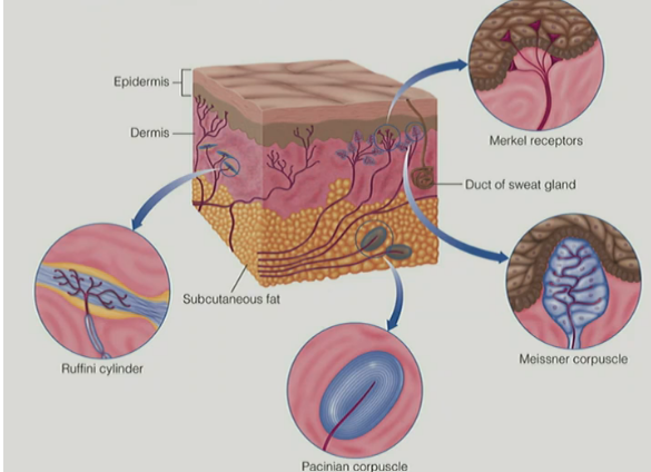
skin
epidermis
the outer layer of the skin
is made up of dead skin cells
dermis
the layer below the epidermis
contains 4 kinds of mechanoreceptors that respond to stimuli, like pressure, stretching, and vibration

for cutaneous system
for the dermis:
describe the structure/function of the mechanoreceptors located w/in the dermis (2)
__
every sensory system has sensory __ where __ cells live, which take outside stimulus/energy & transduce electrical activity that goes into the brain
for the cutaneous system, the __ cells (aka __ __) live in the __
have mechanically-gated ion channels (where it changes shape aka physically/mechanically deforms to let ions pass to make an AP)
different mechanoreceptors in the skin respond to different types of touch forces depending on where force applied on skin & what force is perceived
(aka different mechanoreceptors respond to different touch forces)
__
every sensory system has sensory epithelium where transduction cells live, which take outside stimulus/energy & transduce electrical activity that goes into the brain
for the cutaneous system, the transduction cells (aka sensory receptors) live in the skin
for cutaneous system
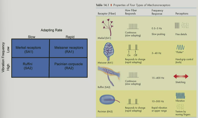
name the 4 types of mechanoreceptors & def. each
^ these are 1 set of __ receptors & 1 set of __ receptors (← don’t have to name yet)
Merkel receptor
disk-shaped receptor located near the border (aka b/w the epidermis and dermis)
Meissner corpuscle
stack of flattened disks in the dermis, specifically just below the epidermis/top layer of dermis
Ruffini cylinder
branched fibers inside a cylindrical capsule
Pacinian corpuscle
onion-like capsule located deep in the skin in the subcutaneous fat (aka layer below the dermis)
(“onion-like” b/c has many layers)
^ 1 set of shallow/surface receptors, 1 set of deep receptors

for mechanoreceptors
name the 2 temporal response properties (aka how quickly/fast adaptation to pressure occurs) of the 4 mechanoreceptors of the cutaneous system
which receptors are involved in which
def. each temporal response property
__
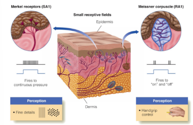
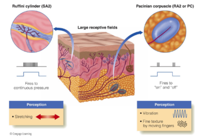
the surface set & deep set of receptors each have a __ and __ __
which ones are surface vs. deep mechanoreceptors (← & include their other name)
slowly adapting (SA) fibers: fire continuously as long as pressure is applied
Merkel receptor
Ruffini cylinder
(slowly adapting fibers have shorter names)
rapidly adapting (RA) fibers: fire at onset and offset for stimulation
Meissner corpuscle
Pacinian corpuscle
(rapidly adapting fibers have longer names)
(ex. of firing for RA fibers: fire when pencil comes in contact w/ finger & fires when pencil is taken off of finger
^ aka from no contact to contact & contact to no contact
where it rapidly adapts to contact in no contact→contact→no contact )
______________
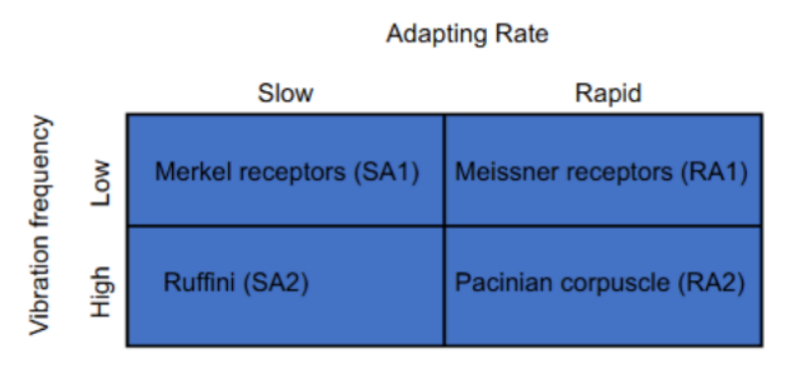
surface and deep sets each have a SA and FA fiber
_
surface mechanoreceptors: ← “1”
Merkel receptors (SA1)
Meissner receptors (RA1)
deep mechanoreceptors: ← “2”
Ruffini cylinders (SA2)
Pacinian corpuscle (RA2)
(SA and FA fibers aren’t relative to location, but surface and deep receptors are grouped relative to location/depth)

for surface mechanoreceptors
name the 2 (w/ 2 names each) & are sensitive to what?
surface mechanoreceptors:
Merkel receptors (SA1) perceives/are sensitive to fine details
(ex: Braille dots)
Meissner corpuscle (RA1) perceives/are sensitive to handgrip control/strength
(strengthening or loosening grip)
__
^ where remember that SA fibers fire continuously, RA fibers fire at onset and offset
for deep mechanoreceptors
name the 2 (w/ 2 names each) & are sensitive to what?
explain 1 of them (2)
deep mechanoreceptors:
Ruffini cylinder (SA2) perceives/are sensitive to stretching of skin
Pacinian corpuscle (RA2 or PC) perceives/are sensitive to vibrations & fine texture felt when moving fingers over object’s surface (aka fine textures felt when moving fingers)
the difference b/w texture felt when moving vs. stationary fingers on object (diff. in perception) is caused by vibrations entering your skin
where vibrations can come from the object itself moving when stationary hand OR your fingers moving across stationary object
__
^ where remember that SA fibers fire continuously, RA fibers fire at onset and offset
for surface vs. deep mechanoreceptors
relate their location/depth, sensitivity, and area of skin
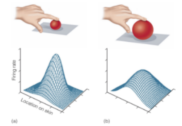
surface mechanoreceptors (“1”) are near the surface → SO is sensitive to small/weaker deformations localized to specific spots on the skin
deep mechanoreceptors (“2”) are deep → SO is sensitive to stronger deformations over a wider range of skin

for perceiving VIBRATIONS
involves which type of mechanoreceptor? that responds to __, but not __ __
if you directly stimulate this nerve fiber after dissecting the corpuscle, it will lead to __ __
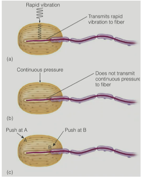
explain the picture (2 parts w/ explanation of each part)
SO the __ __ of the __ __ must be responsible for the __ adaptation (aka __ fibers)
Pacinian corpuscle responds to vibrations, but NOT continuous pressure
(b/c is a RA that fires at onset and offset)
__
if directly stimulate the nerve fiber after dissecting the corpuscle, it will lead to continuous firing
__
(picture)
mechanical stimulation of rapid pressure at location A causes rapid adapting response b/w on and off
b/c stimulates outer area to cause deformation BUT deformation has to travel and will fade before reaching the central nerve ending (in purple) → causing on and off firing with rapid adaptation
mechanical stimulation of continuous pressure at location B does NOT produce rapid adaptation, instead the response is continuous during the entire period of stimulation/cont. pressure
b/c stimulates close to the central nerve ending (in purple), so nerve ending stays continuously deformed → causing continuous firing
^^ SO the onion-like structure of the Pacinian corpuscle must be responsible for the rapid adaptation (aka RA fibers)
for perceiving TEXTURE
perception of texture depends on 2 things (name & they are determined by what?)
the 2 receptors (name them) may be responsible for the process called (1, def.)
depends on:
spatial cues, determined by size, shape, and distribution of coarse textures/elements (i.e. surface elements, like ridges of chair)
temporal cues, determined by rate of vibration as skin moves across finely textured surfaces
__
Meissner & Pacinian corpuscles may be responsible for the duplex theory of texture perception
(theory that texture depends on spatial cues at Meissner corpuscle for coarse textures & temporal cues at Pacinian corpuscle for fine textures)
for perceiving texture
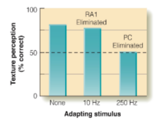
explain experiment in the picture for perception of texture w/ results
adaptation experiment where participants’ skin was adapted to either:
10 Hz stimulus (low frequency) for 6 minutes — to adapt the Meissner corpuscle
250 Hz stimulus (high frequency) for 6 minutes — to adapt the Pacinian corpuscle
__
results showed that ONLY the adaptation to the 250 Hz stimulus affected the perception of FINE textures ==> so Pacinian corpuscle is crucial to texture development
(^ aka Pacinian corpuscle is important for perceiving fine textures using temporal cues)
removing RA1 (Meissner corpuscle) didn’t change much → SO perception of texture doesn’t depend much on Meissner
removing PC/RA2 (Pacinian corpuscle) significantly reduces accuracy of texture perception → SO texture perception really depends on Pacinian
for SPATIAL properties (for resolving/distinguishing fine DETAILS aka small distance b/w 2 stimuli)
state the difference in spatial properties of surface vs. deep receptors (2 each)
_
SO if cutaneous receptor is near surface of skin, will have __ RFs
SO if cutaneous receptor is deeper in dermis OR in subcutaneous fat, will have __ RFs
_____
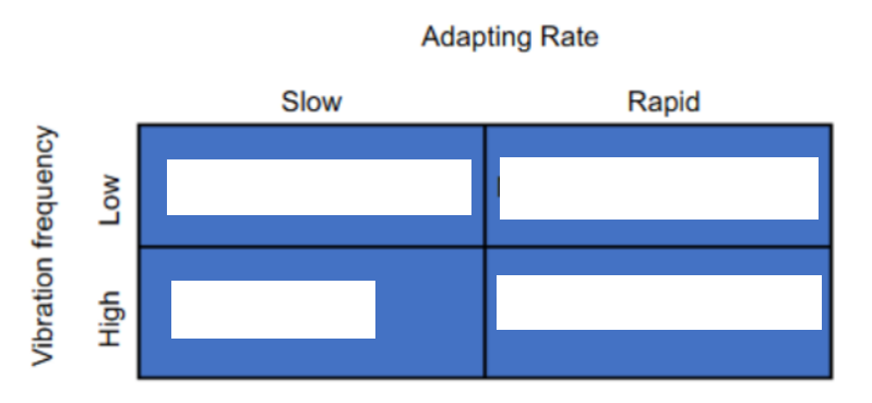
fill in blanks in picture
surface receptors: Merkel receptors (SA1), Meissner corpuscle (RA1)
small RFs
respond to slow vibration rates (aka low frequency vibrations drive surface fibers)
___
deep receptors: Ruffini cylinder (SA2), Pacinian corpuscle (RA2)
large RFs
respond to high vibration rates (aka high frequency vibrations drive deeper fibers)
__
SO if cutaneous receptor is near surface of skin, will have smaller RFs
SO if cutaneous receptor is deeper in dermis OR in subcutaneous fat, will have larger RFs
for each of the 4 mechanoreceptors, state:
if fibers are slow or rapid adapting
if fibers respond continuously or respond to change (on/off)
if fibers respond to low or high frequencies
what each mechanoreceptor perceives/are sensitive to
Merkel receptors (SA1)
slow adapting
fire/respond continuously
respond to low frequency vibrations
perceives fine details
_
Meissner receptors (RA1)
rapid adapting
fire/respond to change (onset/offset)
respond to low frequency vibrations
perceive handgrip control/strength
_
Ruffini cylinders (SA2)
slow adapting
fire/respond continuously
respond to high frequency vibrations
perceives stretching of skin
_
Pacinian corpuscle (RA2)
rapid adapting
fire/respond to change (onset/offset)
respond to high frequency vibrations
perceive vibrations & texture felt when moving fingers
remember (for depth—RFs—frequency vibrations):
surface (1) — small — low
deep (2) — large — high
^ varies a bit for SA vs. RA
for the pathways from the skin to the cortex
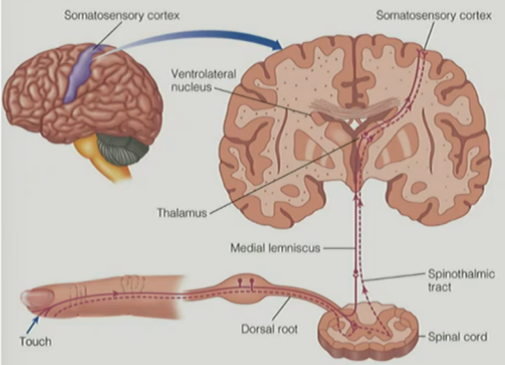
nerve fibers travel in bundles (aka __ __) to the spinal cord
name the 2 major pathways in the spinal cord & describe (1 each)
how do these two pathways continue for transduction
nerve fibers travel in bundles (aka peripheral nerves) to the spinal cord
__
medial lemniscal pathway: made up of large fibers that carry proprioceptive & touch information
spinothalamic pathway: made up of smaller fibers that carry temperature & pain information
then
BOTH pathways cross over to the opposite side of the body & THEN synapse in the ventrolateral nucleus in the thalamus → then to the somatosensory cortex (S1) & S2
specifically, to the somatosensory receiving area (S1) & secondary receiving area (S2) in the parietal lobe
_________________________________________
(^^ remember that touch and pain are the cutaneous senses, while proprioception and temperature are not)
_
(^^ remember visual cortex in occipital lobe, auditory cortex in temporal lobe, and somatosensory cortex in parietal lobe)
_
(^^ know that medial lemniscal pathway crosses over at brainstem, while spinothalamic pathway crosses over at spinal cord)

for the pathways from the skin to the cortex
extra:
state the projection each for the medial lemniscus & spinothalamic tract
__
for traveling of signals in somatosensory transduction, relate SC, brainstem, dorsal root ganglion, ventrolateral nucleus of thalamus, somatosensory primary cortex (S1)
medial lemniscus projects up from SC into brainstem → crosses over in brainstem → projects up into thalamus
__
spinothalamic tract crosses over in SC → projects up into brainstem → projects up into thalamus
__________________
SC connects to the brainstem, which has long fibers going up and down
the fibers going up are sensory fibers, which receive sensory info from parts of the body (including skin surface w/ receptors) → cell bodies of these neurons/fibers gather in the dorsal root ganglion → project up into SC → send to brain SPECIFICALLY ventrolateral nucleus of the thalamus → synapse onto other thalamus neurons → project into somatosensory primary cortex (S1)
…..
(from SC to the brain via either of the two tracts)
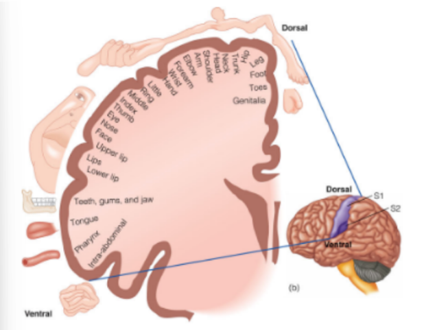
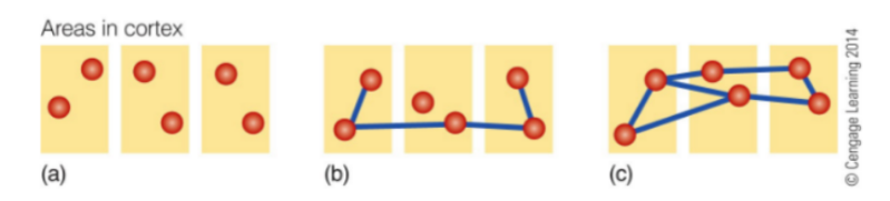
for somatosensory homunculus
def. somatosensory homunculus / body map
discovered by?
cortical space for (3) are large, meaning they are very __
has a whole set of regions representing structures around the __
shows that the legs are connected to the __ and __
__
(extra) for spatial organization:
generally recall what it was for the vision & hearing
what about for touch?
map of the cortex that shows more cortical space being allocated to parts of the body that are more sensitive to detail
by Wilder Penfield
__
cortical space for genitals, hand, and face are large, meaning they are very sensitive
has a whole set of regions representing structures around the mouth
shows that the legs are connected to the back and arms
____
for spatial organization:
for vision: retinotopic organization is preserved from the retina up into the V1
for hearing: frequency tonotopic organization is preserved from the cochlea up into the A1
for touch: somatotopic organization is preserved from the skin up into S1
for somatosensory homunculus
t/f: regardless of the organization, S1 is high plastic
def. plasticity
^^ what does plasticity in neural functioning lead to (2)
true (S1 is high plastic)
plasticity - how experience changes the way your brain represents any type of sensory info
^^ plasticity in neural functioning leads to flexible homunculus & to changes in how cortical cells are allocated to body parts
for somatosensory homunculus
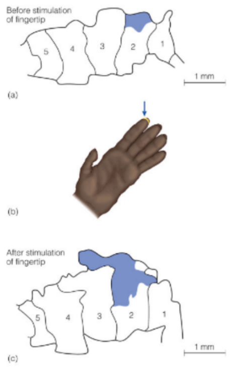
explain the experiment shown in picture
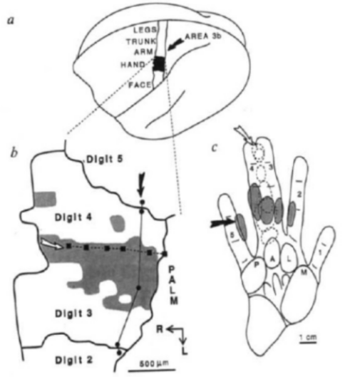
shows the somatosensory cortex response to stimulation at fingers 1-5 of a monkey’s hand
(top) before stimulation, has a small region that responds to stimulation (in blue)
(bottom) after stimulation is done repeatedly or stronger each time, then that region responding to that same stimulus is enlarged
for somatosensory homunculus
explain the experiment shown in picture
experiment where sewed a monkey’s two middle fingers together
monkey learned to adjust to use those fingers as if they were one
result: the specific region underwent extreme plasticity
for somatosensory homunculus
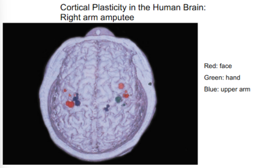
explain the experiment shown in picture
(different colors represent sensory input from the face, hand, or upper arm in an fMRI of a right arm amputee, who still had their upper right arm), stimulated points on a person’s body
face (red) is unaffected/same
upper arm (blue) is overrepresented
on the intact side (L arm), the upper arm has small representation (shown on R side of brain)
on the amputated side (R arm), the upper arm has larger representation (shown on L side of brain)
this shows how S1 has high plasticity
__
^^ remember that L and R sensory signals cross over either at the brainstem or SC, before entering the thalamus of the brain
for phantom limb
t/f: an amputee may feel like their arm is still there
explain 2 possibilities of what may feel (w/ missing arm example)
__
def. phantom limb disorder
true
sometimes, feels like they can move it normally, like if it was still there
sometimes, feels painful, like their hand is clenching so much & that they can’t unclench no matter how hard they try
__
the persistent sensation of an appendage, after the appendage has been removed by amputation or simple denervation
for phantom limb & plasticity relationship / phantom limb sensations
in a patient with missing hand and arm, touching the face produced __ sensations of touch on the (2)
this led to the hypothesis that … (1)
touching the face produced dual sensations of touch on the face & on a SPECIFIC part of the missing limb
(“specific”, like the back of the missing hand or ring finger)
hypothesis:
the brain is “filling in” for missing sensation in the hand & arm by having adjacent cortical regions can activate the cortical region of the missing limb due to proximity of connections in the somatosensory homunculus → which causes sensation in the missing limb
(^^ phantom limb sensations)
for phantom limb sensations
(in the past:)
use to be thought of as a __ __
their solution? effect?
__
(currently:)
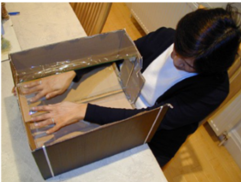
explain the “mirror box” technique by Ramachandran
steps?
what happens with continued practice w/ mirror box?
use to be thought of as a neurological disorder, that they were crazy
solution was to cut off more of the limb, but didn’t get rid of phantom pain
__
“mirror box” simulates/mimics the presence of the amputated hand, which alleviates symptoms in MOST of Ramachandran’s patients
put each arm in different holes, where 1 side is entirely covered & has a mirror that makes it appear like you have two arms
tell the person to clench their working/normal fist & release it, made it look like other hand was also clenching and releasing their fist
this tricks the brain & usually relieves phantom limb pain
continued practice w/ mirror box can sometimes make phantom pain go away completely
for perceiving details
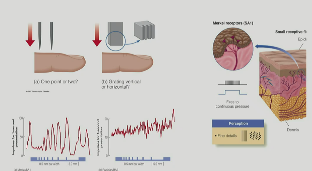
def. tactile acuity
state 2 methods for measuring tactile acuity & def.
^ for each, state context where it may be perceived as 1 or as 2 units/stimuli
__
tactile acuity thresholds for both methods are determined by what type of mechanoreceptor?
tactile acuity - perception of fine (spatial) details from touch
two-point threshold - minimum separation needed b/w 2 points to perceive them as 2 units
grating acuity - placing a grooved stimulus on the skin & asking the participant to indicate the orientation of the grating
__
two-point threshold:
ex: if pencil is touching two areas on the skin close together → may not be able to distinguish it as 2 units
ex: if touching two areas further apart from e/o → can perceive it as 2 units
grating acuity:
the closer the grooves are to e/o, the harder it is to tell that it’s 2 units
(& can get to the point where you are unable to tell that it is a grating WHEN the grooves get really close to e/o)
____
tactile acuity thresholds are determined by Merkel receptors (SA1)
(b/c it perceives/is sensitive to “fine details”)
for receptor mechanisms of tactile acuity
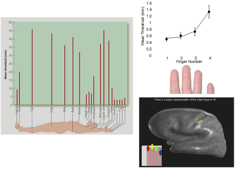
there is a high density of (what type of receptors?) in the fingertips, which means that tactile acuity is __ in the fingertips
(this is similar to high density of cones in the fovea)
__
t/f: tactile acuity changes over the entire body
what 2 things vary across the body?
high density of Merkel receptors (SA1) in the fingertips, meaning tactile acuity is greatest in the fingertips
__
true (where different areas have different sensitivities for two-point acuity/threshold tasks)
receptor density & size of RFs vary across the body
for tactile acuity
tactile acuity is best at the (2)
what does a high vs. low acuity threshold mean?
__
when going from the index to the pinky finger, acuity __ (thresholds __), BUT the (1) is the same across the fingers
explain what this means (aka what 2 things together explain tactile acuity)
the cortical region representing the index finger is __ than for second, third, or fourth fingers (individual)
AND the size of the cortical region relates to __ differences (aka differences b/w __), aka due to __← neuroplasticity
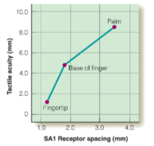
tactile acuity is best at the fingertips & face
high acuity threshold means low acuity/ability to perceive fine details (aka bad at distinguishing if 1 or 2 points)
low acuity threshold means high acuity (aka able to detect if 1 or 2 points)
__
when going from the index to the pinky finger, acuity decreases (thresholds increase), BUT the density of Merkel receptors is the same across the fingers
SO receptor density alone doesn’t explain the limiting/decreasing acuity, it also is explained by receptive field sizes (seen with size of cortical representation in the somatosensory homunculus)
the cortical region representing the index finger is larger than for second, third, or fourth fingers (individual)
AND the size of the cortical region relates to individual differences (aka differences b/w people), aka due to experiences ← neuroplasticity
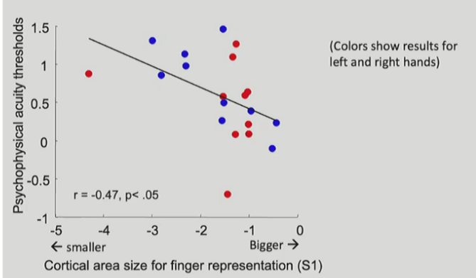
what’s the relationship b/w size of cortical representation in homunculus & size of RF?
lower acuity thresholds have __ cortical representations (v.v)
smaller RF = larger cortical representation of that body part
larger RF = smaller cortical representation
__
lower (better) acuity thresholds have larger cortical representations
b/c lower threshold = higher acuity (good) = smaller RFs = larger cortical representation in homunculus
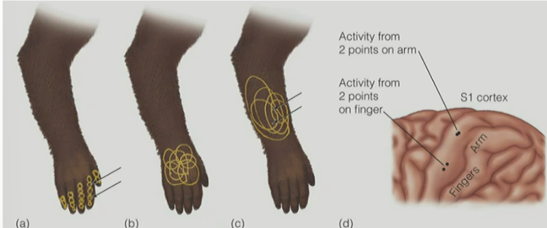
for tactile (spatial) acuity
explain picture
_
why is overlap of RFs crucial for perceptual system?
overall, want …
yellow is size of RFs at fingertips, back of hand, and forearm & black arrows are pointing towards the 2 points of stimulation
(A,D) RFs at fingertips are very small, so will have high acuity ← where will be perceived as separable representations/2 units (perception propagated from the fingertips/skin to S1)
(B,D) RFs at the forearm are much larger, so stimulation of 2 points at the same distance as on the fingertips will have low acuity ← where perceived as inseparable representations/1 unit due to overlap of larger RFs
____________
overlap is crucial b/c:
if every RF was independent and non-overlapping, then cells responding to one area wouldn’t know anything about a different cell responding to another area
(hard to have comprehensive understanding of perception)
^^ SO want to show difference while maintaining the ability to detect similarity/overlap
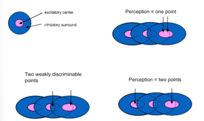
for picture, explain how DENSITY of receptors correlates to acuity
^ when have fixed RF size and density, but changing separation of/distance b/w points
^ OVERALL: __ spacing b/w 2 RFs = __ tactile spatial acuity
(top right)
unable to discriminate b/w 2 points of stimulation b/c both points are stimulating the same excitatory center of the same RF
(bottom left)
can MB weakly discriminate b/w 2 stimuli b/c inhibitory surround inhibits the excitatory response of the same RF
(bottom right)
at maximum ability to discriminate b/w 2 stimuli b/c both points are stimulating the excitatory centers of 2 different RFs
BUT lost the ability to compare/find similarities b/w the 2 stimuli — b/c has less or no overlap of the stimulated RFs
^ OVERALL: less spacing b/w 2 RFs = higher acuity
b/c 2 stimuli close to e/o are more likely to activate different RFs if the RFs are closely packed/dense
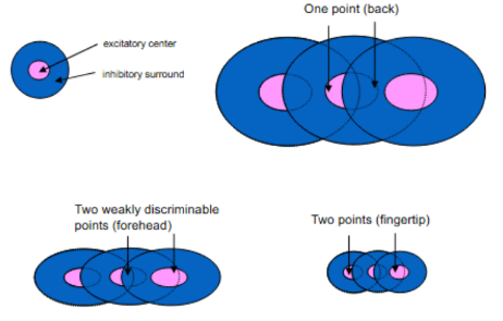
for picture, explain how SIZE of RFs correlates to acuity
^ when have fixed separation of/distance b/w points, but changing RF size (← RF size changes due to stimulation at different parts of body activating differently sized RFs)
^ OVERALL: __ RF size = __ higher tactile spatial acuity
(top right)
at the back, it’s hard to discriminate b/w the responses caused by 2 points of stimuli (b/c large RFs)
(bottom left)
if we shrink the size of the RFs at the forehead, then we get slightly different responses SO can weakly discriminate b/w 2 stimuli
(bottom right)
if we shrink the RFs even smaller at the fingertips, then we can discriminate b/w the 2 responses/points of stimuli
^ OVERALL: smaller RF size = higher acuity
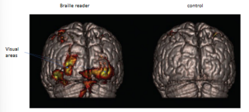
experienced Braille readers can read ~__ words/minutes, compared to __-__ words/minute for visual reading
_
what is the difference in the somatosensory cortex responses b/w experienced Braille readers vs. control/sighted subjects
experienced Braille readers can read ~100 words/minutes, compared to 250-300 words/minute for visual reading
_
experienced Braille readers show large responses in occipital lobe while reading Braille, compared to control/sighted subjects
^ the cortical region for fingertips and face are adjacent, so the perception of viewing words at the face is “taken over” by the cortical region of the fingertips← NO b/c occipital lobe is part of V1, so is not explained by S1 of crossed out info
for perceiving objects (tactile object perception)
t/f: humans use active touch (not passive/stationary/no movement) to interact w/ the environment
__
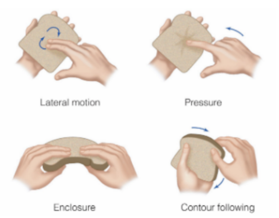
def. haptic exploration/perception
name 3 systems of haptic exploration
people can identify objects haptically in _-_ seconds
people use __ __ to actively interact with objects (name & describe the 4 ways)
true (active touch to interact)
_
haptic exploration:
is the active exploration of 3D objects with the hand (touch)
sensory system (i.e. somatosensory system)
motor system (i.e. moving fingertips)
cognitive system (i.e. decision about what something is, its texture, etc.)
__
people can identify objects haptically/through active touch in 1-2 seconds
__
people use exploratory procedures (EPs) to perceptive objects haptically
lateral motion (side-to-side)
pressure (pressing on objects)
enclosure (wrapping hands/fingers around object)
contour following (following edge/contour of object)
for tactile object perception
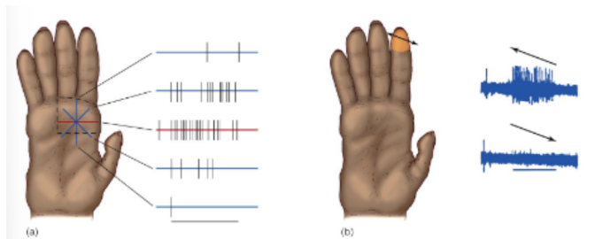
the firing patterns of a group of mechanoreceptors signal __, like the curvature of an object
__
t/f: neurons further upstream become more specialized
(^ upstream is in terms of the up direction of the somatosensory pathway)
explain
the firing patterns of a group of mechanoreceptors signal shape, like the curvature of an object
__
true
the ventrolateral nucleus in the thalamus responds to center-surround RFs, while S1 responds maximally to orientations & direction of movement
(aka thalamus detects contrast from center-surround, while cortex detects features like orientation) ← SO projecting up the somatosensory pathway will make the neurons more specialized
for tactile object perception
the height of the profile indicates …
__
the profile will change when touching a stimulus w/ high-curvature (A) vs. low-curvature (B)
^ explain
conclusion?
height of profile indicates firing rate across the fingertip
__
the profile will change when touching a stimulus w/ high-curvature (A) vs. low-curvature (B), b/c:
each point on the ball’s surface will come in contact with a given point on the surface of the finger for a brief period of time
for (B), the larger ball means the curvature is more shallow (aka more gradual bend) → SO a point on surface of finger comes into contact with a given point on the ball’s surface for a longer period of time
while for (A), the smaller ball means the curvature of the ball is more shallow (sharper bend) → SO a point on the surface of the finger comes into contact with a given point on the ball’s surface for a shorter period of time
__
overall:
the temporal pattern of stimulation & shape of stimulation distribution are different for different objects
(^ aka time they’re in contact & shape of profile are different for different objects)
for tactile object perception in S1 for feature detection: orientation & movement
for L pic, this neuron responds best when the __ oriented edge is presented/stimulated on the monkey’s hand
for R pic, this neuron responds best when the stimulus moves across the fingertip from __ to __
__
t/f: the somatosensory cortex has lateral inhibition (where adjacent activated neurons can inhibit the activity of an activated neuron), as does the other levels of the somatosensory pathway, in order to sharpen contrast of inputs
(L pic) this neuron responds best to the stimulation of the horizontally oriented edge
(seen w/ more continuous firing)
(R pic) this neuron responds best when the stimulus moves across the fingertip from the right to left
(seen w/ higher spiking)
__
true
(in order to sharpen the contrast of inputs → to be better at discriminating if 1 or 2 units & at localization of touch stimuli aka where)
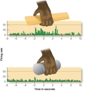
for object specificity
explain why this neuron fires when grasping a ruler, but doesn’t fire when grasping a cylinder
in the parietal cortex (after S1), neurons are even more specialized & respond to action-object interactions (like pressure distribution, position of fingers, contact points, etc.)
→ creates object-specific representation, where neurons respond selectively to interactions b/w object and hand
for attention
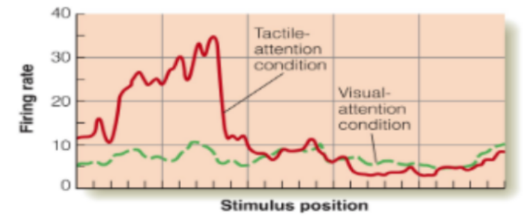
describe picture of monkey’s cortex (1)
conclusion? ← & explain
__
t/f: every movement you make is a combination of some motor action & sensory feedback guiding that motor action
this neuron only responds when the monkey is paying attention to the tactile stimulus (firing rate seen w/ the red peak)
where the green line is monkey focusing its attention on a visual task, so distracting visual attention suppresses tactile perception/tactile firing rate WHILE tactile attention increases firing rate
(^ being distracted away from the touch stimulus vs. paying attention to the touch stimulus)
__
conclusion:
object perception is complex process involving somatosensation, movement, and cognition
………….
^ somatosensation is using the 4 mechanoreceptors for perception of fine detail, handgrip control, stretching of skin, vibrations, and texture felt when moving hand
(detect touch stimuli)
^ movement is haptic exploration
(gather info)
^ cognition is object specificity & attention
(identify and interpret the object)
__
true
for PAIN
state the 3 types of pain & def. (for 1 of them, there’s 2 examples)
inflammatory pain - caused by damage to tissues and joints OR caused by tumor cells
neuropathic pain - caused by damage to CNS
like brain damage caused by stroke
like repetitive movements causing carpal tunnel syndrome, etc.
nociceptive pain - will signal that there is impending/incoming damage to the skin
for nociceptive pain
what do nociceptors (aka pain receptors in skin) respond to (4)
the threshold of causing receptor __ must be balanced SO that will be warned of incoming/possible damage WHILE NOT affecting normal activity and function
__
pain is a multimodal phenomenon, containing (2)
nociceptors respond to heat, cold, chemicals, and severe pressure
__
the threshold of causing receptor responses must be balanced SO that will be warned of incoming/possible damage WHILE NOT affecting normal activity and function
__
pain is a multimodal phenomenon, containing a sensory component & an emotional/affective component
(aka detection of painful stimulus & emotional response/unpleasant feeling to pain)
for nociceptive pain
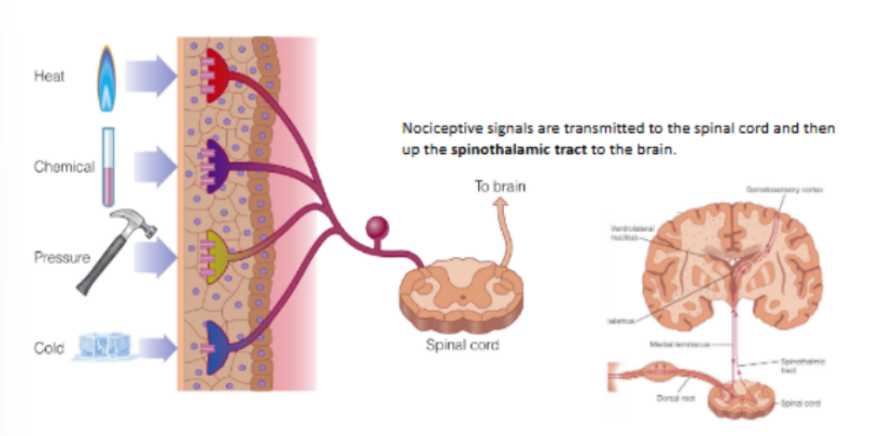
what is nociceptive pain caused by?
nociceptive signals are transmitted through the __ __ __ to the __ __, and then up the __ __ into the __ __ (of the __) into the __ & __
__
t/f: We feel pain not b/c certain receptors are activated, but we feel pain b/c certain receptors are activated in certain contexts and ways
caused by the activation of nociceptors in the skin that respond to different types of stimulation (and that release signals in different contexts and patterns to the brain)
__
nociceptive signals are transmitted through the dorsal root ganglion to the spinal cord, and then up the spinothalamic tract into ventrolateral nucleus (of the thalamus) into the S1 & S2
__
true
activated receptors don’t automatically mean “pain”
(i.e. heat receptors when warm temperature & feel “pain” when temperature gets too high)
“pain” occurs when signals from nociceptors reach the brain in certain patterns & intensities
^ like the brain is asking “does this pattern of sensory activity mean tissue damage or danger?”
very briefly explain the nociceptive pathway ← mentioning the specific tract
all nociceptive receptors go into the dorsal root of SC, where cell bodies reside in the dorsal root ganglion
BUT the output/signals from the dorsal root ganglion project up via spinothalamic tract, NOT the medial lemniscal tract
(^ medial lemniscal tract is for touch & proprioception)
def. “direct pathway model of pain”
name 4 problems of this direct pathway model of pain
__
instead, what works is the “gate control model of pain perception”
“direct pathway model of pain”
early model assuming that pain is caused when nociceptors are stimulated & sends signals to the brain
BUT problems:
pain can be affected by person’s mental state
pain can occur even without stimulation of the skin
pain can be affected by a person’s attention
phantom limbs
(missing limb means missing nociceptors)
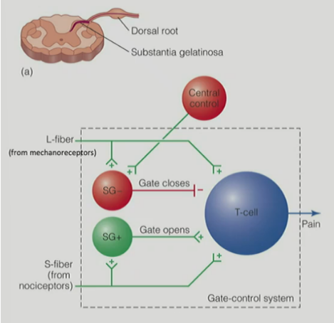
for “gate control model of pain perception”
the “gate” is made of … (AND name the excitatory neurons & inhibitory neurons that regulate pain)
_
input going into the gate comes from 3 things (← name them & def.)
_
name the 2 ways pain does not occur / pain is blocked, relative to the gate control model of pain
do you need both to occur together OR can they happen independently to block pain?
_
name the 1 way pain does occur, relative to the gate control model of pain
________________________
^^ but know that the actual mechanism for pain perception is more complex than shown by the model
the “gate” contains substantia gelatinosa cells in the dorsal horn of the SC (SG- and SG+ are inhibitory or excitatory neurons in dorsal horn of the SC that regulate pain)
__
input going into the gate comes from:
large diameter (L) fibers - information from tactile stimuli via mechanoreceptors
small diameter (S) fibers - information from nociceptors
central control - information from cognitive factors from the cortex
__
pain doesn’t occur / pain is blocked when:
central control → activate SG- cell (inhibitory) → gate closes → inhibits T-cell to block pain
(top-down process)
L-fibers from mechanoreceptors → activate SG- cell (inhibitory) → gate closes → inhibits T-cell to block pain
(bottom-up process)
_
pain does occur when:
S-fibers from nociceptors → activate SG+ cell (excitatory) → gate opens → activates T-cell to induce pain
for “gate control model of pain perception”
t/f: the gate is controlled by both the mechanoreceptor & nociceptors
_
for nociceptive pathway, signals from the nociceptors travel up the spinothalamic path & activate (2)
true
(not at the same time though, but both will activate a SG- or SG+ cell that will close/open the gate)
__
signals from the nociceptors travel up the spinothalamic path & activate:
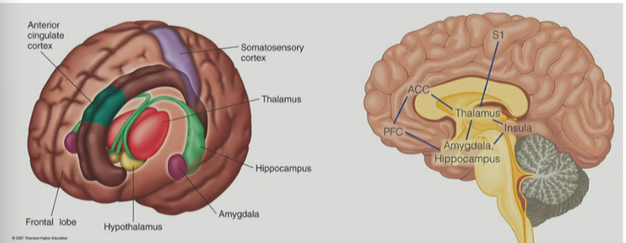
subcortical areas (like hypothalamus, limbic system, and thalamus)
cortical areas (like S1 and S2) in the somatosensory cortex, the insula, and the anterior cingulate cortex (ACC)
& these cortical areas combined make up the pain matrix
for relationship b/w cognition & pain
t/f: rubbing skin after pain will drive/activate mechanoreceptors near nociceptors, which helps reduce pain
__
explain experiment by Derbyshire (w/ the 3 conditions & the result)
_
in another experiment, explain how participants could keep their hands in cold water longer when shown positive pictures
__
explain picture overall (1)
true
painful stimulus projects up through spinothalamic tract & rubbing skin will activate touch mechanoreceptors, which release signals into SC → activate SG- cells in dorsal horn of SC (which suppresses transmission of pain signals)
__
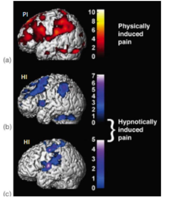
to study hypnotically induced pain (aka only told that they feel pain when hypnotized)
participants had thermal stimulator attached to palm of hand
3 conditions:
physically induced pain (PI)
hypnotically induced pain (HI)
control group that imagined the painful stimulation (IM)
results: hypnosis can produce pain perception without painful stimulus
__
another experiment where participants could keep their hands in cold water longer when shown positive pictures
due to emotional distraction (shows how cognitive system/state can affect pain perception)
(picture)
despite regions in PI, HI, and IM conditions having some overlap in their activated regions, whether they feel pain or not is deter. by their cognitive state
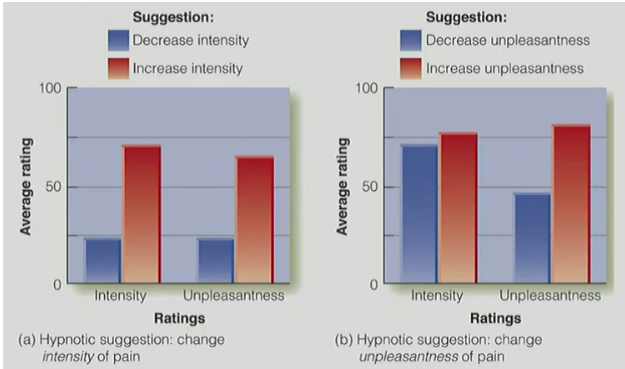
explain experiment where participants were presented w/ potentially painful stimuli & were asked to rate subjective pain intensity and unpleasantness
w/ result
measured brain activity when hands were placed in hot water
hypnosis was used to decrease or increase the sensory & affective/emotional components of pain
result: if told participants that hypnosis changes the subjective intensity of pain → led to changes in ratings & changes in activity in S1
result: if told participants that hypnosis changes the unpleasantness of pain (for better or worse) → led to changes in unpleasantness ratings, but did NOT affect the subjective ratings of pain
for opioids & pain
brain tissue releases NTs called __, where __ reduce pain by binding at __ __ sites
___________________________
__ receptors react to (3)
describe 1 of them
___________________________
another name for placebos & how do they relate to endorphins
_
what happens if both naloxone & SPA/placebos are present?
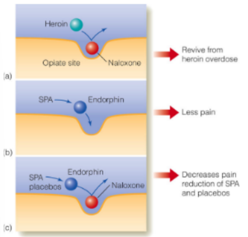
brain tissue releases NTs called endorphins, where endorphins reduce pain by binding at opiate receptor sites
(stimulation of opiate receptor sites will reduce pain)
__
opiate receptors bind to
heroin
naloxone
can revive a victim of heroin overdose — by (binding to &) blocking the receptor sites for heroin
endorphins
__
placebos (stimulation-produced analgesia, SPAs) can lead to release of endorphins → reduces pain
__
if both naloxone & SPA/placebos are present:
naloxone still binds to the opiate receptor site
SPA/placebo releases endorphin, which doesn’t bind to occupied opiate receptor site → SO reduced SPA/placebo’s effect of reducing pain
for pain
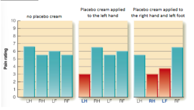
explain the placebo effect using the picture
conclusion?
SO the effects of pain are __ & are blocked by __
before anything, pain firing is similar across all 4 body parts
if place placebo cream on L hand, reports in pain felt in L hand
^ placebo effect
if place placebo cream on R hand and L foot, then report less pain felt in R hand and L foot
__
conclusion: placebo changes the experience even though the stimulus stays the same
SO the effects of pain are location-specific & are blocked by naloxone
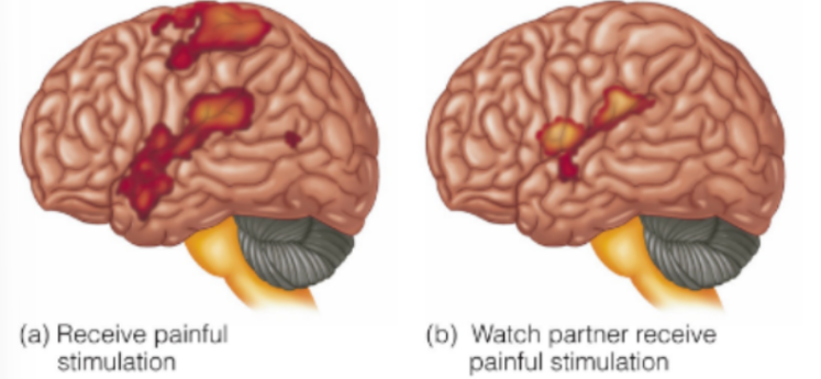
explain the experiment by Singer et al.
participants were romantically involved couples (to maximize the brain activity seen) & measured the woman’s brain activity
presented to 2 conditions: either the woman received shocks OR she watched her partner receive shocks
saw that similar brain areas were activated in both conditions (similar/overlap, but not identical)
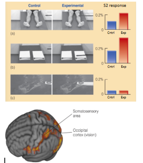
explain the 2 experiments in the picture by 1) Keyser and coworkers, and 2) Meyer and coworkers
Keyser and coworkers (top pic)
watched videos of people or objects being touched
generally saw that the experimental condition of pain being caused has higher S2 response, while lower S2 response for control where no pain is caused yet
(exception is bottom picture)
__
Meyer and coworkers
watched videos of a person’s hands haptically exploring objects
saw that both the visual & somatosensory areas were activated
for smell/olfaction
def. olfaction
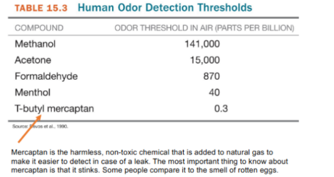
rats are _-_ times more sensitive to odors than humans, while dogs are _-_ times more sensitive to odors than humans
despite animals having different sensitivities to smell, individual receptors for all these animals are __ __
explain ONE factor the partially explains this difference b/w animals’ sensitivities to smell (& w/ number in humans vs. dogs)
olfaction
the sense of smell, usually from stimulation of receptors in the olfactory mucosa
__
rats 8-50 times more sensitive than humans to smell
dogs 300-10,000 times sensitive than humans to smell
__
despite animals having different sensitivities to smell, individual receptors for all these animals are equally sensitive
the difference b/w animal’s sensitivities to smell are due to the # of olfactory receptors they each have (density of receptors in the olfactory mucosa)
in which humans have 100 million olfactory receptors, while dogs have 1 billion olfactory receptors
for olfaction
many animals are __ (def.)
humans are __ (def.)
__
you detect odors by measuring the __ __
state, def., and describe the 2 ways to measure this
many animals are macrosmatic - have keen sense of smell necessary for survival
humans are microsmatic - have a less keen sense of smell that’s NOT necessary for survival
__
detect odors by measuring the detection threshold
yes/no procedure - participants are given trials w/ odors & given “blank”/no odor trials
either respond yes (odor is present) or no
can result in bias b/c depends on the participant
forced-choice - participants are given trials w/ odors & given “blank”/no odor trials
participants indicate which trial smells the strongest
for olfaction
what does a smaller odor detection threshold mean?
smaller odor threshold = high sensitivity to that specific odor
(aka is easier to detect the odor when present at small amounts in the air)
for measuring difference threshold for olfaction
def. the “just noticeable difference” / JND
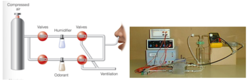
measuring the JND for olfaction needs to be done carefully using an __
__
relate olfaction w/ Weber’s law and Weber’s fraction
JND
the smallest difference in concentration that can be detected b/w 2 samples
measuring the JND for olfaction needs to be done carefully using an olfactometer
__
olfaction follows Weber’s law and has a Weber fraction of ~11%
(review: Weber’s law is that the difference threshold/JND is proportional to the baseline value)
for olfaction: identifying odors
def. recognition threshold
t/f: humans can discriminate among 100,000 odors, but cannot label them accurately
this happens b/c … NOT b/c …
can you improve this? how?
recognition threshold
concentration (of odor in air) needed to determine the quality of an odorant
(i.e. what the odor is, not just being able to detect it)
__
true
B/C is an inability to retrieve the name from memory, NOT due to a lack of sensitivity
yes, can improve labeling odors w/ training/experience
for olfaction
t/f: researchers have found it difficult to map perceptual experience onto physical attributes of odorants
__
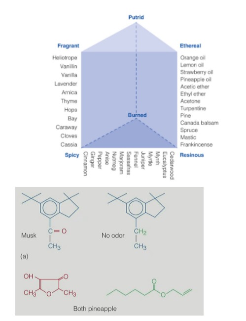
describe Henning’s odor prism (3)
__
what were the 2 difficulties with linking chemical structures to the types of smells?
def. odor objects
def. specificity coding vs. distributed coding
true (difficult to map perception w/ odors)
__
Henning’s odor prism
6 corners with the qualities: putrid, ethereal, resinous, spicy, fragrant, burned
other odors are located on the prism relative to the perception of the corner qualities
HOWEVER, Henning’s prism isn’t very useful in olfactory research
__
linking chemical structure to types of smells had problems where:
some molecules w/ similar shapes have different smells
some similar smells come from molecules w/ different shapes
__
odor objects - the source of an odor (i.e. coffee, bacon, car exhaust)
specificity coding - each type of receptor produces its own sensation
(1:1 for type of receptor:sensation)
distributed coding - sensations are determined by combinations of many types of receptors
(many:1 for type of receptor:sensation)
AND (1 receptor may code for multiple odors)
for olfaction: structure of the olfactory system
where is olfactory mucosa located?
what occurs here?
where are the olfactory receptors contained/located?
_
describe the 5 general steps for transduction of olfaction
olfactory mucosa located at top of the nasal cavity
odorants are carried along the mucosa & comes into contact with sensory neurons
olfactory receptors are located on the cilia of these sensory neurons
__
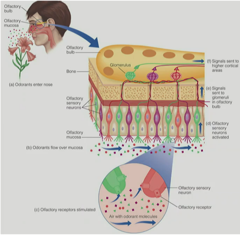
transduction of olfaction
odorants enter the nose & onto olfactory epithelium
odorants flow over the olfactory mucosa
where the odorants come in contact w/ olfactory sensory neurons/ORNs on the mucosa & stimulate/activate the olfactory receptors on ORNs
all ORNs of a specific type will send signals to 1 or 2 glomeruli on the olfactory bulb
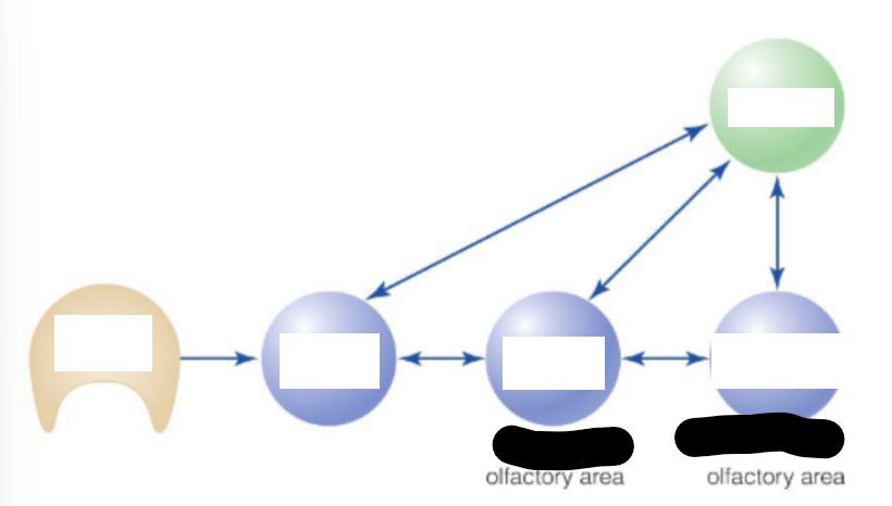
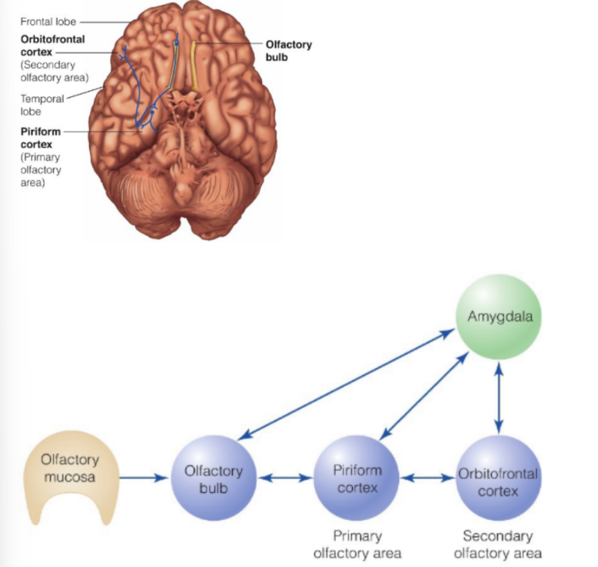
these signals are sent to the cortex:
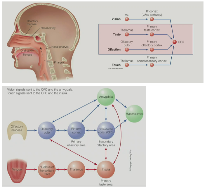
primary olfactory cortex (piriform cortex) in the temporal lobe & amygdala
→ secondary olfactory cortex (orbitofrontal cortex, OFC) in frontal lobe
for olfactory mucosa
the mucosa contains __ different types of olfactory receptors in the mucosa, where each ORN has about __ receptors of 1 receptor type
all ORNs of a __ type will send their signals to __ or __ glomeruli in the __ __
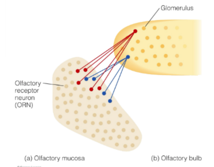
the mucosa contains 350 different types of olfactory receptors in the mucosa, where each ORN has about 10,000 receptors of 1 receptor type
all ORNs of a particular type will send their signals to 1 or 2 glomeruli in the olfactory bulb
aka
the olfactory mucosa has many ORNs
where each ORN has about 10,000 receptors of 1 receptor type (there are 350 possible types of olfactory receptors)
the olfactory bulb has many glomeruli
where 1 or 2 glomeruli will receive inputs from 10,000 receptors of ONE receptor/ORN type
(aka 1-2 glomeruli to 1 receptor/ORN type of 10,000 receptors)
for olfactory mucosa & bulb
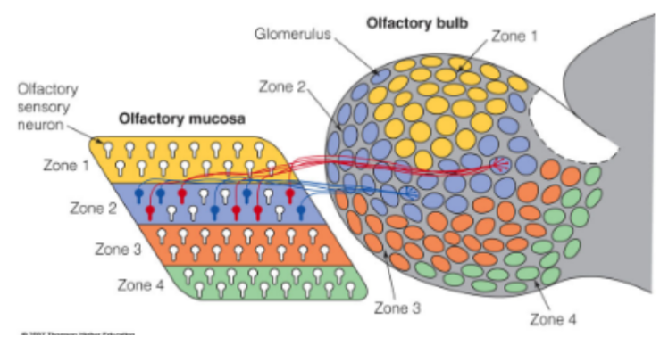
olfactory mucosa is divided into _ zones
where each zone has many __ __ of receptors/ORNs
& specific types of receptors (on ORNs) are found in only __ zone
odorants tend to activate specific __ within a specific zone
(b/c specific zones have specific types of ORNs containing specific types of receptors)
__
t/f: specific types of ORNs synapse to only 1 or 2 glomeruli on the olfactory bulb
(aka 1 or 2 glomeruli will receive inputs from 10,000 receptors of ONE receptor/ORN type)
olfactory mucosa is divided into 4 zones
where each zone has many different types of receptors/ORNs
& specific types of receptors (on ORNs) are found in only ONE zone
odorants tend to activate specific ORNs (OM neurons/olfactory mucosa neurons) within a specific zone
^^ specific zones have specific types of ORNs containing specific types of receptors
__
true
(1 or 2 glomeruli will receive inputs from 10,000 receptors of ONE receptor/ORN type)
for olfactory mucosa & bulb
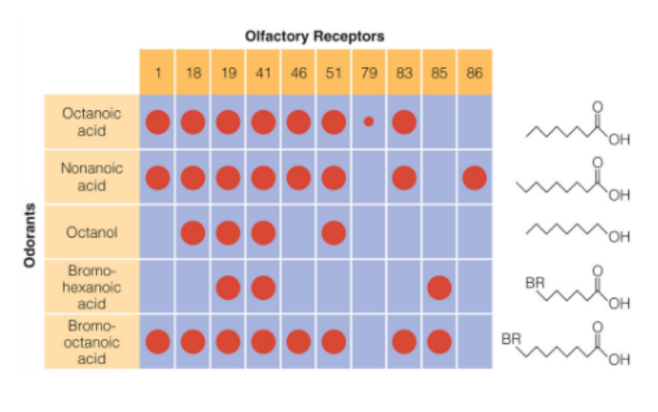
def. recognition profiles
t/f: different odors have different recognition profiles
t/f: specific receptors on ORNs may be part of the code for multiple odorants (aka specific receptors may be involved in the coding of multiple odorants) ← distributed coding
recognition profiles
a combination/pattern of activated ORNs that code for each odorant
__
true
(different odors have different recognition profiles)
_
true
(specific receptors on ORNs may be involved in the coding of multiple odorants)
for activating olfactory bulb: methods to view activation of olfactory bulb
describe the optimal imaging method (4)
conclusion?
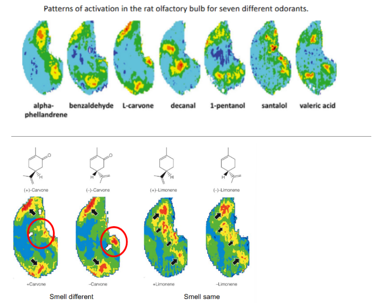
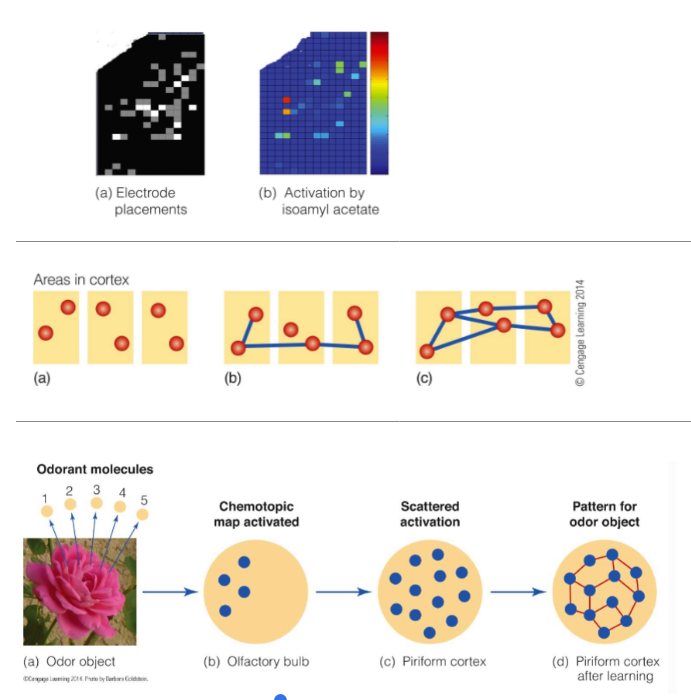
def. chemotopic map
__
describe the 2DG technique (3)
what does 2DG stand for?
conclusion?
describe the picture
optimal imaging method
cortical cells consume oxygen when activated
so use red light to see the amount of oxygen in these cells
where less oxygen reflects less light
measuring the amount of light reflected tells us which areas of the cortex are most active
^ less amount of light reflected (looks less red) = more oxygen consumed = more active cortical cells (SO area of cortex containing those cells are most active)
chemotopic map - different odors activate different areas on the olfactory bulb
__
2DG (2-deoxyglucose) technique
2DG (containing glucose) is injected into an animal
then, animal is exposed to different chemicals
measure neural activation by the amount of radioactivity present
^ the pattern of olfactory bulb activation is related to both chemical structure & perception
__
(picture)
the more active regions are in red, less active regions in blue
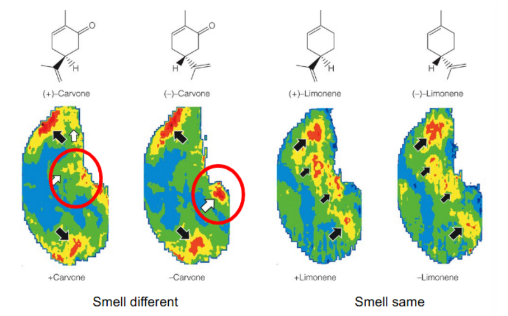
for “smells different”, have similar patterns of activity EXCEPT for the area circled in red, where different areas are activated (red) vs. nonactivated (blue)
for “smells same”, have similar patterns of activity (where red region in one structure is still red in the other structure, though in different amounts)
for odors from olfactory bulb → cortex
describe transduction from the olfactory bulb to cortex
_
the amygdala plays a role in __ reactions to odors (i.e. …)
_
fill in blanks of picture
signals from olfactory bulb
→ project to primary olfactory cortex (piriform cortex) in the temporal lobe & amygdala
→ then project to secondary olfactory cortex (orbitofrontal cortex, OFC) in the frontal lobe
__
the amygdala plays a role in emotional reactions to odors (i.e. decide if scent is pleasant or unpleasant)
for odors in cortex
general statement for: representation of odorants in piriform cortex
__
use picture to explain how memories are formed in the cortex
orderly maps (organized odor maps) of the olfactory bulb are NOT preserved when signals reach the piriform cortex, as seen by scattering and spreading out activated neurons from bulb to piriform cortex
__
(a) initially, incoming info/odorants activate certain areas in the PIRIFORM cortex ← where the rectangles are different cortical areas/cells & red circles are activated areas
chemotopic map activated when odorants activate ORNs of mucosa, which will send signals to the glomeruli of bulb (in olfactory bulb)
scattered activation (from bulb → piriform cortex)
(b) as time passes, the neural activity is replayed, which starts to form connections b/w the activated areas
(c) eventually, the activated areas for a certain memory are ALL linked together, which stabilizes the memory
forms a pattern of activation for that specific odor
for taste
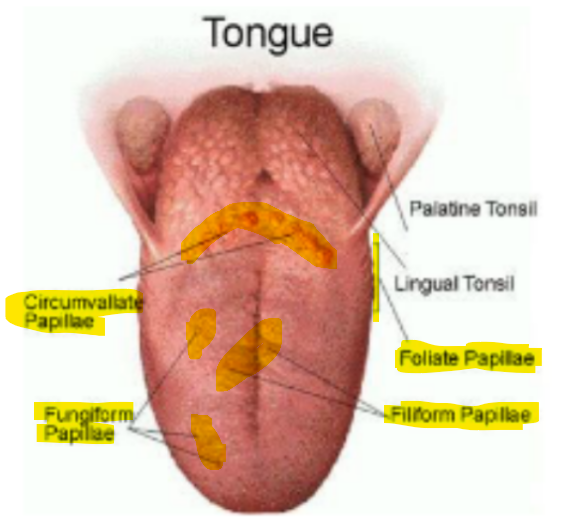
def. tongue
def. papillae
name & def. the 4 types of papillae on the tongue
tongue - the sensory epithelium for taste that contains taste receptors
papillae - structures that give the tongue its rough appearance
__
filiform papillae - cone-shaped & located over the entire surface of the tongue BUT has no taste buds
fungiform papillae - mushroom-shaped (look like red dots) & located on sides and tip of tongue
foliate papillae - the series of folds on the back and sides of tongue
circumvallate papillae - shaped like flat mounds in a trench & located at the back of the tongue
for taste
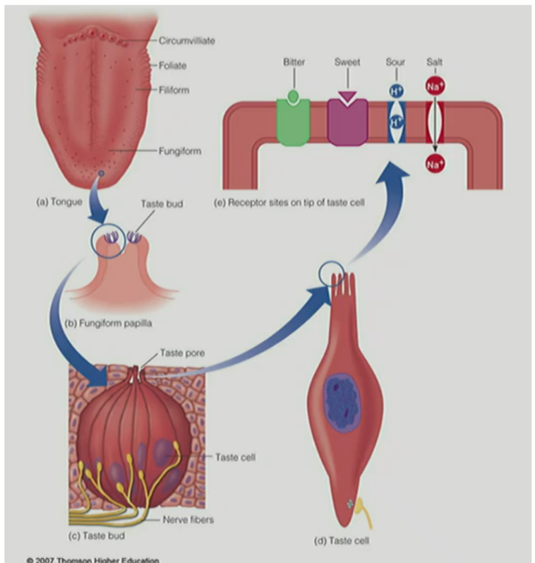
where are taste buds located?
the tongue has about __ taste buds, where each taste bud has …
taste transduction occurs when __ contact the receptor sites on the __/__ of __ __ in the __ __ on the __
taste buds are located on the papillae, except for on filiform papillae (contains no taste buds)
_
tongue has ~10,000 taste buds, where each taste bud has taste cells w/ tips/microvilli that extend into the taste pore
__
transduction occurs when chemicals contact the receptor site on tips/microvilli of taste cells in the taste buds on the tongue
for taste
name the 5 basic taste qualities
(def. 1 of them)
(describe 4 of them)
is there or is there not a perfect connection b/w taste & function of substances?
salty
indicates presence of sodium
sour
sweet
associated with substances that have nutritive value
bitter
associated with potentially harmful substances
umami (meaty, brothy, savory)
associated with MSG
__
there is not a perfect connection b/w taste & function of substances
(the sensory experience of taste, like sweet, doesn’t always correctly signal the function/association, like nutritive value)
for individual/people’s differences in taste
people (genetically) have different responses to the taste of __ and __ (← these are for __), where:
name the 3 types of individuals relative to taste sensitivity
compare (3)
for 1 of them, “this comparison” can affect (2)
__
t/f: sensations of different qualities are NOT strictly localized to specific parts of the tongue
this means that …
__
t/f: taste can usually be described as a combination of basic taste qualities
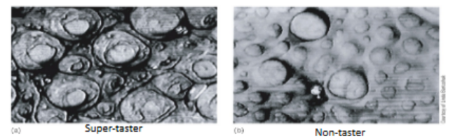
people (genetically) have different responses to the taste of PTC (phenylthiocarbamide) and PROP (6-n-propyltiouracil) (← both are for bitterness), where:
tasters, supertasters, nontasters:
tasters have more taste buds than nontasters
tasters have specialized receptors for these chemical compounds
supertasters appear more sensitive to bitter substances than tasters
which can affect diet (dislike bitter veggies) & susceptibility to pain (burning mouth syndrome)
__
true
all taste qualities can be detected across most areas of the tongue (except places on the tongue w/ filiform papillae)
__
true
for structure of taste system/pathway
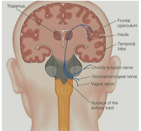
taste buds are innervated by __ that carry taste information to the __
__
after taste transduction where chemical signals are converted into neural signals, explain the pathway of neural signals to the brain (~3 parts)
taste buds are innervated by axons that carry taste information to the brain
__
after taste transduction where chemical signals are converted into neural signals, the pathway of neural signals to the brain is:
neural signals project up via 4 nerves:
chorda tympani nerve (VII) — from the front & sides of the tongue (fungiform & filiform papillae)
glossopharyngeal nerve (IX) — from back of tongue (circumvallate & foliate papillae)
vagus nerve (X) — from mouth & throat
superficial petronasal nerve — from soft palate
these nerves connect at the nucleus of solitary tract (NST) in the SC
to thalamus
to areas of the frontal lobe:
insula
frontal operculum cortex
orbital frontal cortex (OFC)
for taste coding
for distributed coding: (L pic)
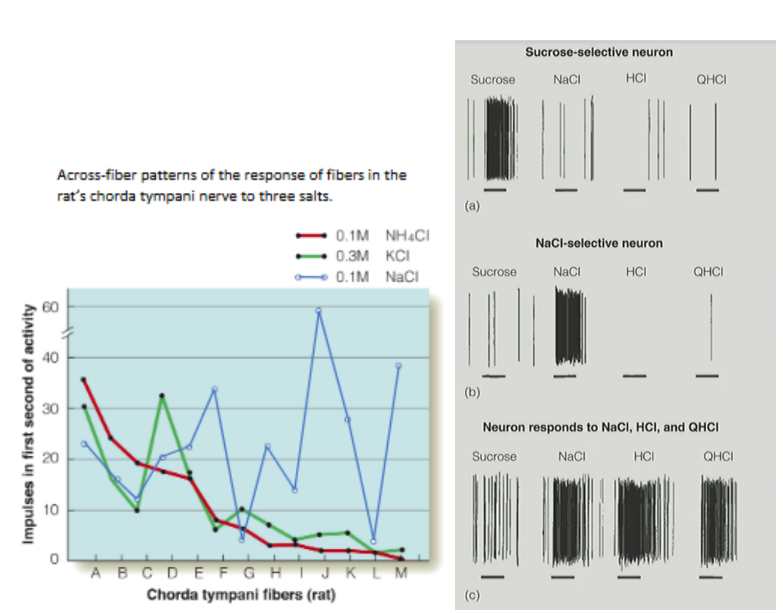
describe the experiment by Erickson w/ rats
for specificity coding: (R pic)
describe the same experiment by Erickson w/ rats
conclusion?
3 different taste stimuli were presented to rats & looked at recordings of the chorda tympani nerve fibers
patterns across the chorda tympani nerve fibers showed that 2 substances were similar to e/o (potassium chloride & ammonium chloride), but 1 substance was different from the rest (sodium chloride)
then, rats were trained by shocking them when they drank potassium chloride
afterwards, when rats were given the choice b/w the 3, the rats avoided ammonium chloride
shows distributed coding b/c many types of receptors control 1 odor & a receptor may code for multiple odors (← latter explains why this happens)
^ this experiment shows physiological & behavioral evidence for distributed coding
__
looked at responses of 3 neurons & recorded the chorda tympani nerve fibers in the rat
4 solutions: sucrose, salt, HCl, and QHCl, were flowed over rat’s tongue for 15 seconds (horizontal lines)
results:
top neuron responded selectively to sweet stimulus (sucrose)
middle neuron responded selectively to salt
bottom neuron responded to salty, sour, and bitter stimuli (didn’t respond to only sucrose/sweet stim.)
^ specific fibers respond selectively/favorably to specific chemicals

for taste coding
for specificity coding:
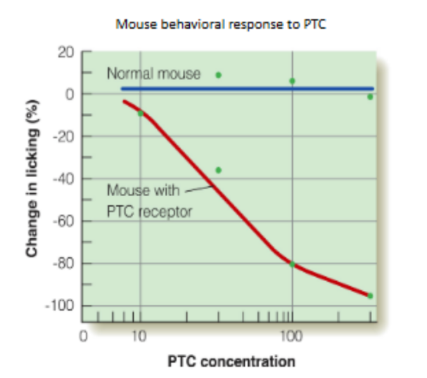
describe the experiment w/ mice and PTC
overall: evidence suggests that taste involves both __ & __ __
did genetic cloning to see if mice could be created to have the human receptor that responds to PTC
usually, mice don’t have this receptor OR respond to PTC
was successful, where cloned mice that had the human receptor for substance PTC
overall: evidence suggests that taste involves BOTH specificity & distributed coding
for flavor
def. flavor
describe (2)
flavor is the product of many __ __
__
explain how the OFC is involved in flavor & in general for the 4 sensory systems
the firing of these bimodal neurons of the OFC affects the amount of __/__ for a specific food due to the food-related odor
flavor - the combination of smell, taste, and other sensations
odor stimuli (smell stimuli) from food in the mouth will reach the olfactory mucosa through the retronasal route (from mouth → back of throat → up to nasal cavity)
the taste of most compounds is influenced by olfaction, with a few exceptions like MSG
__
flavor is the product of many sensory inputs
__
responses from taste & smell first combine in the OFC (orbital frontal cortex)
the OFC also receives input from S1 (touch) & from the IT cortex in the visual “what” pathway (vision)
..
the firing of these bimodal neurons of the OFC affects the amount of hunger/satiety for a specific food due to the food-related odor
for flavor
flavor is influenced by (2)
def. sensory-specific satiety
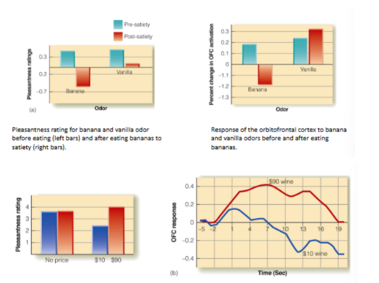
flavor is influenced by food intake & expectation
__
(top) for food intake → influence pleasantness rating & amount of OFC activation
satiety influences how pleasant a food-related odor is & the brain’s response to the odor (aka amount of OFC activation)
__
(bottom) for expectation → influence pleasantness rating & amount of OFC activation
ex: the higher the price on the wine label, the more pleasant the rating wave AND the more OFC activation there is
which of the 4 sensory systems discussed overall is the only sensory system to not involve the thalamus?
olfactory system doesn’t involve the thalamus
instead, signals don’t have to pass through the thalamus to project into the cortex
signals go directly from the olfactory bulb to the cortex (converged signals on glomeruli on olfactory bulb → primary/piriform cortex → secondary/orbitofrontal cortex)
How does brief exposure to physical energy lead to a vivid perceptual experience of the world around you?
It’s NOT about the physical energy we care about, but the __ it carries
Perception is __ & __
These processes are __ by the physiology of each sensory system
…
Perception is __ & __
It’s not about the physical energy we care about, but the information it carries
Perception is active & inferential
These processes are constrained by the physiology of each sensory system
…
Perception is reliable & malleable
(able to control, improve, impair, and change perception)
state the whole pathway for nociception/pain stimulus
signals from all nociceptors
→ into dorsal root ganglion of SC/into SC
→ SPINOTHALAMIC TRACT (crosses over at SC)
→ up into subcortical areas (ventrolateral nucleus of thalamus, hypothalamus, limbic systems)
→ into cortical areas (S1 and S2 of somatosensory cortex, insula, anterior cingulate cortex)
perception of pain is influenced by (4)
cognition (hypnosis)
emotion (hand in hot water, hypnosis to change unpleasantness of pain)
expectation (placebo effect)
seeing others in pain (Singer et al.)
for specificity coding
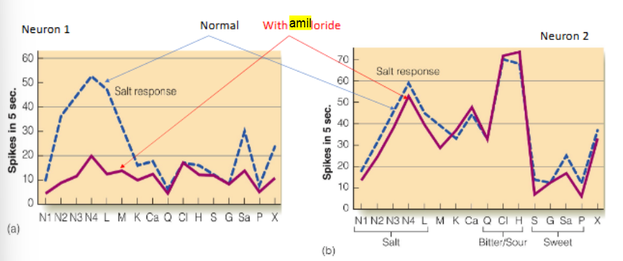
relate the experiment of Amiloride w/ salty compounds
applying Amiloride (sodium channel blocker) to the tongue will inhibit response in NST (nucleus of solitary tract) neurons that selectively respond to salty compounds, not for other NST neurons that are less/not selective to salty compounds