Protozoa Identification
1/123
Earn XP
Description and Tags
Prelims to midterm (SOURCE: CDC) | jcc
Name | Mastery | Learn | Test | Matching | Spaced | Call with Kai |
|---|
No analytics yet
Send a link to your students to track their progress
124 Terms
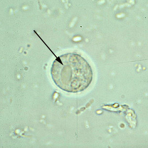
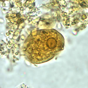
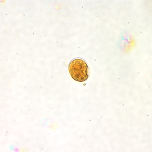
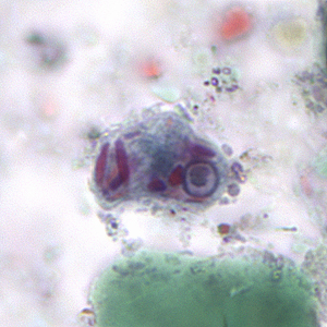
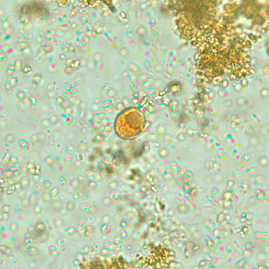
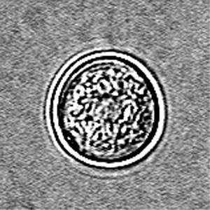
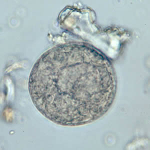
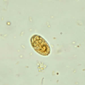
Entamoeba histolytica/ Entamoeba dispar
CYST
chromatoid bodies with blunt, rounded ends (arrow)
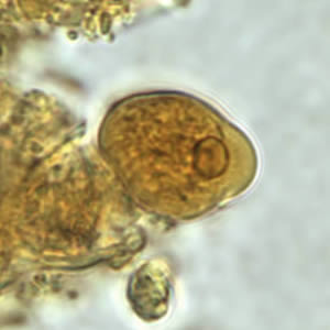
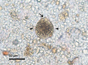
Entamoeba histolytica/ Entamoeba dispar
CYST
chromatoid body with blunt, rounded ends (arrow).
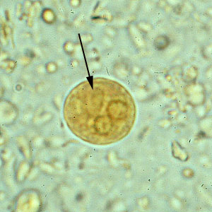
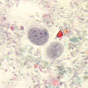
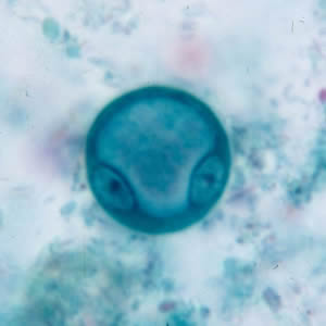
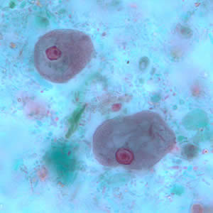
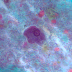
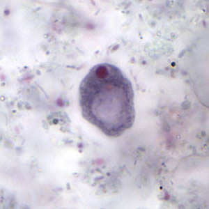
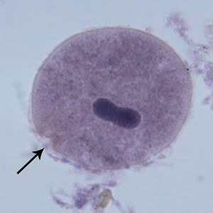
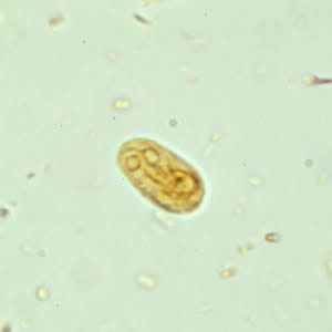
Entamoeba histolytica/ Entamoeba dispar
CYST
3 nuclei visible, chromatoid body present
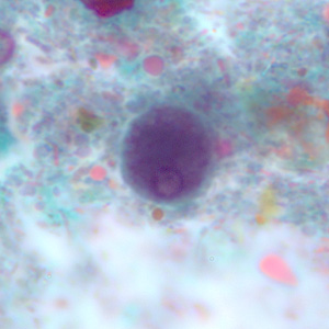
Entamoeba histolytica/ Entamoeba dispar
IMMATURE CYST
cyst has large vacuoles and the chromatin around the nucleus is clumpy.
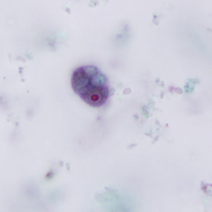
Entamoeba histolytica/ Entamoeba dispar
TROPHOZOITE
DFS, Iodine
Entamoeba histolytica/ Entamoeba dispar
TROPHOZOITE
DFS, Iodine
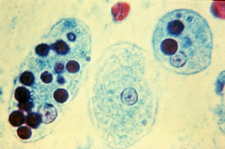
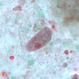
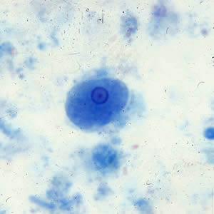
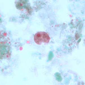
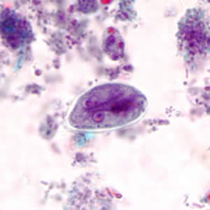
Entamoeba histolytica
TROPHOZOITE
with ingested erythrocytes (dark inclusions)
Entamoeba histolytica
TROPHOZOITE
with ingested erythrocytes (arrow), DIC microscopy
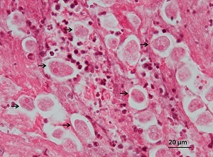
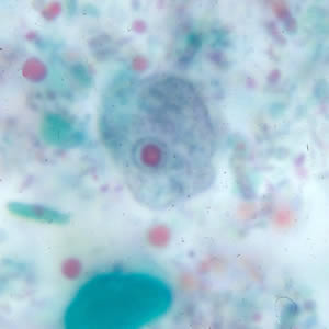
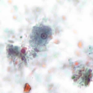
Entamoeba histolytica
TROPHOZOITE
rectal biopsy
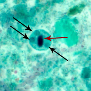
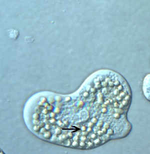
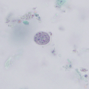
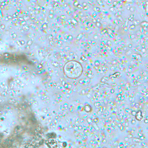
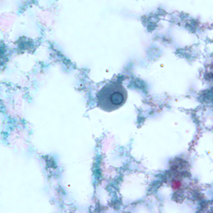
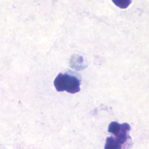
Endolimax nana
CYST
Iodine stained, nuclei visible
Endolimax nana
CYST
unstained, nuclei not visible
Endolimax nana
CYST
trichrome stained
Endolimax nana
CYST
trichrome stained
Endolimax nana
TROPHOZOITE
trichrome

Endolimax nana
TROPHOZOITE
trichrome
Endolimax nana
TROPHOZOITE
trichrome
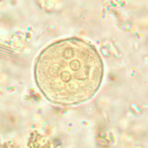
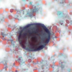
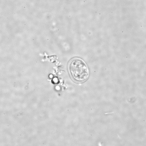
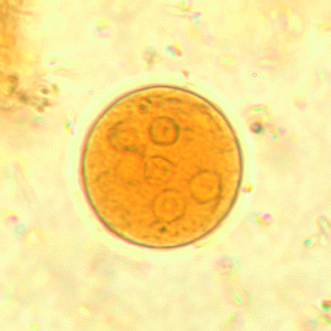
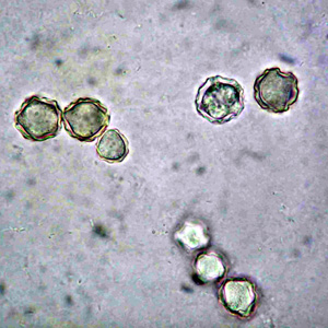
Entamoeba coli
CYST
unstained. six nuclei visible
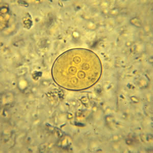
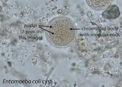
Entamoeba coli
CYST
iodine stained, seven nuclei visible
Entamoeba coli
CYST
iodine stained, five nuclei visible
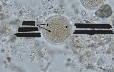
Entamoeba coli
CYST
chromatoidal body visible
Entamoeba coli
IMMATURE CYST
two nuclei visible, large glycogen vacuole
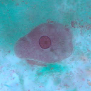
Entamoeba coli
TROPHOZOITE
trichrome
Entamoeba coli
TROPHOZOITE
trichrome
Entamoeba coli
TROPHOZOITE
trichrome
Entamoeba coli
TROPHOZOITE
trichrome
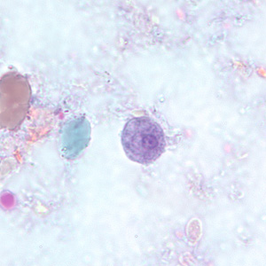
Entamoeba hartmanni
CYST
Iodine
Entamoeba hartmanni
CYST
trichrome
Entamoeba hartmanni
CYST
trichrome
Entamoeba hartmanni
TROPHOZOITE
trichrome
Entamoeba hartmanni
TROPHOZOITE
trichrome
Entamoeba hartmanni
TROPHOZOITE
trichrome
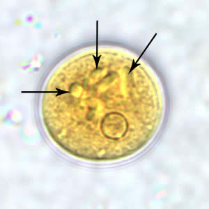
Entamoeba polecki
CYST
Iodine, numerous chromatoid bodies (arrows)
Entamoeba polecki
CYST
trichrome, large nucleus with a pleomorphic karyosome
numerous chromatoid bodies
Entamoeba polecki
TROPHOZOITE
trichrome
Entamoeba polecki
TROPHOZOITE
trichrome
Iodamoeba butschlii
CYST
unstained, glycogen vacuole seen
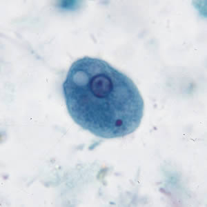
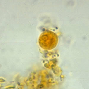
Iodamoeba butschlii
CYST
Iodine stained, glycogen vacuole seen as a dark-staining mass
Iodamoeba butschlii
CYST
trichrome stained, nucleus and large vacuole seen
Iodamoeba butschlii
TROPHOZOITE
trichrome,
Entamoeba gingivalis
TROPHOZOITE
Entamoeba gingivalis
TROPHOZOITE
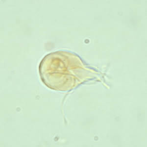
Naegleria fowleri
CYST
Naegleria fowleri
AMEBOFLAGELLATE TROPHOZOITE
Naegleria fowleri
TROPHOZOITE
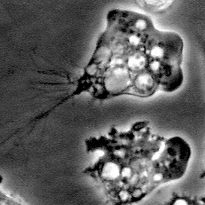
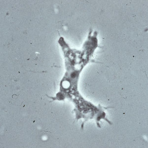
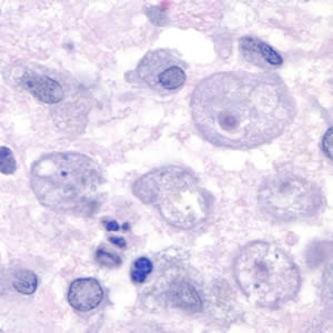
Acanthamoeba spp.
CYST
Acanthamoeba spp.
CYST
brain tissue
Acanthamoeba spp.
TROPHOZOITE
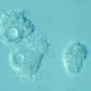
Acanthamoeba spp.
TROPHOZOITE
Acanthamoeba spp.
TROPHOZOITE
corneal scraping
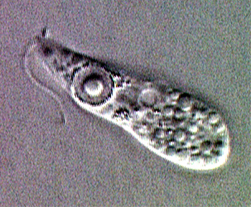
Balantidium coli
CYST

Balantidium coli
TROPHOZOITE
visible cilia
Balantidium coli
TROPHOZOITE
cytostome (black arrow)
Balantidium coli
TROPHOZOITE
cytostome (arrow), cilia (darts)
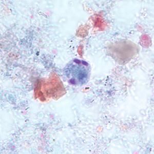
Giardia duodenalis
CYST
iodine
Giardia duodenalis
CYST
iodine
Giardia duodenalis
CYST
trichrome
Giardia duodenalis
TROPHOZOITE
Iodine, wet mount
Giardia duodenalis
TROPHOZOITE
geimsa
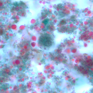
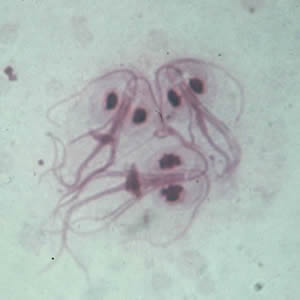
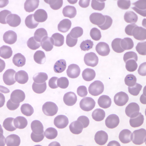
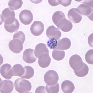
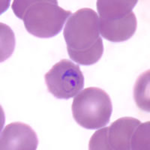
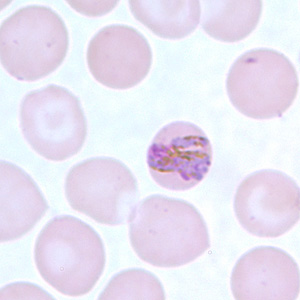
Babesia spp.
TETRAD
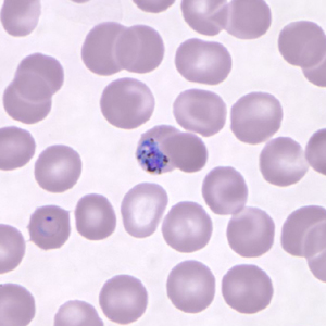
Babesia spp.
TETRAD, AMEBOID TROPHOZOITE
Babesia spp.
TETRAD
PAIRS ALIGNED
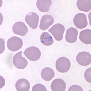
Babesia spp.
EXTRACELLULAR FORM
indicative of Babesia
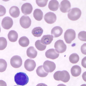
Babesia spp.
RING, TETRAD
vacuole
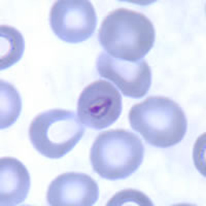
Leishmania spp.
AMASTIGOTES
Leishmania spp.
AMASTIGOTES
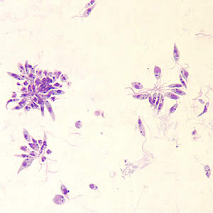
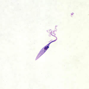
Leishmania spp.
PROMASTIGOTES
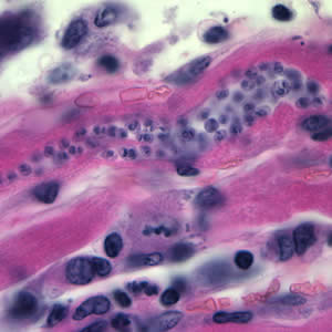
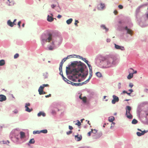
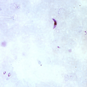
Trypanosoma cruzi
AMASTIGOTES
heart tissue
Trypanosoma cruzi
TRYPOMASTIGOTE
CSF sample
Trypanosoma cruzi
EPIMASTIGOTE
kinetoplast anterior to the nucleus
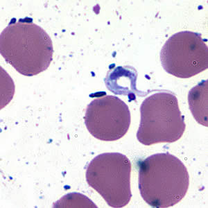
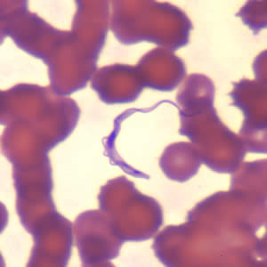
Trypanosoma cruzi
TRYPOMASTIGOTE
Trypanosoma cruzi
TRYPOMASTIGOTE
typical C-shape of the trypomastigote that characterizes T. cruzi in fixed blood smears
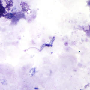
Trypanosoma brucei subsp.
TRYPOMASTIGOTE
Trypanosoma brucei subsp.
TRYPOMASTIGOTE
Trypanosoma brucei subsp.
TRYPOMASTIGOTE
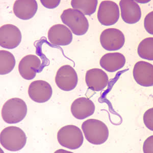
Trypanosoma brucei subsp.
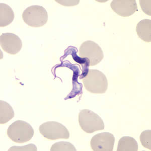
TRYPOMASTIGOTE
beginning to divide
dividing forms are seen in African trypanosomes, but not in American trypanosomes
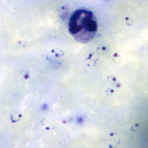
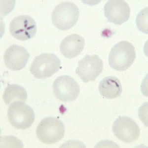
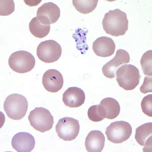
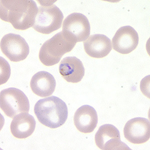
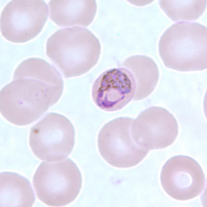
Plasmodium falciparum
RINGS
Plasmodium falciparum
RINGS
exhibiting Maurer's clefts
Plasmodium falciparum
TROPHOZOITES
Plasmodium falciparum
TROPHOZOITES
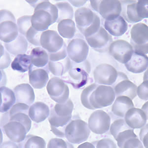
Plasmodium falciparum
GAMETOCYTES
Plasmodium falciparum
GAMETOCYTES
Plasmodium falciparum
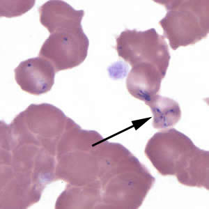
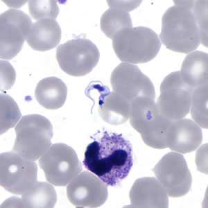
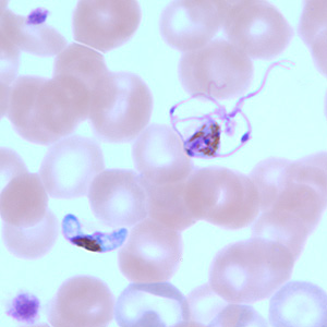
GAMETOCYTES
The gametocyte in the upper right is undergoing exflagellation, a process that normally occurs in the mid-gut of the mosquito host.
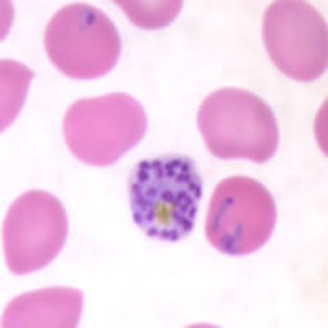
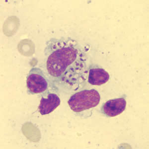
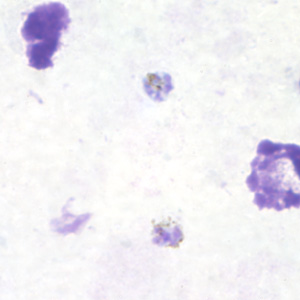
Plasmodium falciparum
SCHIZONTS
Trophozoites are also seen
Plasmodium falciparum
SCHIZONTS
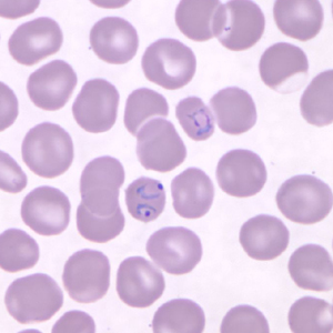
Plasmodium knowlesi
RINGS
RBC multiply-infected
Plasmodium knowlesi
RINGS
RBC multiply-infected
Plasmodium knowlesi
TROPHOZOITES
Plasmodium knowlesi
TROPHOZOITES, RING
Plasmodium knowlesi
GAMETOCYTES

Plasmodium knowlesi
GAMETOCYTES
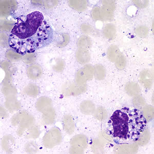
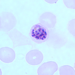
Plasmodium knowlesi
SCHIZONTS
Plasmodium knowlesi
SCHIZONTS
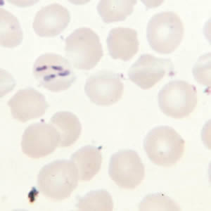
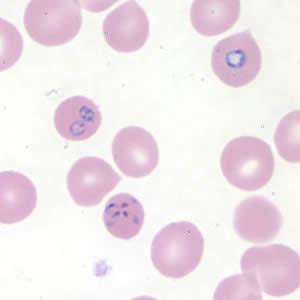
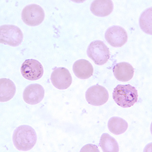
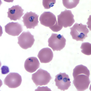
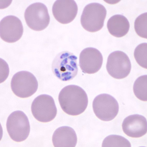
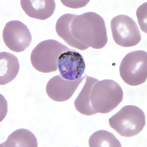
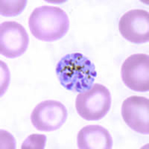
Plasmodium malariae
RING FORM TROPHOZOITE
Birds-eye
Plasmodium malariae
RING FORM TROPHOZOITE
Plasmodium malariae
DEVELOPING TROPHOZOITE
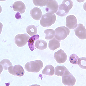
Plasmodium malariae
MATURE TROPHOZOITE/ BAND FORM
Plasmodium malariae
MATURE TROPHOZOITE/ BASKET FORM
Plasmodium malariae
GAMETOCYTES
Plasmodium malariae
GAMETOCYTES
Plasmodium malariae
SCHIZONTS
rosette cluster